43
views
0
recommends
+1 Recommend
0 collections
    0
    shares
      • Record: found
      • Abstract: found
      • Article: found
      Is Open Access

      GATA5 mutation homozygosity linked to a double outlet right ventricle phenotype in a Lebanese patient

      research-article

      Read this article at

      Bookmark
          There is no author summary for this article yet. Authors can add summaries to their articles on ScienceOpen to make them more accessible to a non-specialist audience.

          Abstract

          Background

          GATA transcription factors are evolutionary conserved zinc finger proteins with multiple roles in cell differentiation/proliferation and organogenesis. GATA5 is only transiently expressed in the embryonic heart, and the inactivation of both Gata5 alleles results in a partially penetrant bicuspid aortic valve ( BAV) phenotype in mice. We hypothesized that only biallelic mutations in GATA5 could be disease causing.

          Methods

          A total of 185 patients with different forms of congenital heart disease ( CHD) were screened along 150 healthy individuals for GATA4, 5 , and 6. All patients' phenotypes were diagnosed with echocardiography.

          Results

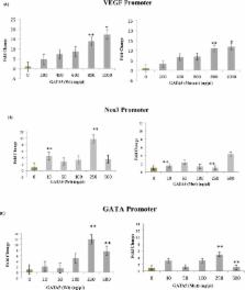
          Sequencing results revealed eight missense variants (three of which are novel) in cases with various conotruncal and septal defects. Out of these, two were inherited in recessive forms: the p.T67P variant, which was found both in patients and in healthy individuals, and the previously described p.Y142H variant which was only found in a patient with a double outlet right ventricle ( DORV). We characterized the p.Y142H variant and showed that it significantly reduced the transcriptional activity of the protein over cardiac promoters by 30–40%.

          Conclusion

          Our results do prove that p.Y142H is associated with DORV and suggests including GATA5 as a potential gene to be screened in patients with this phenotype.

          Related collections

          Most cited references43

          • Record: found
          • Abstract: found
          • Article: not found

          Genetics of congenital heart disease: the glass half empty.

          Congenital heart disease (CHD) is the most common congenital anomaly in newborn babies. Cardiac malformations have been produced in multiple experimental animal models, by perturbing selected molecules that function in the developmental pathways involved in myocyte specification, differentiation, or cardiac morphogenesis. In contrast, the precise genetic, epigenetic, or environmental basis for these perturbations in humans remains poorly understood. Over the past few decades, researchers have tried to bridge this knowledge gap through conventional genome-wide analyses of rare Mendelian CHD families, and by sequencing candidate genes in CHD cohorts. Although yielding few, usually highly penetrant, disease gene mutations, these discoveries provided 3 notable insights. First, human CHD mutations impact a heterogeneous set of molecules that orchestrate cardiac development. Second, CHD mutations often alter gene/protein dosage. Third, identical pathogenic CHD mutations cause a variety of distinct malformations, implying that higher order interactions account for particular CHD phenotypes. The advent of contemporary genomic technologies including single nucleotide polymorphism arrays, next-generation sequencing, and copy number variant platforms are accelerating the discovery of genetic causes of CHD. Importantly, these approaches enable study of sporadic cases, the most common presentation of CHD. Emerging results from ongoing genomic efforts have validated earlier observations learned from the monogenic CHD families. In this review, we explore how continued use of these technologies and integration of systems biology is expected to expand our understanding of the genetic architecture of CHD.
            Bookmark
            • Record: found
            • Abstract: found
            • Article: not found

            Requirement of the transcription factor GATA4 for heart tube formation and ventral morphogenesis.

            The zinc finger transcription factor GATA4 has been implicated in heart development based on its early expression in precardiogenic splanchnic mesoderm and its ability to activate the expression of a number of cardiac-specific genes. To determine the role of GATA4 in embryogenesis, we generated mice homozygous for a GATA4 null allele. Homozygous GATA4 null mice arrested in development between E7.0 and E9.5 because of severe developmental abnormalities. Mutant embryos most notably lacked a primitive heart tube and foregut and developed partially outside the yolk sac. In the mutants, the two bilaterally symmetric promyocardial primordia failed to migrate ventrally but instead remained lateral and generated two independent heart tubes that contained differentiated cardiomyocytes. We show that these deformities resulted from a general loss in lateral to ventral folding throughout the embryo. GATA4 is most highly expressed within the precardiogenic splanchnic mesoderm at the posterior lip of the anterior intestinal portal, corresponding to the region of the embryo that undergoes ventral fusion. We propose that GATA4 is required for the migration or folding morphogenesis of the precardiogenic splanchnic mesodermal cells at the level of the AIP.
              Bookmark
              • Record: found
              • Abstract: found
              • Article: not found

              GATA6 regulates HNF4 and is required for differentiation of visceral endoderm in the mouse embryo.

              GATA6 belongs to a family of zinc finger transcription factors that play important roles in transducing nuclear events that regulate cellular differentiation and embryonic morphogenesis in vertebrate species. To examine the function of GATA6 during embryonic development, gene targeting was used to generate GATA6-deficient (GATA6(-/-)) ES cells and mice harboring a null mutation in GATA6. Differentiated embryoid bodies derived from GATA6(-/-) ES cells lack a covering layer of visceral endoderm and severely attenuate, or fail to express, genes encoding early and late endodermal markers, including HNF4, GATA4, alpha-fetoprotein (AFP), and HNF3beta. Homozygous GATA6(-/-) mice died between embryonic day (E) 6.5 and E7. 5 and exhibited a specific defect in endoderm differentiation including severely down-regulated expression of GATA4 and absence of HNF4 gene expression. Moreover, widespread programmed cell death was observed within the embryonic ectoderm of GATA6-deficient embryos, a finding also observed in HNF4-deficient embryos. Consistent with these data, forced expression of GATA6 activated the HNF4 promoter in nonendodermal cells. Finally, to examine the function of GATA6 during later embryonic development, GATA6(-/-)-C57BL/6 chimeric mice were generated. lacZ-tagged GATA6(-/-) ES cells contributed to all embryonic tissues with the exception of the endodermally derived bronchial epithelium. Taken together, these data suggest a model in which GATA6 lies upstream of HNF4 in a transcriptional cascade that regulates differentiation of the visceral endoderm. In addition, these data demonstrate that GATA6 is required for establishment of the endodermally derived bronchial epithelium.
                Bookmark

                Author and article information

                Journal
                Mol Genet Genomic Med
                Mol Genet Genomic Med
                10.1002/(ISSN)2324-9269
                MGG3
                Molecular Genetics & Genomic Medicine
                John Wiley and Sons Inc. (Hoboken )
                2324-9269
                20 December 2015
                March 2016
                : 4
                : 2 ( doiID: 10.1002/mgg3.2016.4.issue-2 )
                : 160-171
                Affiliations
                [ 1 ] Department of Biochemistry and Molecular GeneticsAmerican University of Beirut BeirutLebanon
                [ 2 ] Department of BiochemistryUniversity of Ottawa Ottawa OntarioCanada
                [ 3 ] Department of Genetics Harvard Medical School and Department of Internal MedicineMassachusetts General Hospital Boston Massachusetts
                [ 4 ] Department of SurgeryAmerican University of Beirut BeirutLebanon
                [ 5 ] Department of Pediatrics and Adolescent MedicineAmerican University of Beirut BeirutLebanon
                Author notes
                [*] [* ] Correspondence

                Georges Nemer, Department of Biochemistry and Molecular Genetics, American University of Beirut, Bliss Street, P.O. Box: 11‐0236, Beirut, Lebanon. Tel: +9611350000; Fax: +9611744474; E‐mail: gn08@ 123456aub.edu.lb

                [†]

                These authors contributed equally to the work.

                Article
                MGG3190
                10.1002/mgg3.190
                4799877
                27066509
                98653a54-35e1-45ad-9f2e-2fcf5b4cf1b3
                © 2015 The Authors. Molecular Genetics & Genomic Medicine published by Wiley Periodicals, Inc.

                This is an open access article under the terms of the Creative Commons Attribution License, which permits use, distribution and reproduction in any medium, provided the original work is properly cited.

                History
                : 07 October 2015
                : 04 November 2015
                : 04 November 2015
                Page count
                Pages: 12
                Funding
                Funded by: Medical Practice Plan (MPP)
                Funded by: Universiy Research Board (URB)
                Funded by: American University of Beirut
                Categories
                Original Article
                Original Articles
                Custom metadata
                2.0
                mgg3190
                March 2016
                Converter:WILEY_ML3GV2_TO_NLMPMC version:4.8.5 mode:remove_FC converted:20.03.2016

                congenital,gata5,heart,homozygous,recessive,transcription
                congenital, gata5, heart, homozygous, recessive, transcription

                Comments

                Comment on this article