0
views
0
recommends
+1 Recommend
0 collections
    0
    shares
      • Record: found
      • Abstract: found
      • Article: found
      Is Open Access

      Masson's Tumor as an Uncommon Cause of Neck Mass: A Case Presentation

      case-report

      Read this article at

      Bookmark
          There is no author summary for this article yet. Authors can add summaries to their articles on ScienceOpen to make them more accessible to a non-specialist audience.

          Abstract

          Background

          Masson’s tumor, commonly referred to as intravascular papillary endothelial hyperplasia (IPEH), is an uncommon growth of endothelial cells within a vessel wall that is frequently assumed to indicate an abnormal resolution of thrombosis. IPEH is most typically found in the extremities however it is rare for IPEH to appear as a neck tumor. The issue with IPEH is that it could clinically, radiologically, and pathologically imitate some malignant neoplasms such as angiosarcomas creating a diagnostic challenge.

          Case Report

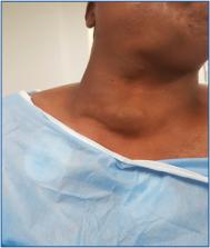
          We describe a 21-year-old male patient who presented with right anterolateral neck swelling for 12 months. Ultrasound revealed a 9.0 × 8.0 cm well-defined echogenic hyper-vascular lesion. The contrast computed tomography (CT) scan of the neck revealed an oval, well-defined subcutaneous mass, measuring 9 × 4.5 cm, situated over and separable from the right sternocleidomastoid muscle with no significant enhancement in the post-contract study. T1-weighted and T2-weighted MRI revealed a 10 × 9 × 7 cm well-defined subcutaneous lobulated lesion superficial to the sternocleidomastoid expanding upward to the Rt. side of the cheek and below to the suprasternal region, eliciting an intermediate signal in T1 and a heterogenous bright signal (mostly fluid) in T2 with low signal foci within the mass. The decision had been reached to entirely excise the lesion surgically with safety margins for histological evaluation. Histological examination indicated thrombosed variable-sized ectatic vascular spaces with papillary formations related to the thrombus, covered with a single layer of flat endothelium, and no features suggestive of malignancy. There was no recurrence at 18 months follow-up post-surgery.

          Conclusion

          Masson's tumor is a benign intravascular disease with an unclear origin and no confirmed inheritance pattern. Presentation of Masson’s tumor as a neck mass is incredibly uncommon. Masson's tumor lacks a distinct or distinguishing clinical and radiological appearance. Histopathologic examination is the sole definitive way for diagnosing the disease and the only tool for distinguishing it from angiosarcoma. Surgical excision is the best treatment for IPEH. Recurrence is extremely rare.

          Related collections

          Most cited references17

          • Record: found
          • Abstract: found
          • Article: not found

          Intravascular papillary endothelial hyperplasia. A clinicopathologic study of 91 cases.

          Ninety-one cases of intravascular papillary endothelial hyperplasia were studied clinically and histologically. This peculiar benign process, occasionally resembling hemangiosarcoma, was subgrouped in the following categories: a pure form that occurs within a dilated vascular space (30 cases), a mixed form that appears as a focal change in a hemangioma (55 cases), a third form (six cases) that belongs to neither of the first two. In the pure form, the lesions were most frequently situated in the subcutis of fingers (14 cases), of the head and neck (seven cases), and in the region between the elbows and hands (six cases). In the mixed form, half of the accompanying hemangiomas were intramuscular in no particular predilective sites. Papillary proliferation of endothelial cells was commonly found to be closely associated with thrombotic material and seemed to be an unusual feature of a thrombus undergoing organization.
            Bookmark
            • Record: found
            • Abstract: found
            • Article: not found

            Intravascular papillary endothelial hyperplasia.

            Papillary endothelial hyperplasia is a peculiar benign intravascular process that bears a remarkable resemblance to a hemangiosarcoma. In 44 cases of this lesion studied from the files of the Armed Forces Institute of Pathology, the process manifested as a small tumor-like lesion that occurred most frequently in the subcutis of the fingers (14 cases), the head and neck region (ten), and the trunk (seven). Microscopically, the tuft-like or papillary proliferation of endothelial cells was nearly always intimately associated with a thrombus and seemed to represent a peculiar variant of an organizing process. Features that aided in recognition and differential diagnosis from a hemangio-sarcoma included the intraluminal location of the lesion, the absence of tissue necrosis, and the intimate association of the proliferated tuft-like structures with thrombotic material. Follow-up information obtained in 31 cases indicated a benign clinical course despite the sarcoma-like microscopic appearance of this condition.
              Bookmark
              • Record: found
              • Abstract: found
              • Article: not found

              Masson's "vegetant intravascular hemangioendothelioma:" a lesion often mistaken for angiosarcoma: study of seventeen cases located in the skin and soft tissues.

              The occurrence of Masson's "hemangio-endotheliome vegetant intravasculaire" (Masson's pseudoangiosarcoma) in the skin and soft tissues is illustrated with 17 surgically excised specimens. Two forms are recognized; it may appear either as a pure lesion or as a focal condition in a pre-existing vascular process, such as pyogenic granuloma or hemangioma. The clinical appearance is not specific and the diagnosis can only be established by microscopic examination. It shows a predilection for the head and extremities. Its characteristic morphologic appearance makes possible its differentiation from a group of benign and malignant vascular proliferations. The key microscopic feature is the presence of a papillary growth composed of hyperplastic endothelial cells supported by delicate fibrous stalks entirely confined within the vascular lumen. The lesion should not be mistaken for angiosarcoma, since its clinical behavior is invariably benign.
                Bookmark

                Author and article information

                Journal
                Vasc Endovascular Surg
                Vasc Endovascular Surg
                spves
                VES
                Vascular and Endovascular Surgery
                SAGE Publications (Sage CA: Los Angeles, CA )
                1538-5744
                1938-9116
                14 November 2023
                April 2024
                : 58
                : 4
                : 405-409
                Affiliations
                [1 ]Department of Vascular Surgery, Faculty of Medicine (for Girls), Ringgold 68820, universityAl-Azhar University; , Cairo, Egypt
                [2 ]Pathology Department, Faculty of Medicine (for Girls), Ringgold 68820, universityAl-Azhar University; , Cairo, Egypt
                [3 ]Department of Radiodiagnosis, Faculty of Medicine (for girls), Ringgold 68820, universityAl-Azhar University; , Cairo, Egypt
                [4 ]Vascular Surgery Department, Ringgold 195495, universityAl-Azhar University Hospitals; , Assiut, Egypt
                Author notes
                [*]Ehab M. Abdo, MD, Department of Vascular Surgery, Faculty of Medicine (for Girls), Al-Azhar University, Youssef Abbas Street, Nasr City, Cairo 11884, Egypt. Email: ehababdo48@ 123456yahoo.com
                Author information
                https://orcid.org/0000-0001-6190-1230
                https://orcid.org/0000-0002-2025-6172
                Article
                10.1177_15385744231215102
                10.1177/15385744231215102
                10996301
                37962479
                9858c55a-a407-4b58-bbca-db03dab1c5f8
                © The Author(s) 2023

                This article is distributed under the terms of the Creative Commons Attribution-NonCommercial 4.0 License ( https://creativecommons.org/licenses/by-nc/4.0/) which permits non-commercial use, reproduction and distribution of the work without further permission provided the original work is attributed as specified on the Sage and Open Access page ( https://us.sagepub.com/en-us/nam/open-access-at-sage).

                History
                Categories
                Case Reports
                Custom metadata
                ts10

                masson's tumor,intravascular papillary endothelial hyperplasia,vascular tumor,cervical tumor,neck tumor

                Comments

                Comment on this article