42
views
0
recommends
+1 Recommend
0 collections
    0
    shares
      • Record: found
      • Abstract: found
      • Article: found
      Is Open Access

      Implications of False Negative and False Positive Diagnosis in Lymph Node Staging of NSCLC by Means of 18F-FDG PET/CT

      research-article

      Read this article at

      Bookmark
          There is no author summary for this article yet. Authors can add summaries to their articles on ScienceOpen to make them more accessible to a non-specialist audience.

          Abstract

          Background

          Integrated 18F-fluorodeoxyglucose positron emission tomography/computed tomography ( 18F-FDG PET/CT) is widely performed in hilar and mediastinal lymph node (HMLN) staging of non-small cell lung cancer (NSCLC). However, the diagnostic efficiency of PET/CT remains controversial. This retrospective study is to evaluate the accuracy of PET/CT and the characteristics of false negatives and false positives to improve specificity and sensitivity.

          Methods

          219 NSCLC patients with systematic lymph node dissection or sampling underwent preoperative PET/CT scan. Nodal uptake with a maximum standardized uptake value (SUVmax) >2.5 was interpreted as PET/CT positive. The results of PET/CT were compared with the histopathological findings. The receiver operating characteristic (ROC) curve was generated to determine the diagnostic efficiency of PET/CT. Univariate and multivariate analysis were conducted to detect risk factors of false negatives and false positives.

          Results

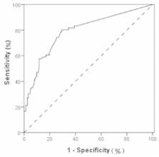
          The sensitivity, specificity, positive predictive value (PPV), negative predictive value (NPV), and accuracy of PET/ CT in detecting HMLN metastases were 74.2% (49/66), 73.2% (112/153), 54.4% (49/90), 86.8% (112/129), and 73.5% (161/219). The ROC curve had an area under curve (AUC) of 0.791 (95% CI 0.723-0.860). The incidence of false negative HMLN metastases was 13.2% (17 of 129 patients). Factors that are significantly associated with false negatives are: concurrent lung disease or diabetes (p<0.001), non-adenocarcinoma (p<0.001), and SUVmax of primary tumor >4.0 (p=0.009). Postoperatively, 45.5% (41/90) patients were confirmed as false positive cases. The univariate analysis indicated age > 65 years old (p=0.009), well differentiation (p=0.002), and SUVmax of primary tumor ≦4.0 (p=0.007) as risk factors for false positive uptake.

          Conclusion

          The SUVmax of HMLN is a predictor of malignancy. Lymph node staging using PET/CT is far from equal to pathological staging account of some risk factors. This study may provide some aids to pre-therapy evaluation and decision-making.

          Related collections

          Most cited references16

          • Record: found
          • Abstract: found
          • Article: not found

          Clinical applications of PET in oncology.

          Positron emission tomography (PET) provides metabolic information that has been documented to be useful in patient care. The properties of positron decay permit accurate imaging of the distribution of positron-emitting radiopharmaceuticals. The wide array of positron-emitting radiopharmaceuticals has been used to characterize multiple physiologic and pathologic states. PET is used for characterizing brain disorders such as Alzheimer disease and epilepsy and cardiac disorders such as coronary artery disease and myocardial viability. The neurologic and cardiac applications of PET are not covered in this review. The major utilization of PET clinically is in oncology and consists of imaging the distribution of fluorine 18 fluorodeoxyglucose (FDG). FDG, an analogue of glucose, accumulates in most tumors in a greater amount than it does in normal tissue. FDG PET is being used in diagnosis and follow-up of several malignancies, and the list of articles supporting its use continues to grow. In this review, the physics and instrumentation aspects of PET are described. Many of the clinical applications in oncology are mature and readily covered by third-party payers. Other applications are being used clinically but have not been as carefully evaluated in the literature, and these applications may not be covered by third-party payers. The developing applications of PET are included in this review.
            Bookmark
            • Record: found
            • Abstract: found
            • Article: not found

            Patterns of disease recurrence after stereotactic ablative radiotherapy for early stage non-small-cell lung cancer: a retrospective analysis.

            Stereotactic ablative radiotherapy (SABR) is increasingly used in the treatment of medically inoperable early stage non-small-cell lung cancer (NSCLC). Because patterns of late disease recurrence after SABR are not well characterised, we aimed to assess these outcomes in a cohort of patients with NSCLC. Patients with (18)F-fluorodeoxyglucose ((18)F-FDG)-PET confirmed stage 1-2 NSCLC who were treated with SABR at the VU University Medical Center (Amsterdam, Netherlands) were identified from an institutional database. SABR doses were 54-60 Gy, delivered in three to eight once-daily fractions, depending on tumour size and location. Clinical follow-up and CT scans were done at 3, 6, and 12 months, then yearly thereafter. (18)F-FDG-PET restaging was only done when clinically indicated. Initial sites of recurrence were classified as local, regional, and distant, and were differentiated from second primary tumours in the lung at multidisciplinary tumour board review. Between April 4, 2003, and Dec 5, 2011, 676 patients were treated with SABR and were eligible for assessment of recurrence. The median follow-up was 32·9 months (IQR 14·9-50·9 months). 124 (18%) of 676 patients had disease recurrence. Actuarial 2-year rates of local, regional, and distant recurrence were 4·9% (95% CI 2·7-7·1), 7·8% (5·3-10·3), and 14·7% (11·4-18·0), respectively. Corresponding 5-year rates were 10·5% (95% CI 6·4-14·6), 12·7% (8·4-17·0), and 19·9% (14·9-24·6), respectively. Of the 124 recurrences, 82 (66%) were distant recurrences and 57 (46%) were isolated distant recurrences. Isolated locoregional recurrences occurred in the remaining 42 patients with disease recurrence (34%), 35 (83%) of whom did not develop subsequent distant recurrence. The median times to local, regional, and distant recurrence were 14·9 months (95% CI 11·4-18·4), 13·1 months (7·9-18·3), and 9·6 months (6·8-12·4), respectively. New pulmonary lesions characterised as second primary tumours in the lung developed in 42 (6%) of 676 patients at a median of 18·0 months (95% CI 12·5-23·5) after SABR. Late recurrences after SABR are infrequent and two distinct patterns account for most cases. The predominant pattern is out-of-field, isolated distant recurrence presenting early, despite initial PET staging. A third of patients develop isolated locoregional recurrence; for these patients standardised follow-up is important to ensure that appropriate salvage treatments are considered. None. Copyright © 2012 Elsevier Ltd. All rights reserved.
              Bookmark
              • Record: found
              • Abstract: found
              • Article: not found

              Non-small cell lung cancer: prospective comparison of integrated FDG PET/CT and CT alone for preoperative staging.

              To evaluate prospectively the accuracy of integrated positron emission tomography (PET) and computed tomography (CT) with use of fluorodeoxyglucose (FDG), compared with that of stand-alone CT, for the preoperative staging of non-small cell lung cancer, with surgical and histologic findings used as the reference standard. Institutional review board approval and patient informed consent were obtained. From November 2003 to February 2004, 106 patients (78 men, 28 women; mean age, 56 years) with non-small cell lung cancer underwent curative surgical resection (tumor resection and lymph node dissection) after stand-alone CT followed by integrated FDG PET/CT. Tumor stages were determined by using the TNM and American Joint Committee on Cancer staging systems. Histopathologic results served as the reference standard. Statistically significant differences in tumor staging between integrated PET/CT and stand-alone CT were determined with P < .05 obtained by using the McNemar test or with a generalized estimating equation. The primary tumor was correctly staged in 84 patients (79%) at stand-alone CT and in 91 patients (86%) at integrated FDG PET/CT (P = .25). For the depiction of malignant nodes, the sensitivity, specificity, and accuracy of CT were 70% (23 of 33 nodal groups), 69% (248 of 360), and 69% (271 of 393), respectively, whereas those of PET/CT were 85% (28 of 33), 84% (302 of 360), and 84% (330 of 393) (P = .25, P < .001, and P < .001, respectively). There were 112 false-positive interpretations at CT for 54 hilar, 16 subcarinal, 29 paratracheal, 10 subaortic, and two pulmonary ligament nodal groups and one upper paratracheal group, compared with only 58 false-positive interpretations at PET/CT for 32 hilar, seven subcarinal, 13 lower paratracheal, and six subaortic nodal groups. There were 10 false-negative interpretations at CT for four hilar, two lower paratracheal, and two subcarinal nodal groups, one prevascular and retrotracheal group, and one inferior pulmonary group, but only five false-negative interpretations at PET/CT (one each for paratracheal, subaortic, subcarinal, inferior pulmonary, and hilar nodal groups). Integrated FDG PET/CT is significantly better than stand-alone CT for lung cancer staging and provides enhanced accuracy and specificity in nodal staging.
                Bookmark

                Author and article information

                Contributors
                Role: Editor
                Journal
                PLoS One
                PLoS ONE
                plos
                plosone
                PLoS ONE
                Public Library of Science (San Francisco, USA )
                1932-6203
                2013
                25 October 2013
                : 8
                : 10
                : e78552
                Affiliations
                [1]Key Laboratory of Carcinogenesis and Translational Research (Ministry of Education), Thoracic Surgery II, Peking University Cancer Hospital & Institute, Beijing, China
                Faculty of Medicine, University of Porto, Portugal
                Author notes

                Competing Interests: The authors have declared that no competing interests exist.

                Conceived and designed the experiments: SL QZ YY. Performed the experiments: SL QZ YW YF YY. Analyzed the data: SL QZ YM BZ YY. Contributed reagents/materials/analysis tools: YY. Wrote the manuscript: SL QZ YY.

                Article
                PONE-D-13-19765
                10.1371/journal.pone.0078552
                3808350
                24205256
                984657b5-e4e8-4c40-b380-c94fc8f8df35
                Copyright @ 2013

                This is an open-access article distributed under the terms of the Creative Commons Attribution License, which permits unrestricted use, distribution, and reproduction in any medium, provided the original author and source are credited.

                History
                : 13 May 2013
                : 19 September 2013
                Funding
                This work was supported partially by Beijing Natural Science Foundation (Grant Nos 11G2633), the National High Technology Research and Development Program of China (863 Program, No. 2012AA02A502), and Beijing Municipal Science & Technology Commission (No. Z111107067311018). The funders had no role in study design, data collection and analysis, decision to publish, or preparation of the manuscript.
                Categories
                Research Article

                Uncategorized
                Uncategorized

                Comments

                Comment on this article