Inviting an author to review:
Find an author and click ‘Invite to review selected article’ near their name.
Search for authorsSearch for similar articles
8
views
0
recommends
+1 Recommend
0 collections
    0
    shares
      • Record: found
      • Abstract: found
      • Article: found
      Is Open Access

      Conformational specificity of opioid receptors is determined by subcellular location irrespective of agonist

      research-article

      Read this article at

      Bookmark
          There is no author summary for this article yet. Authors can add summaries to their articles on ScienceOpen to make them more accessible to a non-specialist audience.

          Abstract

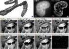
          The prevailing model for the variety in drug responses is that different drugs stabilize distinct active states of their G protein-coupled receptor (GPCR) targets, allowing coupling to different effectors. However, whether the same ligand generates different GPCR active states based on the immediate environment of receptors is not known. Here we address this question using spatially resolved imaging of conformational biosensors that read out distinct active conformations of the δ-opioid receptor (DOR), a physiologically relevant GPCR localized to Golgi and the surface in neuronal cells. We have shown that Golgi and surface pools of DOR both inhibit cAMP, but engage distinct conformational biosensors in response to the same ligand in rat neuroendocrine cells. Further, DOR recruits arrestins on the surface but not on the Golgi. Our results suggest that the local environment determines the active states of receptors for any given drug, allowing GPCRs to couple to different effectors at different subcellular locations.

          Related collections

          Most cited references78

          • Record: found
          • Abstract: found
          • Article: not found

          Fiji: an open-source platform for biological-image analysis.

          Fiji is a distribution of the popular open-source software ImageJ focused on biological-image analysis. Fiji uses modern software engineering practices to combine powerful software libraries with a broad range of scripting languages to enable rapid prototyping of image-processing algorithms. Fiji facilitates the transformation of new algorithms into ImageJ plugins that can be shared with end users through an integrated update system. We propose Fiji as a platform for productive collaboration between computer science and biology research communities.
            Bookmark
            • Record: found
            • Abstract: found
            • Article: found
            Is Open Access

            ImageJ2: ImageJ for the next generation of scientific image data

            Background ImageJ is an image analysis program extensively used in the biological sciences and beyond. Due to its ease of use, recordable macro language, and extensible plug-in architecture, ImageJ enjoys contributions from non-programmers, amateur programmers, and professional developers alike. Enabling such a diversity of contributors has resulted in a large community that spans the biological and physical sciences. However, a rapidly growing user base, diverging plugin suites, and technical limitations have revealed a clear need for a concerted software engineering effort to support emerging imaging paradigms, to ensure the software’s ability to handle the requirements of modern science. Results We rewrote the entire ImageJ codebase, engineering a redesigned plugin mechanism intended to facilitate extensibility at every level, with the goal of creating a more powerful tool that continues to serve the existing community while addressing a wider range of scientific requirements. This next-generation ImageJ, called “ImageJ2” in places where the distinction matters, provides a host of new functionality. It separates concerns, fully decoupling the data model from the user interface. It emphasizes integration with external applications to maximize interoperability. Its robust new plugin framework allows everything from image formats, to scripting languages, to visualization to be extended by the community. The redesigned data model supports arbitrarily large, N-dimensional datasets, which are increasingly common in modern image acquisition. Despite the scope of these changes, backwards compatibility is maintained such that this new functionality can be seamlessly integrated with the classic ImageJ interface, allowing users and developers to migrate to these new methods at their own pace. Conclusions Scientific imaging benefits from open-source programs that advance new method development and deployment to a diverse audience. ImageJ has continuously evolved with this idea in mind; however, new and emerging scientific requirements have posed corresponding challenges for ImageJ’s development. The described improvements provide a framework engineered for flexibility, intended to support these requirements as well as accommodate future needs. Future efforts will focus on implementing new algorithms in this framework and expanding collaborations with other popular scientific software suites. Electronic supplementary material The online version of this article (doi:10.1186/s12859-017-1934-z) contains supplementary material, which is available to authorized users.
              Bookmark
              • Record: found
              • Abstract: found
              • Article: not found

              Membrane lipids: where they are and how they behave.

              Throughout the biological world, a 30 A hydrophobic film typically delimits the environments that serve as the margin between life and death for individual cells. Biochemical and biophysical findings have provided a detailed model of the composition and structure of membranes, which includes levels of dynamic organization both across the lipid bilayer (lipid asymmetry) and in the lateral dimension (lipid domains) of membranes. How do cells apply anabolic and catabolic enzymes, translocases and transporters, plus the intrinsic physical phase behaviour of lipids and their interactions with membrane proteins, to create the unique compositions and multiple functionalities of their individual membranes?
                Bookmark

                Author and article information

                Contributors
                Role: Reviewing Editor
                Role: Senior Editor
                Journal
                eLife
                Elife
                eLife
                eLife
                eLife Sciences Publications, Ltd
                2050-084X
                20 May 2021
                2021
                : 10
                : e67478
                Affiliations
                [1 ]Cellular and Molecular Biology Program, University of Michigan Ann ArborUnited States
                [2 ]Department of Pharmacology University of Michigan Medical School Ann ArborUnited States
                Fred Hutchinson Cancer Research Centre United States
                Fred Hutchinson Cancer Research Center United States
                Fred Hutchinson Cancer Research Centre United States
                University of California, San Francisco United States
                Augusta University United States
                Oregon Health and Science University United States
                Author notes
                [†]

                Department of Pharmacology and Physiology, University of Rochester Medical Center, Rochester, United States.

                [‡]

                Department of Biochemistry and Biophysics, California Institute for Quantitative Biosciences, University of California, San Francisco, San Francisco, United States.

                Author information
                https://orcid.org/0000-0002-8151-290X
                https://orcid.org/0000-0001-7176-038X
                https://orcid.org/0000-0002-3177-4231
                Article
                67478
                10.7554/eLife.67478
                8208814
                34013886
                97ff6bc9-5256-41fa-923e-ea1908cecd7d
                © 2021, Crilly et al

                This article is distributed under the terms of the Creative Commons Attribution License, which permits unrestricted use and redistribution provided that the original author and source are credited.

                History
                : 12 February 2021
                : 19 May 2021
                Funding
                Funded by: FundRef http://dx.doi.org/10.13039/100000001, National Science Foundation;
                Award ID: DGE 1256260
                Award Recipient :
                Funded by: FundRef http://dx.doi.org/10.13039/100000057, National Institute of General Medical Sciences;
                Award ID: GM117425
                Award Recipient :
                Funded by: FundRef http://dx.doi.org/10.13039/100000001, National Science Foundation;
                Award ID: 1935926
                Award Recipient :
                The funders had no role in study design, data collection and interpretation, or the decision to submit the work for publication.
                Categories
                Research Article
                Cell Biology
                Custom metadata
                A GPCR activated by the same agonist engages different conformational biosensors at different subcellular locations, suggesting that location-based conformations drive spatial encoding of signaling.

                Life sciences
                gpcr,spatial encoding,signaling,organelle,rat
                Life sciences
                gpcr, spatial encoding, signaling, organelle, rat

                Comments

                Comment on this article

                scite_
                0
                0
                0
                0
                Smart Citations
                0
                0
                0
                0
                Citing PublicationsSupportingMentioningContrasting
                View Citations

                See how this article has been cited at scite.ai

                scite shows how a scientific paper has been cited by providing the context of the citation, a classification describing whether it supports, mentions, or contrasts the cited claim, and a label indicating in which section the citation was made.

                Similar content138

                Cited by9

                Most referenced authors1,339