0
views
0
recommends
+1 Recommend
0 collections
    0
    shares
      • Record: found
      • Abstract: found
      • Article: found
      Is Open Access

      Sulindac selectively induces autophagic apoptosis of GABAergic neurons and alters motor behaviour in zebrafish

      research-article

      Read this article at

      Bookmark
          There is no author summary for this article yet. Authors can add summaries to their articles on ScienceOpen to make them more accessible to a non-specialist audience.

          Abstract

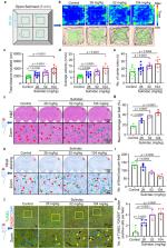
          Nonsteroidal anti-inflammatory drugs compose one of the most widely used classes of medications, but the risks for early development remain controversial, especially in the nervous system. Here, we utilized zebrafish larvae to assess the potentially toxic effects of nonsteroidal anti-inflammatory drugs and found that sulindac can selectively induce apoptosis of GABAergic neurons in the brains of zebrafish larvae brains. Zebrafish larvae exhibit hyperactive behaviour after sulindac exposure. We also found that akt1 is selectively expressed in GABAergic neurons and that SC97 (an Akt1 activator) and exogenous akt1 mRNA can reverse the apoptosis caused by sulindac. Further studies showed that sulindac binds to retinoid X receptor alpha (RXRα) and induces autophagy in GABAergic neurons, leading to activation of the mitochondrial apoptotic pathway. Finally, we verified that sulindac can lead to hyperactivity and selectively induce GABAergic neuron apoptosis in mice. These findings suggest that excessive use of sulindac may lead to early neurodevelopmental toxicity and increase the risk of hyperactivity, which could be associated with damage to GABAergic neurons.

          Abstract

          Nonsteroidal anti-inflammatory drugs (NSAIDs) are widely used but their risks for early neurodevelopment remain controversial. Here, the authors showed in zebrafish larvae that sulindac induces GABAergic neuron apoptosis through autophagy activation that leads to hyperactive behavior.

          Related collections

          Most cited references97

          • Record: found
          • Abstract: found
          • Article: not found

          Integrating single-cell transcriptomic data across different conditions, technologies, and species

          Computational single-cell RNA-seq (scRNA-seq) methods have been successfully applied to experiments representing a single condition, technology, or species to discover and define cellular phenotypes. However, identifying subpopulations of cells that are present across multiple data sets remains challenging. Here, we introduce an analytical strategy for integrating scRNA-seq data sets based on common sources of variation, enabling the identification of shared populations across data sets and downstream comparative analysis. We apply this approach, implemented in our R toolkit Seurat (http://satijalab.org/seurat/), to align scRNA-seq data sets of peripheral blood mononuclear cells under resting and stimulated conditions, hematopoietic progenitors sequenced using two profiling technologies, and pancreatic cell 'atlases' generated from human and mouse islets. In each case, we learn distinct or transitional cell states jointly across data sets, while boosting statistical power through integrated analysis. Our approach facilitates general comparisons of scRNA-seq data sets, potentially deepening our understanding of how distinct cell states respond to perturbation, disease, and evolution.
            Bookmark
            • Record: found
            • Abstract: found
            • Article: not found

            Fast, sensitive, and accurate integration of single cell data with Harmony

            The emerging diversity of single cell RNAseq datasets allows for the full transcriptional characterization of cell types across a wide variety of biological and clinical conditions. However, it is challenging to analyze them together, particularly when datasets are assayed with different technologies. Here, real biological differences are interspersed with technical differences. We present Harmony, an algorithm that projects cells into a shared embedding in which cells group by cell type rather than dataset-specific conditions. Harmony simultaneously accounts for multiple experimental and biological factors. In six analyses, we demonstrate the superior performance of Harmony to previously published algorithms. We show that Harmony requires dramatically fewer computational resources. It is the only currently available algorithm that makes the integration of ~106 cells feasible on a personal computer. We apply Harmony to PBMCs from datasets with large experimental differences, 5 studies of pancreatic islet cells, mouse embryogenesis datasets, and cross-modality spatial integration.
              Bookmark
              • Record: found
              • Abstract: found
              • Article: not found
              Is Open Access

              The zebrafish reference genome sequence and its relationship to the human genome.

              Zebrafish have become a popular organism for the study of vertebrate gene function. The virtually transparent embryos of this species, and the ability to accelerate genetic studies by gene knockdown or overexpression, have led to the widespread use of zebrafish in the detailed investigation of vertebrate gene function and increasingly, the study of human genetic disease. However, for effective modelling of human genetic disease it is important to understand the extent to which zebrafish genes and gene structures are related to orthologous human genes. To examine this, we generated a high-quality sequence assembly of the zebrafish genome, made up of an overlapping set of completely sequenced large-insert clones that were ordered and oriented using a high-resolution high-density meiotic map. Detailed automatic and manual annotation provides evidence of more than 26,000 protein-coding genes, the largest gene set of any vertebrate so far sequenced. Comparison to the human reference genome shows that approximately 70% of human genes have at least one obvious zebrafish orthologue. In addition, the high quality of this genome assembly provides a clearer understanding of key genomic features such as a unique repeat content, a scarcity of pseudogenes, an enrichment of zebrafish-specific genes on chromosome 4 and chromosomal regions that influence sex determination.
                Bookmark

                Author and article information

                Contributors
                shenjuan0412@126.com
                mczhangwq@scut.edu.cn
                huangzhb1986@scut.edu.cn
                Journal
                Nat Commun
                Nat Commun
                Nature Communications
                Nature Publishing Group UK (London )
                2041-1723
                2 September 2023
                2 September 2023
                2023
                : 14
                : 5351
                Affiliations
                [1 ]GRID grid.79703.3a, ISNI 0000 0004 1764 3838, Division of Cell, Developmental and Integrative Biology, School of Medicine, , South China University of Technology, ; Guangzhou, 510006 China
                [2 ]GRID grid.79703.3a, ISNI 0000 0004 1764 3838, National Engineering Research Center for Tissue Restoration and Reconstruction, Key Laboratory of Biomedical Engineering of Guangdong Province, Key Laboratory of Biomedical Materials and Engineering of the Ministry of Education, Innovation Center for Tissue Restoration Reconstruction, , South China University of Technology, ; Guangzhou, 510006 China
                [3 ]GRID grid.411847.f, ISNI 0000 0004 1804 4300, Guangdong Provincial Key Laboratory of Pharmaceutical Bioactive Substances, , Guangdong Pharmaceutical University, ; Guangzhou, 510006 China
                [4 ]GRID grid.24515.37, ISNI 0000 0004 1937 1450, Division of Life Science, State Key Laboratory of Molecular Neuroscience and Center of Systems Biology and Human Health, the Hong Kong University of Science and Technology, , Clear Water Bay, Kowloon, ; Hong Kong, People’s Republic of China
                [5 ]GRID grid.510951.9, ISNI 0000 0004 7775 6738, Greater Bay Biomedical Innocenter, Shenzhen Bay Laboratory, , Shenzhen Peking University-Hong Kong University of Science and Technology Medical Center, ; Shenzhen, 518055 China
                [6 ]GRID grid.510951.9, ISNI 0000 0004 7775 6738, Greater Bay Biomedical Innocenter, , Shenzhen Bay Laboratory, ; Shenzhen, 518055 China
                Author information
                http://orcid.org/0000-0002-1150-0794
                http://orcid.org/0000-0001-8561-6362
                http://orcid.org/0000-0001-6970-6173
                http://orcid.org/0009-0003-4864-4155
                http://orcid.org/0000-0002-9892-4521
                http://orcid.org/0000-0003-3831-5166
                http://orcid.org/0000-0003-1248-245X
                http://orcid.org/0000-0002-9402-3523
                http://orcid.org/0000-0002-6840-1359
                http://orcid.org/0000-0001-7803-3054
                http://orcid.org/0000-0002-8735-8771
                http://orcid.org/0000-0002-4260-7682
                http://orcid.org/0000-0003-3015-4070
                http://orcid.org/0000-0002-3636-7133
                http://orcid.org/0000-0003-4286-4002
                Article
                41114
                10.1038/s41467-023-41114-y
                10475106
                37660128
                96e3d8c4-ee0f-45b6-b98b-9790a1c15a33
                © Springer Nature Limited 2023

                Open Access This article is licensed under a Creative Commons Attribution 4.0 International License, which permits use, sharing, adaptation, distribution and reproduction in any medium or format, as long as you give appropriate credit to the original author(s) and the source, provide a link to the Creative Commons license, and indicate if changes were made. The images or other third party material in this article are included in the article’s Creative Commons license, unless indicated otherwise in a credit line to the material. If material is not included in the article’s Creative Commons license and your intended use is not permitted by statutory regulation or exceeds the permitted use, you will need to obtain permission directly from the copyright holder. To view a copy of this license, visit http://creativecommons.org/licenses/by/4.0/.

                History
                : 16 October 2022
                : 22 August 2023
                Funding
                Funded by: the National Key R&D Program of China (2018YFA0801000), National Natural Science Foundation of China (32170830), Natural Science Foundation of Guangdong Province, China (2021A1515010422), Fundamental Research Funds for the Central Universities (2022ZYGXZR031).
                Categories
                Article
                Custom metadata
                © Springer Nature Limited 2023

                Uncategorized
                toxicology,development of the nervous system,phenotypic screening
                Uncategorized
                toxicology, development of the nervous system, phenotypic screening

                Comments

                Comment on this article