- Record: found
- Abstract: found
- Article: found
Lamins at the crossroads of mechanosignaling
Read this article at
Abstract
The intermediate filament proteins, A- and B-type lamins, form the nuclear lamina scaffold adjacent to the inner nuclear membrane. Lamins also contribute to chromatin regulation and various signaling pathways affecting gene expression. In this review, Osmanagic-Myers et al. focus on the role of nuclear lamins in mechanosensing and also discuss how disease-linked lamin mutants may impair the response of cells to mechanical stimuli and influence the properties of the extracellular matrix.
Abstract
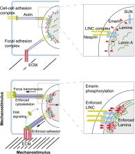
The intermediate filament proteins, A- and B-type lamins, form the nuclear lamina scaffold adjacent to the inner nuclear membrane. B-type lamins confer elasticity, while A-type lamins lend viscosity and stiffness to nuclei. Lamins also contribute to chromatin regulation and various signaling pathways affecting gene expression. The mechanical roles of lamins and their functions in gene regulation are often viewed as independent activities, but recent findings suggest a highly cross-linked and interdependent regulation of these different functions, particularly in mechanosignaling. In this newly emerging concept, lamins act as a “mechanostat” that senses forces from outside and responds to tension by reinforcing the cytoskeleton and the extracellular matrix. A-type lamins, emerin, and the linker of the nucleoskeleton and cytoskeleton (LINC) complex directly transmit forces from the extracellular matrix into the nucleus. These mechanical forces lead to changes in the molecular structure, modification, and assembly state of A-type lamins. This in turn activates a tension-induced “inside-out signaling” through which the nucleus feeds back to the cytoskeleton and the extracellular matrix to balance outside and inside forces. These functions regulate differentiation and may be impaired in lamin-linked diseases, leading to cellular phenotypes, particularly in mechanical load-bearing tissues.
Related collections
Most cited references185
- Record: found
- Abstract: found
- Article: not found
Mechanotransduction and extracellular matrix homeostasis.
- Record: found
- Abstract: found
- Article: not found
Nuclear lamin-A scales with tissue stiffness and enhances matrix-directed differentiation.
- Record: found
- Abstract: found
- Article: not found