6
views
0
recommends
+1 Recommend
0 collections
    0
    shares
      • Record: found
      • Abstract: found
      • Article: found
      Is Open Access

      Clinical classification and individualized design for the treatment of basicranial artery injuries

      research-article

      Read this article at

      Bookmark
          There is no author summary for this article yet. Authors can add summaries to their articles on ScienceOpen to make them more accessible to a non-specialist audience.

          Abstract

          This study aims to explore the principles of clinical classification and individualized treatment of basicranial artery injuries based on its anatomical correlation.

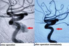
          The data of 172 patients with various types of basicranial artery injuries were retrospectively analyzed. Among these patients, 128 patients were male and 44 patients were female, and the average age of these patients was 28.3 years old. All patients underwent computed tomography, some patients underwent computed tomography angiography or magnetic resonance angiography, and all the diagnoses were confirmed by digital subtraction angiography (DSA). According to anatomical correlation, the injuries were classified into 5 types: vascular wall injury (type I), intradural injury (type II), epidural injury (type III), sinus injury (type IV), and skull base bone injury (type V). Individualized treatment was adopted based on the different types and characteristics of injuries.

          The percentages of basicranial artery injuries were as follows: type I, 4.6%; type II, 5.8%; type III, 3.5%; type IV, 77.9%; and type V, 8.1%. All 172 patients underwent DSA to demonstrate the classification. The lesion elimination rate revealed by DSA was 99.4% immediately after the operation, 98.3% at 1 week after the operation, and 98.8% at 3 months after the operation. The follow-up after 6 months revealed that the percentage of patients in whom clinical symptoms or signs completely disappeared was 97.7%, the percentage of patients with limited eye movement or visual impairment was 1.2%, and the percentage of patients with mild limb dysfunction was 0.6%.

          Basicranial artery injuries can be classified into 5 types. Individualized design of embolization therapy based on different characteristics might be applicable for basicranial artery injuries treatment.

          Related collections

          Most cited references19

          • Record: found
          • Abstract: found
          • Article: not found

          Incidence trends of traumatic spinal cord injury and traumatic brain injury in Spain, 2000-2009.

          The aim of the present study was to estimate the incidence of hospital discharges for traumatic spinal cord injury (TSCI) and traumatic brain injury (TBI) in Spain by injury circumstances (traffic crashes and others), injury severity, gender and age group and to describe its trends over the period 2000-2009.
            Bookmark
            • Record: found
            • Abstract: found
            • Article: not found

            Direct traumatic carotid cavernous fistula: angiographic classification and treatment strategies. Study of 172 cases.

            We report our experience in treatment of traumatic direct carotid cavernous fistula (CCF) via endovascular intervention. We hereof recommend an additional classification system for type A CCF and suggest respective treatment strategies. Only type A CCF patients (Barrow's classification) would be recruited for the study. Based on the angiographic characteristics of the CCF, we classified type A CCF into three subtypes including small size, medium size and large size fistula depending on whether there was presence of the anterior carotid artery (ACA) and/or middle carotid artery (MCA). Angiograms with opacification of both ACA and MCA were categorized as small size fistula. Angiograms with opacification of either ACA or MCA were categorized as medium size fistula and those without opacification of neither ACA nor MCA were classified as large size fiatula. After the confirm angiogram, endovascular embolization would be performed impromptu using detachable balloon, coils or both. All cases were followed up for complication and effect after the embolization. A total of 172 direct traumatic CCF patients were enrolled. The small size fistula was accountant for 12.8% (22 cases), medium size 35.5% (61 cases) and large size fistula accountant for 51.7% (89 cases). The successful rate of fistula occlusion under endovascular embolization was 94% with preservation of the carotid artery in 70%. For the treatment of each subtype, a total of 21/22 cases of the small size fistulas were successfully treated using coils alone. The other single case of small fistula was defaulted. Most of the medium and large size fistulas were cured using detachable balloons. When the fistula sealing could not be obtained using detachable balloon, coils were added to affirm the embolization of the cavernous sinus via venous access. There were about 2.9% of patient experienced direct carotid artery puncture and 0.6% puncture after carotid artery cut-down exposure. About 30% of cases experienced sacrifice of the parent vessels and it was associated with sizes of the fistula. Total severe complication was about 2.4% which included 1 death (0.6%) due to vagal shock; 1 transient hemiparesis post-sacrifice occlusion of the carotid artery but the patient had recovered after 3 months; 1 acute thrombus embolism and the patient was completely saved with recombinant tissue plaminogen activator (rTPA); 1 balloon dislodgement then got stuck at the anterior communicating artery but the patient was asymptomatic. Endovascular intervention as the treatment of direct traumatic CCF had high cure rate and low complication with its ability to preserve the carotid artery. It also can supply flexible accesses to the fistulous site with various alternative embolic materials. The new classification of type A CCF based on angiographic features was helpful for planning for the embolization. Coil should be considered as the first embolic material for small size fistula meanwhile detachable balloons was suggested as the first-choice embolic agent for the medium and large size fistula.
              Bookmark
              • Record: found
              • Abstract: found
              • Article: not found

              Ophthalmologic outcome of direct and indirect carotid cavernous fistulas.

              Carotid cavernous fistulas (CCFs) can be classified as direct and indirect, depending on their flow rates and their etiology. Both forms can cause the same characteristic ophthalmological symptoms and signs. We analyzed these ocular characteristics and determined the prognostics factors associated with treatment outcome. Forty-seven patients with an angiographically confirmed diagnosis of CCF, a preoperative ophthalmic evaluation and at least one ophthalmic sign or symptom at the initial presentation were retrospectively evaluated. The patients were followed-up ophthalmically until the end of treatment, and the complications and the remaining ophthalmological signs and symptoms were then recorded. The patients' ages ranged from 13 to 89 years, with an average of 55.78 (±20.73) years, and a predominance of 28 female (57.8 %) patients. The patients with a direct CCF had a lower average age (p = 0.02). The most common symptoms were blurred vision in 17 (36.2 %) and proptosis in 37 (78.7 %) patients. Elevated intraocular pressure (IOP) was more prevalent in patients with an indirect CCF (p = 0.02). Thrill was more prevalent in patients with direct CCF (p = 0.01). The presence of an initial decrease of visual acuity at the first ophthalmic evaluation was significantly associated with the persistence of ocular symptoms after fistula treatment (odds ratio 3.33). In conclusion our study shows a slight difference in ophthalmic symptoms among patients with different types of fistula. Elevated IOP was significantly associated with indirect fistulas, whereas thrill was significantly associated with direct fistulas. The presence of an initial decrease of visual acuity was significantly associated with a worse ophthalmic prognosis.
                Bookmark

                Author and article information

                Journal
                Medicine (Baltimore)
                Medicine (Baltimore)
                MEDI
                Medicine
                Wolters Kluwer Health
                0025-7974
                1536-5964
                March 2019
                15 March 2019
                : 98
                : 11
                : e14732
                Affiliations
                Department of Neurosurgery, The Affiliated Hospital of Guizhou Medical University, Guiyang, China.
                Author notes
                []Correspondence: Jian Liu, Department of Neurosurgery, The Affiliated Hospital of Guizhou Medical University, No. 28 Guiyi Street, Yunyan District, Guiyang 550004, China (e-mail: jianliudr@ 123456163.com ).
                Article
                MD-D-18-04933 14732
                10.1097/MD.0000000000014732
                6426552
                30882640
                969ba245-c0d0-422b-abe4-8aa979fec6d4
                Copyright © 2019 the Author(s). Published by Wolters Kluwer Health, Inc.

                This is an open access article distributed under the terms of the Creative Commons Attribution-Non Commercial-No Derivatives License 4.0 (CCBY-NC-ND), where it is permissible to download and share the work provided it is properly cited. The work cannot be changed in any way or used commercially without permission from the journal. http://creativecommons.org/licenses/by-nc-nd/4.0

                History
                : 17 July 2018
                : 1 February 2019
                : 7 February 2019
                Categories
                5300
                Research Article
                Observational Study
                Custom metadata
                TRUE

                artery injuries,basicranial artery,embolization therapy

                Comments

                Comment on this article