27
views
0
recommends
+1 Recommend
0 collections
    0
    shares
      • Record: found
      • Abstract: found
      • Article: not found

      Bovine lactoferrin improves bone mass and microstructure in ovariectomized rats via OPG/RANKL/RANK pathway

      research-article

      Read this article at

      ScienceOpenPublisherPMC
      Bookmark
          There is no author summary for this article yet. Authors can add summaries to their articles on ScienceOpen to make them more accessible to a non-specialist audience.

          Abstract

          Aim:

          Lactoferrin (LF), an 80-kDa iron-binding glycoprotein, is a pleiotropic factor found in colostrum, milk, saliva and epithelial cells of the exocrine glands. The aim of this study was to evaluate the effects of LF on the bones in ovariectomized (Ovx) rats and to identify the pathways that mediate the anabolic action of LF on the bones.

          Methods:

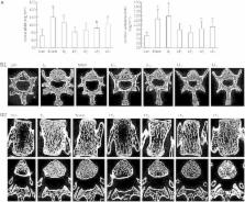
          Female Sprague-Dawley rats (6-month-old) underwent ovariectomy, and were treated with different doses of LF (10, 100, 1000, and 2000 mg·kg −1·d −1, po) or with 7β-estradiol (0.1 mg·kg −1, im, each week) as the positive control. By the end of 6 month-treatments, the bone mass and microstructure in the rats were scanned by micro-computed tomography (micro-CT), and the bone metabolism was evaluated with specific markers, and the mRNA levels of osteoprotegerin (OPG) and the receptor-activator of nuclear factor κB ligand (RANKL) in femur were measured using qRT-PCR.

          Results:

          LF treatment dose-dependently elevated the bone volume (BV/TV), trabecular thickness (TbTh) and trabecular number (TbN), and reduced the trabecular separation (TbSp) in Ovx rats. Furthermore, higher doses of LF (1000 and 2000 mg·kg −1·d −1) significantly increased the bone mineral density (BMD) compared with the untreated Ovx rats. The higher doses of LF also significantly increased the serum levels of OC and BALP, and decreased the serum levels of β-CTx and NTX. LF treatment significantly increased the OPG mRNA levels, and suppressed the RANKL mRNA levels, and the RANKL/OPG mRNA ratio in Ovx rats.

          Conclusion:

          Oral administration of LF preserves the bone mass and improves the bone microarchitecture. LF enhances bone formation, reduces bone resorption, and decreases bone mass loss, possibly through the regulation of OPG/RANKL/RANK pathway.

          Related collections

          Most cited references15

          • Record: found
          • Abstract: found
          • Article: not found

          Single-step method of RNA isolation by acid guanidinium thiocyanate-phenol-chloroform extraction.

          A new method of total RNA isolation by a single extraction with an acid guanidinium thiocyanate-phenol-chloroform mixture is described. The method provides a pure preparation of undegraded RNA in high yield and can be completed within 4 h. It is particularly useful for processing large numbers of samples and for isolation of RNA from minute quantities of cells or tissue samples.
            Bookmark
            • Record: found
            • Abstract: found
            • Article: not found

            Bone marrow, cytokines, and bone remodeling. Emerging insights into the pathophysiology of osteoporosis.

            Both osteoblasts and osteoclasts are derived from progenitors that reside in the bone marrow; osteoblasts belong to the mesenchymal lineage of the marrow stroma, and osteoclasts to the hematopoietic lineage. The development of osteoclasts from their progenitors is dependent on stromal-osteoblastic cells, which are a major source of cytokines that are critical in osteoclastogenesis, such as interleukin-6 and interleukin-11. The production of interleukin-6 by stromal osteoblastic cells, as well as the responsiveness of bone marrow cells to cytokines such as interleukin-6 and interleukin-11, is regulated by sex steroids. When gonadal function is lost, the formation of osteoclasts as well as osteoblasts increases in the marrow, both changes apparently mediated by an increase in the production of interleukin-6 and perhaps by an increase in the responsiveness of bone marrow progenitor cells not only to interleukin-6 but also to other cytokines with osteoclastogenic and osteoblastogenic properties. The cellular activity of the bone marrow is also altered by the process of aging. Specifically, senescence may decrease the ability of the marrow to form osteoblast precursors. The association between the dysregulation of osteoclast or osteoblast development in the marrow and the disruption of the balance between bone resorption and bone formation, resulting in the loss of bone, leads to the following notion. Like homeostasis of other regenerating tissues, homeostasis of bone depends on the orderly replenishment of its cellular constituents. Excessive osteoclastogenesis and inadequate osteoblastogenesis are responsible for the mismatch between the formation and resorption of bone in postmenopausal and age-related osteopenia. The recognition that changes in the numbers of bone cells, rather than changes in the activity of individual cells, form the pathogenetic basis of osteoporosis is a major advance in understanding the mechanism of this disease.
              Bookmark
              • Record: found
              • Abstract: found
              • Article: not found

              Transcription factors controlling osteoblastogenesis.

              J-L Marié (2008)
              The recent development of molecular biology and mouse genetics and the analysis of the skeletal phenotype induced by genetic mutations in humans led to a better understanding of the role of transcription factors that govern bone formation. This review summarizes the role of transcription factors in osteoblastogenesis and provides an integrated perspective on how the activities of multiple classes of factors are coordinated for the complex process of developing the osteoblast phenotype. The roles of Runx2, the principal transcriptional regulator of osteoblast differentiation, Osterix, beta-Catenin and ATF which act downstream of Runx2, and other transcription factors that contribute to the control of osteoblastogenesis including the AP1, C/EBPs, PPARgamma and homeodomain, helix-loop-helix proteins are discussed. This review also updates the regulation of transcription factor expression by signaling factors and hormones that control osteoblastogenesis.
                Bookmark

                Author and article information

                Journal
                Acta Pharmacol Sin
                Acta Pharmacol. Sin
                Acta Pharmacologica Sinica
                Nature Publishing Group
                1671-4083
                1745-7254
                October 2012
                20 August 2012
                : 33
                : 10
                : 1277-1284
                Affiliations
                [1 ]Provincial Clinical Medical College of Fujian Medical University , Fuzhou 350004, China
                Author notes
                [#]

                These authors contributed equally to this work.

                Article
                aps201283
                10.1038/aps.2012.83
                4002710
                22902986
                96680d93-2d91-4eda-9b0a-eb216fb0f895
                Copyright © 2012 CPS and SIMM
                History
                : 17 February 2012
                : 30 May 2012
                Categories
                Original Article

                Pharmacology & Pharmaceutical medicine
                lactoferrin,bone,bone mineral density,opg/rankl/rank pathway,bone turnover

                Comments

                Comment on this article