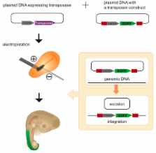
Introduction The natural medaka fish hAT gene family element Tol2 [1–3], the engineered Tc1/mariner transposons Sleeping Beauty (SB) [4] and Frog Prince [5], and the insect-derived natural element PiggyBac [6] represent transposons potentially suitable as DNA transfer tools for gene discovery and gene delivery applications in vertebrates. During the past decade, we have been studying the Sleeping Beauty transposable element for molecular genetic applications in vertebrates. While exceptionally active in higher vertebrates [7], the SB system does have two significant shortcomings: relatively modest cargo-capacity and decreased activity under high transposase concentrations (overexpression inhibition) [8,9]. The availability of an alternative, active transposon system devoid of these disadvantages and adapted for use in higher vertebrates would offer tremendous potential uses in a variety of molecular genetic and biotechnological fields. There is also a potential for synergism between multiple transposon systems, both in gene discovery and in gene transfer applications. Tol2 is widely used as a transgenesis tool in the zebrafish Danio rerio due to its high level of activity in this organism's germline [10,11]. However, gene transfer into cultured human cells or live mammals using Tol2 has not been previously reported, nor have the transposon sequence requirements for Tol2-based transposition been previously well characterized. Results While testing green fluorescent protein (GFP)-marked Tol2 vectors in zebrafish, we noticed an unusually high number of GFP-positive cells in embryos injected with Tol2 transposon and synthetic transposase mRNA derived from an updated Tol2 transcription vector (Figure 1), suggesting that this transposon is highly active in somatic tissues. The difference was particularly striking (over 10-fold) when percentages of embryos exhibiting eye fluorescence were evaluated (Figure 1A, right panel, red arrows). This effect was not observed when a Sleeping Beauty transposon with the same expression cassette was previously used in zebrafish (Figure 1 and [12,13]). To determine if this increase in overall and particularly eye fluorescence was a result of increased transposition, we conducted a PCR-based assay for one of the molecular markers of the transposition reaction—generation of a transposon-excision footprint [14]. We found that increased GFP fluorescence in Tol2-injected embryos versus SB-injected embryos correlated with transposon excision. The Tol2 excision band was more robust than the SB excision band and appeared much earlier, as soon as 2 h after injection (Figure 1B). This may reflect a kinetic difference between the two systems; SB is thought to function as a tetramer [4,15] and may take longer to form an active complex than Tol2, which may function like another hAT family member Hermes, i.e., as a dimer [16]. We also note that the high Tol2 somatic transposition rate correlates with germline transposition: 18 of 25 tested fish gave GFP-positive progeny (a 72% transgenesis and expression rate), with an average founder fish transmitting an estimated five independent insertion events (unpublished data). This increase in GFP fluorescence together with generation of a robust excision footprint provides a rapid Tol2 activity assay using zebrafish embryos. We used this assay to address a significant drawback of Tol2—minimal sequences required for transposition had not been previously defined. The commonly used Tol2 vector [11,17] has deletions encompassing part of exon-2, all of exon-3, and part of exon-4 with intervening introns. While these deletions render the transposase inactive, they leave intact a significant fraction of the transposase open reading frame with several splice sites, as well as promoter and polyadenylation sequences. The presence of these sequences hinders vector design and compromises vector utility in its application to gene discovery, gene transfer, and gene therapy. For gene therapy applications in particular, they pose an increased risk of insertional mutagenesis. We made a systematic series of deletions of the Tol2 vector and tested them in our zebrafish somatic transposition assay (Figure 1C). Surprisingly, the first four deletions were all active in this assay, even though two of them removed sequences previously thought to be required for transposon excision [18]. A combination of 5′ and 3′ deletions resulted in the smallest active element containing 261 nucleotides of 5′ Tol2 DNA and 202 nucleotides of Tol2 3′ DNA (“miniTol2”). A further truncation of the Tol2 sequences (“Tol2 clipped”) by PCR inactivated the element, however, indicating that there are sequence requirements beyond the minimal 17-base inverted terminal repeats. The miniTol2 vector was subsequently tested in human cells in vitro (Figure 2) and for gene transfer in vivo (Figure 3) and demonstrated activity statistically indistinguishable from that of the full-length vector in each case. In initial experiments in mammalian cells, modest gene transfer activity was noted for Tol2 in mouse ES cells when very high amounts of transposase-encoding plasmid were used [10]. Tol2 has also been shown to excise in HeLa and NIH/3T3 cells [19]; however, Tol2 transposase-mediated gene transfer was notably not described in latter studies using human cells. These observations combined with high activity of Tol2 in zebrafish somatic tissues prompted us to compare the activity of Tol2 to that of the best-studied and highly active vertebrate transposon, SB. We found that in HeLa cells, the activity of 5-kb Tol element was indistinguishable from that of a 2-kb SB element (Figure 2A). We then tested the activity of Tol2 as a function of transposase concentration in HT1080 cells by varying the amount of transposase-encoding plasmid. Sleeping Beauty is known to exhibit decreased activity in presence of excess transposase, a phenomenon termed “overexpression inhibition” [8]. We found that the Tol2 transposon system does not exhibit overexpression inhibition within the tested transposase concentration range, while peak gene transfer rates of the two systems are very similar despite the difference in transposon size (Figure 2B). Restrictions in cargo-capacity (less than 8 kb) have been reported for integrating viral vectors including retroviruses and lentiviruses [20]. The SB transposon system exhibits a similar but less absolute capacity restriction, such that 10-kb elements are notably less mobile than 2-kb–sized SB transposons [8]. The larger native size of Tol2 (5 kb) versus the presumed size of the SB progenitor (2 kb) might explain the surprisingly high activity that was observed for the 5-kb Tol2 element in human cells (Figure 2A). We tested a larger, greater-than-10-kb Tol2 element and found that it mediated gene transfer nearly as efficiently as the 5-kb (Figure 2C) element. Using a smaller, 2-kb miniTol2 element, we found Tol2 activity to be largely independent of reduced cargo load. Tol2 thus exhibits an effective, high-capacity gene transfer capability in human cells. Large cargo-capacity and gene transfer efficiency in human cells make Tol2 an excellent candidate for molecular medicine applications. We therefore tested whether Tol2 can be used for gene delivery in live animals. We first generated a Tol2 transposon encoding a firefly luciferase expression cassette to facilitate in vivo imaging of live animals. We then injected 5 μg of pTol2/luc with varying amounts of pCMV-Tol2 to achieve gene transfer into mouse liver using a rapid, high-volume (hydrodynamic) injection method [21,22]. Luciferase expression was assayed by bioluminescence imaging at day 1 and subsequently followed through 28 wk. While we did not observe overexpression inhibition by using large amounts of pCMV-Tol2 (transposon:transposase plasmid ratios of up to 1:10), the minimal amount of transposase plasmid required to achieve optimal, long-term expression was with transposon:transposase plasmids at a ratio of 1:1 (unpublished data). We next investigated the ability of Tol2 to correct a mouse model of hereditary tyrosinemia type 1 (HT1). HT1 is caused by deficiency of fumarylacetoacetate hydrolase (FAH) leading to the accumulation of fumarylacetoacetate, which is toxic to hepatocytes. HT1 patients can be treated with 2-(2-nitro-4-trifluoro-methylbenzoyl)-1,3 cyclohexanedione (NTBC), a compound that blocks an enzyme upstream of FAH, thus preventing toxicity [23]. Mice deficient in FAH develop symptoms of HT1, providing a model to study the disease [24]. To test the effectiveness of Tol2 in this gene therapy application, FAH knockout mice maintained on NTBC were treated by hydrodynamic injection of either standard or minimal (mini) versions of pTol2/FAHIL, a transposon that encodes both murine FAH and firefly luciferase as products of a single transcription unit (Figure 3) (A. Wilber, K. J. Wangnesteen, Y. Chen, L. Zhuo, J. L. Frandsen, J. B. Bell, Z. J. Chen, S. C. Ekker, R. S. McIvor, and X. Wang, unpublished data). One day after injection, luciferase expression was assayed by in vivo bioluminescence imaging to provide a relative assessment of gene transfer efficiency for each animal (Figure 3D). NTBC was then withdrawn to induce tyrosinemia. For subsequent images taken during the first 4 wk, all animals were administered NTBC for the 24 h preceding the time of imaging to minimize any adverse effect resulting from administration of the anesthetic, which is metabolized by the liver. Previous studies have found body weight after withdrawal of NTBC is a good measure of therapeutic effect ([25]; A. Wilber, K. J. Wangnesteen, Y. Chen, L. Zhuo, J. L. Frandsen, J. B. Bell, Z. J. Chen, S. C. Ekker, R. S. McIvor, and X. Wang, unpublished data). Therefore, body weight measurements were taken for each animal to assess therapeutic efficacy, expressed as the percentage of weight at the beginning of the experiment (Figure 3C). Animals infused with either version of pTol2/FAHIL plus pCMV-GFP required readministration of NTBC in the drinking water for 5 d (starting at the time indicated by an asterisk in Figure 3B) to survive. Importantly, animals coinjected with Tol2 transposase-encoding plasmid did not require additional NTBC, except just prior to anesthesia for imaging, and maintained weight during the subsequent period of study, indicating that the metabolic deficiency in these animals had been corrected [25,26]. Because the pTol2/FAHIL and pminiTol2/FAHIL transposons include the luciferase-coding sequence on the same transcript as the FAH-coding sequence, we were able to evaluate the extent of liver repopulation resulting from random recombination compared to co-delivery of transposase in individual animals over time by bioluminescence imaging (Figure 3C). Emitted light was measured after luciferin injection as an indication of stable gene expression at several times over 2 mo. Figure 3C represents luciferase expression over time after injection of pTol2/FAHIL and pminiTol2/FAHIL with or without pCMV-Tol2. Figure 3D depicts the images obtained for a representative animal co-infused with pminiTol2/FAHIL plus pCMV-Tol2, showing the increase in luciferase activity observed during the course of repopulation. We observed a notable decrease in transient gene expression (day 1) for pTol2/FAHIL compared to pminiTol2/FAHIL, possibly resulting from lower rates of uptake of the larger pTol2/FAHIL plasmids compared to pminiTol2/FAHIL, which are 3.1 kb smaller. Thus, the reduction in gene transfer rate does not necessarily reflect a lower transposition rate of pTol2/FAHIL. Alternatively, sequences found within the Tol2 transposon but not within miniTol2 may reduce transient expression of luciferase from nonintegrated plasmid DNA. After 2 mo, FAH-deficient animals infused with pCMV-Tol2 exhibited about a 100-fold increase in luciferase expression over animals that did not receive pCMV-Tol2 (Figure 3D). In general, the kinetics of liver repopulation using either the longer version or mini derivative of the Tol2 transposon were similar, suggesting that Tol2 is capable of efficiently integrating transposons as large as 8.5 kb in vivo. To confirm that the observed stable gene expression was due to transposition and not increased random integration, we used inverse PCR to recover Tol2 insertions from mouse and human chromosomes. A total of 36 integration sites were analyzed: 24 from HT1080 cell colonies, two from HT1080 transient transfections, and ten from mouse liver (Table 1). All had hallmarks suggesting transposition; 22 of the integration sites displayed the expected 8-bp target-site duplication, while the remaining two had 7-bp target-site duplications. These results confirm that Tol2 mediates authentic transposition in mammalian cells and tissues. We thus conclude that miniTol2 is an effective, large cargo-capacity transposase-mediated gene transfer tool suitable for gene therapy and other vertebrate gene transfer applications. Discussion We have demonstrated that the Tol2 transposable element from medaka fish shows high potential as a mammalian gene transfer agent. Tol2 is highly active in human cells in vitro and in mouse liver in vivo, displaying activity comparable to that of the most active vertebrate transposon, Sleeping Beauty. The gene transfer rate of Tol2 in vivo is sufficient to achieve correction of the mouse model of hereditary tyrosinemia type 1. Tol2 exhibits two additional features that make it a very attractive gene transfer system: apparent absence of overexpression inhibition and the ability to ferry large DNA cargo. We demonstrate that a 10-kb Tol2 transposon is nearly as active as a 2-kb element. We defined a minimized set of inverted terminal repeat sequences required for Tol2 transposition and generated a high-utility miniTol2 vector for gene transfer that can be used from zebrafish to clinical applications. The ability to efficiently move cargo as large as 10-kb sets Tol2 apart from most other integrating gene transfer vectors. The best-studied vertebrate transposon system, Sleeping Beauty, retains only about 20% activity with such large cargo [8]. Integrating viruses, on the other hand, can only pack about 8 kb of exogenous DNA into their capsids, structurally limiting cargo-capacity [20]. Bacteriophage-derived integrases are capable of integrating such large cargo into vertebrate genomes [27,28]. Integrases tend to integrate into several pseudosites in a vertebrate genome, which can be either an advantage or a drawback depending on application. It appears that these preferred sites are not only different between species but also differ depending on the cell type used [29–32]. Recent studies demonstrate that phiC31 integrase induces chromosomal translocations in mammalian cells, which is highly undesirable for any application [32,33]. The frequency of such events needs to be thoroughly investigated to determine if this poses a significant safety risk in gene therapy applications. One feature of Sleeping Beauty transposon system that makes it especially attractive in a wide variety of applications ranging from gene discovery to gene therapy is its near random integration capabilities. SB displays little preference for specific sequence flanking the target TA dinucleotide or for genomic region, such as genes versus intergenic regions [34–36]. From analysis of the current and limited Tol2 integration site data, Tol2 seems to be equally unbiased (Table 1 and [17]). A much larger sample similar to the ones analyzed for SB [34–36] will be required to know if Tol2 exhibits any notable insertion-site preferences. The ability to move large cargo opens new possibilities for applications in gene therapy, gene discovery, and transgenesis. For gene therapy, increased cargo-capacity accommodates large therapeutically relevant genes such as DNA-PKcs for X-linked severe combined immune deficiency [37] or factor VIII for hemophilia A [38]. This also allows for more complex vector design, including tissue-specific promoters, insulators, suicide genes, and other features that may improve both efficacy and safety of such vectors. In addition, lack of overexpression inhibition demonstrated by Tol2 will be advantageous in designing cis vectors for co-delivery,of transposon and transposase, something that has required a great deal of optimization for SB. High cargo-capacity will facilitate adaptation of Tol2 for gene discovery applications by allowing implementation of vectors that consist of multiple functional cassettes, for example, a 5′ gene-trap and a 3′ gene-trap, with different reporters. Tol2 has been successfully adapted for enhancer identification in zebrafish [39], and the newly documented ability of Tol2 to carry large cargos will facilitate such scanning of genomic sequences for regulatory elements. Animal biotechnology, an emerging area of transgenic research, will benefit from a large cargo-capacity vector that lacks overexpression inhibition. SB and Tol2 belong to different families of transposable elements: SB to the Tc1/mariner family and Tol2 to the hAT family. These two highly active and very different transposon systems should have a strong synergistic effect in many areas of research. Different transgenes can be delivered to the same cell at the same or different time points without a concern of cross-mobilization. One such example is generation of transposase-expressing animals for cancer gene discovery. So far, transposase-expressing mice were generated either using plasmid transgenesis or by knock-in approaches [40–42]. It will now be possible to easily generate lines of animals that express SB transposase using Tol2 transposons and vice versa. Since the original publication of Sleeping Beauty transposon system [4], considerable research has been devoted to characterizing and improving transposition, and multiple laboratories have identified and published hyperactive variants of SB transposase [8,43–45]. Without any modifications, Tol2 is already much more active than SB in zebrafish, but the activity of the two systems is comparable in mammalian cells. It remains to be determined if the Tol2 hyperactivity potential seen in zebrafish can be mimicked in mammalian cells. Other members of the same hAT family to which Tol2 belongs display additional beneficial characteristics that strongly suggest host factors are not necessary for transposition. Maize Ac is active in yeast [46] and Drosophila Hermes is active in vitro [16]. These observations suggest that Tol2 can be further developed quite rapidly and thereby contribute insight into mechanisms of transposition as well as serve as a tool for many other biological applications. Materials and Methods Transposase RNA and DNA expression vectors. The Tol2 open reading frame was amplified by PCR using primers Tol2-F3 (5′-GCTGGATCCACCATGGAGGAAGTATGTGATTC-3′) and Tol2-R1 (5′-CAGACTAGTCTACTCAAAGTTGTAAAACC-3′) and the pBluescript-based Tol2 transposase vector described by Parinov et al. (2004) as template. The PCR fragment was first cloned into pCRIItopo vector (Invitrogen, Carlsbad, California, United States), resulting in pDB595. For RNA expression, the Tol2 ORF was subcloned from pDB595 as BamHI-SpeI fragment into BglII-SpeI digested transcription vector pT3TS [47], resulting in pT3TS-Tol2 (pDB600). pDB600 was cleaved with XbaI and transcribed with T3 polymerase using Ambion mMessage Machine kit (Austin, Texas, United States) to produce Tol2 mRNA. To make pCMV-Tol2, the transposase ORF was amplified from pDB595 using Tol2-NotI-F (5′-ATATGCGGCCGCCACCATGGAGGAAGTATG-3′) and M13R primers, cloned into pCR4topo vector (Invitrogen), and then a Tol2 NotI fragment was excised and cloned into the NotI site of pCMV-bgal vector (Clontech, Palo Alto, California, United States) in the forward orientation. The SB10 ORF [4] was PCR subcloned into pT3TS to generate pT3TS-SB10. Tol2 transposon vectors. To construct pTol2/S2EF1a-GM2 (pDB591), the S2EF1a-GM2 expression cassette [13] was cloned as an SphI-NruI fragment of pDB371 between EcoRV sites of pGEM-T/Tol2 [11] in reverse orientation. To generate pTol2/SVneo (pDB625), the G418 resistance cassette of pT2/SVneo [8] was subcloned as HindIII fragment into EcoRV-digested pGemT-Tol2. To generate pminiTol2/SVneo (pDB678), the same fragment was cloned between HindIII and SwaI sites of pGEM-T/Tol2. The same fragment was cloned into the Eco105I site of pDB633 (see below) to make pTol2/FAHIL-SVneo (pDB691). To define minimal sequences required for Tol2 transposition in zebrafish somatic tissues, two strategies were used. Tol2 sequences between SwaI and XcmI sites of pDB591 were deleted to make pDB665 (delta exon1), and sequences between XcmI and EcoO109I sites were deleted to make pDB664 (delta repeats). Also, S2EF1a-GM2 cassette was cloned into pGEM-T/Tol2 in reverse orientation between the following restriction enzyme sites: SwaI and EcoRV (pDB668, delta exon 1 and repeats), EcoRV and HindIII (pDB667, delta exon 4), and SwaI and HindIII (pDB674, Tol2mini). To make a “clipped” version of Tol2 transposon, S2EF1a-GM2 expression cassette was amplified from pDB371 [13] using primers Tol2-sph-F (5′-CAGAGGTGTAAAGTACTTGAGTAATTTTACTTGATTACTGTACTTAAGTATGCATGCAAGCTAGTACAAGACG-3′) and Tol2-nru-R (5′-CAGAGGTGTAAAAAGTACTCAAAAATTTTACTCAAGTGAAAGTACAAGTACTTATCGCGATGATAATCAGCCATACC-3′). The PCR fragment was cloned into pCR4topo vector (Invitrogen) and sequenced. To express FAH and firefly luciferase in mouse, bicistronic expression cassette consiting of miniCAGGS promoter, mouse FAH cDNA, encephalomyocarditis virus internal ribosome entry site, firefly luciferase, and rabbit β-globin polyadenylation sequences was subcloned as an NheI-PmeI fragment from pKT2/FAHIL (A. Wilber, K. J. Wangnesteen, Y. Chen, L. Zhuo, J. L. Frandsen, J. B. Bell, Z. J. Chen, S. C. Ekker, R. S. McIvor, and X. Wang, unpublished data) into EcoRV-digested pGEM-T/Tol2 [11] (pTol2/FAHIL) or SwaI-HindIII digested pGEM-T/Tol2 (pminiTol2/FAHIL). In vitro transposition assays. A colony-forming assay was conducted as described [8]. Briefly, 3 × 10⁵ HT1080 cells were plated onto 6-cm plates and 24 h later were transfected with 1 to 2 μg of DNA using FuGene 6 reagent (Roche, Basel, Switzerland). Two days later, the cells were trypsinized and 3 × 10⁴ cells were plated onto 10-cm plates with complete medium supplemented with 600 μg/ml Geneticin (G418; Invitrogen). After 10 d of selection, the cells were stained with methylene blue and counted. Three plates of cells were transfected for each data point using different transposon DNA preps. For Tol2/SB activity comparison (Figure 2B), colonies were visually counted. For comparison of different-size Tol2 elements (Figure 2C), colonies were counted using ImageJ software (http://rsb.info.nih.gov/ij). The number of colonies obtained in transfection including transposase was divided by the number of colonies obtained in control transfection to compute gene transfer efficiency. The efficiency of miniTol2/SVneo was set as 100%. Molecular analysis of genomic integration sites. DNA was isolated from cultured cells or mouse liver using the Puregene DNA purification system (Gentra Systems, Minneapolis, Minnesota, United States). DNA was digested and used in an adapted inverse PCR (iPCR) protocol [12]. Tol2-specific iPCR primers were Tol2-R3 (5′-ACTGGGCATCAGCGCAATTCAATTG-3′) and Tol2-F7 (5′-AGCAGGATAAAACCTTGTATGCATT-3′) for the first round and Tol2-R4 (5′-ATAATACTTAAGTACAGTAATCAAG-3′) and Tol2-F8 (5′-CTCAAGTAAGATTCTAGCCAGATAC-3′) for the nested reaction. For iPCR of cell clones, Neo-resistant colonies were picked and amplified 2 wk after tranfection with pDB625 and pCMV-Tol2. Genomic DNA was extracted, digested with NheI, SpeI, and XbaI and ligated in a dilute reaction. The first round of iPCR was performed with Tol2-R3 and Tol2-F7, and a second with Tol2-R4 and Tol2-F7. The PCR products were isolated by gel electrophoresis, and sequenced using primers Tol2-R4 or Tol2-F7. For cloning of miniTol2 insertion sites, HT1080 cells were transfected with miniTol2SVneo and pCMV-Tol2 and grown for 4 d without selection. Genomic DNA was extracted and digested with AflIII (which cuts the vector twice outside of the transposon and leaves incompatible ends) SpeI and XbaI, ligated in a dilute reaction, and one round of PCR was performed with Tol2-R3 and Tol2-F8. The resultant products were used in a Topo-cloning reaction (Invitrogen), and two insertions were identified by sequencing using M13-F and M13-R primers. For the mouse insertion sites, genomic DNA was extracted from each of five liver lobes of one FAH-deficient mouse 2 mo after injection with pTol2-FAHIL. The DNA was digested with NheI, BamHI, XhoI, and BsiWI, which do not cut vector sequences, blunted with Klenow fragment, and ligated in a dilute reaction. Two rounds of iPCR were performed and sequenced as described above for cell clones. Zebrafish somatic transposition assays. For SB/Tol2 comparison in the GFP fluorescence analysis (Figure 1), one-cell stage zebrafish embryos were injected with a mixture of 20 pg of pT2/S2EF1a/GM2 (pDB371) and pTol2/S2EF1a/GM2 (pDB591) each. Some of the embryos were then injected with 25 pg of T3TS-Tol2 or 25 pg of T3TS-SB RNA. Dead and grossly abnormal embryos were removed, and GFP fluorescence was scored at 3 d postfertilization. Randomly chosen embryos were photographed using Zeiss Axioscope II with Zeiss Axiocam using Zeiss Axiovision software. For excision analysis, embryos were injected with either a mixture of 20 pg of pT2/S2EF1a/GM2 (pDB371) DNA, 20 pg of pTol2/S2EF1a/GM2 (pDB591) DNA, and 25 pg of T3TS-Tol2 RNA or a mixture of 20 pg of pT2/S2EF1a/GM2 (pDB371) DNA, 20 pg of pTol2/S2EF1a/GM2 (pDB591) DNA, and 25 pg of T3TS-SB10 RNA. Twenty embryos were pooled at each indicated time point, and the DNA was prepared using a modified genomic DNA preparation protocol [48] and used for excision PCR [14]. To compare different Tol2 truncations, embryos were first injected with 25 pg of appropriate plasmid solution, and then all plasmid-injected embryos were injected with 25 pg of T3TS-Tol2 RNA using the same needle. Surviving and grossly normal embryos were scored for GFP fluorescence at 3 d postfertilization. Twenty GFP-positive embryos were used for DNA preparation. DNA was dissolved in 50 μl of TE and diluted 1:1 in water; 1 μl of this solution was subsequently used for excision PCR (see above). Animals and plasmid injections. Normal C57BL/6 mice and FAHDexon 5 transgenic (tg) mice [24], 18 to 25 g, were bred in a specific pathogen-free environment at the University of Minnesota. FAH-deficient mice were maintained on drinking water supplemented with NTBC at a concentration of 7.5 mg/ml. Plasmids were delivered primarily to the liver using a rapid, high-volume tail vein injection procedure [21,22] as previously described [49]. Only animals that received a complete injection in less than 8 s were used for subsequent studies. In vivo bioluminescent imaging. At various times after injection of DNA, animals were anesthetized by intraperitoneal injection of 160 mg/kg ketamine plus 0.8 mg/kg acepromazine and 0.08 mg/kg butorphanol. One hundred milliliters of 28.5 mg/ml luciferin substrate was then injected intraperitoneally. At 4 to 5 min after injection of substrate, the live anesthetized mice were imaged for 1 s to 2 min using an intensified, charge-coupled device camera (Series 100; Xenogen, Hopkinton, Massachusetts, United States) as described [49]. Raw values of luciferase activity were recorded as photons of light emitted per second.