55
views
0
recommends
+1 Recommend
0 collections
    0
    shares
      • Record: found
      • Abstract: found
      • Article: found
      Is Open Access

      Attainment of polarity promotes growth factor secretion by retinal pigment epithelial cells: Relevance to age-related macular degeneration

      research-article

      Read this article at

      Bookmark
          There is no author summary for this article yet. Authors can add summaries to their articles on ScienceOpen to make them more accessible to a non-specialist audience.

          Abstract

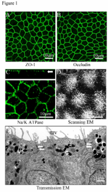
          The antiangiogenic and neurotrophic growth factor, pigment epithelial derived factor (PEDF), and the proangiogenic growth factor, vascular endothelial growth factor-A (VEGF), are released from retinal pigment epithelial (RPE) cells where they play a critical role in the pathogenesis of age-related macular degeneration (AMD). Since RPE polarity may be altered in advanced AMD, we studied the effect of polarization of differentiated, human RPE monolayer cultures on expression and secretion of PEDF and VEGF. Polarized RPE demonstrated apical microvilli, expression of tight junction proteins, apical localization of Na/K- ATPase, and high transepithelial resistance (490 ± 17 Ω•cm 2). PEDF secretion was about 1000 fold greater than that for VEGF in both polarized and non-polarized cultures. Polarization of the RPE monolayer increased PEDF secretion, which was predominantly apical, by 34 fold (p<0.02) and VEGF secretion, which was predominantly basolateral, by 5.7 fold (p<0.02). Treatment of non-polarized RPE cultures with bone morphogenetic protein-4 (BMP-4) had no effect on PEDF or VEGF secretion, but resulted in a dose-dependent >2-fold increase in basolateral VEGF secretion (p<0.05) in polarized cultures. Our data show that polarity is an important determinant of the level of PEDF and VEGF secretion in RPE and support the contention that loss of polarity of RPE in AMD results in marked loss of neurotrophic and vascular support for the retina potentially leading to photoreceptor loss and blindness.

          Related collections

          Most cited references50

          • Record: found
          • Abstract: found
          • Article: not found

          Increased vascular endothelial growth factor levels in the vitreous of eyes with proliferative diabetic retinopathy.

          The vitreous levels of the angiogenic polypeptide vascular endothelial growth factor (also known as vascular permeability factor) were measured and compared in eyes with and without proliferative diabetic retinopathy. Undiluted vitreous samples from 20 eyes were collected at the time of vitrectomy, and vascular endothelial growth factor levels were determined by using a time-resolved immunofluorometric assay. Vitreous vascular endothelial growth factor levels were significantly higher in eyes with proliferative diabetic retinopathy than in eyes without proliferative diabetic retinopathy (P = .006; Wilcoxon Rank Sum Test). The median vitreous concentration in the eyes with proliferative diabetic retinopathy was 29.1 pM and exceeded the known concentration required for the maximal proliferation of vascular endothelial cells in vitro. These data are consistent with vascular endothelial growth factor serving as a physiologically relevant angiogenic factor in proliferative diabetic retinopathy.
            Bookmark
            • Record: found
            • Abstract: found
            • Article: not found

            Confluent monolayers of cultured human fetal retinal pigment epithelium exhibit morphology and physiology of native tissue.

            Provide a reproducible method for culturing confluent monolayers of hfRPE cells that exhibit morphology, physiology, polarity, and protein expression patterns similar to native tissue. Human fetal eyes were dissected on arrival, and RPE cell sheets were mechanically separated from the choroid and cultured in a specifically designed medium comprised entirely of commercially available components. Physiology experiments were performed with previously described techniques. Standard techniques were used for immunohistochemistry, electron microscopy, and cytokine measurement by ELISA. Confluent monolayers of RPE cell cultures exhibited epithelial morphology and heavy pigmentation, and electron microscopy showed extensive apical membrane microvilli. The junctional complexes were identified with immunofluorescence labeling of various tight junction proteins. The mean transepithelial potential (TEP) was 2.6 +/- 0.8 mV, apical positive, and the mean transepithelial resistance (R(T)) was 501 +/- 138 Omega . cm(2) (mean +/- SD; n = 35). Addition of 100 microM adenosine triphosphate (ATP) to the apical bath increased net fluid absorption from 13.6 +/- 2.6 to 18.8 +/- 4.6 microL . cm(-2) per hour (mean +/- SD; n = 4). In other experiments, VEGF was mainly secreted into the basal bath (n = 10), whereas PEDF was mainly secreted into the apical bath (n = 10). A new cell culture procedure has been developed that produces confluent primary hfRPE cultures with morphological and physiological characteristics of the native tissue. Epithelial polarity and function of these easily reproducible primary cultures closely resemble previously studied native human fetal and bovine RPE-choroid explants.
              Bookmark
              • Record: found
              • Abstract: found
              • Article: not found

              Polarized vascular endothelial growth factor secretion by human retinal pigment epithelium and localization of vascular endothelial growth factor receptors on the inner choriocapillaris. Evidence for a trophic paracrine relation.

              The retinal pigment epithelium (RPE) maintains the choriocapillaris (CC) in the normal eye and is involved in the pathogenesis of choroidal neovascularization in age-related macular degeneration. Vascular endothelial growth factor-A (VEGF) is produced by differentiated human RPE cells in vitro and in vivo and may be involved in paracrine signaling between the RPE and the CC. We investigated whether there is a polarized secretion of VEGF by RPE cells in vitro. Also, the localization of VEGF receptors in the human retina was investigated. We observed that highly differentiated human RPE cells, cultured on transwell filters in normoxic conditions, produced two- to sevenfold more VEGF toward their basolateral side as compared to the apical side. In hypoxic conditions, VEGF-A secretion increased to the basal side only, resulting in a three- to 10-fold higher basolateral secretion. By immunohistochemistry in 30 human eyes and in two cynomolgus monkey eyes, KDR (VEGFR-2) and flt-4 (VEGFR-3) were preferentially localized at the side of the CC endothelium facing the RPE cell layer, whereas flt-1 (VEGFR-1) was found on the inner CC and on other choroidal vessels. Our results indicate that RPE secretes VEGF toward its basal side where its receptor KDR is located on the adjacent CC endothelium, suggesting a role of VEGF in a paracrine relation, possibly in cooperation with flt-4 and its ligand. This can explain the known trophic function of the RPE in the maintenance of the CC and its fenestrated permeable phenotype and points to a role for VEGF in normal eye functioning. Up-regulated basolateral VEGF secretion by RPE in hypoxia or loss of polarity of VEGF production may play a role in the pathogenesis of choroidal neovascularization.
                Bookmark

                Author and article information

                Journal
                Aging (Albany NY)
                ImpactJ
                Aging (Albany NY)
                Impact Journals LLC
                1945-4589
                January 2010
                27 December 2009
                : 2
                : 1
                : 28-42
                Affiliations
                1 Department of Ophthalmology, Keck School of Medicine of the University of Southern California, Los Angeles, CA 90089, USA
                2 Department of Pathology, Keck School of Medicine of the University of Southern California, Los Angeles, CA 90089, USA
                3 Arnold and Mabel Beckman Macular Research Center at the Doheny Eye Institute, Los Angeles, CA 90033, USA
                4 Department of Ophthalmology, Kagoshima University, Graduate School of Medical and Dental Sciences, Kagoshima, Japan
                5These two authors contributed equally to this work
                Author notes
                Correspondence: David R. Hinton, MD, FARVO, Department of Pathology, Keck School of Medicine of the University of Southern California, 2011 Zonal Ave. HMR 209, Los Angeles, CA 90089, USA dhinton@ 123456usc.edu
                Article
                10.18632/aging.100111
                2837203
                20228934
                963962ad-16e9-41f5-8dc0-f22da92f8710
                Copyright: ©2010 Sonoda et al.

                This is an open-access article distributed under the terms of the Creative Commons Attribution License, which permits unrestricted use, distribution, and reproduction in any medium, provided the original work is properly cited.

                History
                : 20 December 2009
                : 26 December 2009
                Categories
                Research Article

                Cell biology
                retinal pigment epithelial cell,pedf,vegf-a,bmp-4,age-related macular degeneration,cell polarity

                Comments

                Comment on this article