20
views
0
recommends
+1 Recommend
0 collections
    0
    shares
      • Record: found
      • Abstract: found
      • Article: found
      Is Open Access

      Segmentation of the Proximal Femur from MR Images using Deep Convolutional Neural Networks

      research-article

      Read this article at

      Bookmark
          There is no author summary for this article yet. Authors can add summaries to their articles on ScienceOpen to make them more accessible to a non-specialist audience.

          Abstract

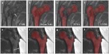
          Magnetic resonance imaging (MRI) has been proposed as a complimentary method to measure bone quality and assess fracture risk. However, manual segmentation of MR images of bone is time-consuming, limiting the use of MRI measurements in the clinical practice. The purpose of this paper is to present an automatic proximal femur segmentation method that is based on deep convolutional neural networks (CNNs). This study had institutional review board approval and written informed consent was obtained from all subjects. A dataset of volumetric structural MR images of the proximal femur from 86 subjects were manually-segmented by an expert. We performed experiments by training two different CNN architectures with multiple number of initial feature maps, layers and dilation rates, and tested their segmentation performance against the gold standard of manual segmentations using four-fold cross-validation. Automatic segmentation of the proximal femur using CNNs achieved a high dice similarity score of 0.95 ± 0.02 with precision = 0.95 ± 0.02, and recall = 0.95 ± 0.03. The high segmentation accuracy provided by CNNs has the potential to help bring the use of structural MRI measurements of bone quality into clinical practice for management of osteoporosis.

          Related collections

          Most cited references67

          • Record: found
          • Abstract: found
          • Article: not found

          Deep learning.

          Deep learning allows computational models that are composed of multiple processing layers to learn representations of data with multiple levels of abstraction. These methods have dramatically improved the state-of-the-art in speech recognition, visual object recognition, object detection and many other domains such as drug discovery and genomics. Deep learning discovers intricate structure in large data sets by using the backpropagation algorithm to indicate how a machine should change its internal parameters that are used to compute the representation in each layer from the representation in the previous layer. Deep convolutional nets have brought about breakthroughs in processing images, video, speech and audio, whereas recurrent nets have shone light on sequential data such as text and speech.
            Bookmark
            • Record: found
            • Abstract: found
            • Article: not found

            A survey on deep learning in medical image analysis

            Deep learning algorithms, in particular convolutional networks, have rapidly become a methodology of choice for analyzing medical images. This paper reviews the major deep learning concepts pertinent to medical image analysis and summarizes over 300 contributions to the field, most of which appeared in the last year. We survey the use of deep learning for image classification, object detection, segmentation, registration, and other tasks. Concise overviews are provided of studies per application area: neuro, retinal, pulmonary, digital pathology, breast, cardiac, abdominal, musculoskeletal. We end with a summary of the current state-of-the-art, a critical discussion of open challenges and directions for future research.
              Bookmark
              • Record: found
              • Abstract: not found
              • Article: not found

              Index for rating diagnostic tests

                Bookmark

                Author and article information

                Contributors
                cem.deniz@nyulangone.org
                Journal
                Sci Rep
                Sci Rep
                Scientific Reports
                Nature Publishing Group UK (London )
                2045-2322
                7 November 2018
                7 November 2018
                2018
                : 8
                : 16485
                Affiliations
                [1 ]ISNI 0000 0004 1936 8753, GRID grid.137628.9, Department of Radiology, , New York University School of Medicine, ; New York, NY 10016 USA
                [2 ]ISNI 0000 0004 1936 8753, GRID grid.137628.9, Bernard and Irene Schwartz Center for Biomedical Imaging, , New York University School of Medicine, ; New York, NY 10016 USA
                [3 ]ISNI 0000 0004 1936 8753, GRID grid.137628.9, Center for Data Science, , New York University, ; New York, NY 10012 USA
                [4 ]ISNI 000000041936754X, GRID grid.38142.3c, Harvard College, ; Cambridge, MA 02138 USA
                [5 ]ISNI 0000 0001 2109 4251, GRID grid.240324.3, Osteoporosis Center, Hospital for Joint Diseases, , New York University Langone Medical Center, ; New York, NY 10003 USA
                [6 ]ISNI 0000 0004 1936 8753, GRID grid.137628.9, Courant Institute of Mathematical Science, , New York University, ; New York, NY 10012 USA
                Author information
                http://orcid.org/0000-0001-8809-5945
                Article
                34817
                10.1038/s41598-018-34817-6
                6220200
                30405145
                9621fe27-7603-4f50-a77e-c44345467948
                © The Author(s) 2018

                Open Access This article is licensed under a Creative Commons Attribution 4.0 International License, which permits use, sharing, adaptation, distribution and reproduction in any medium or format, as long as you give appropriate credit to the original author(s) and the source, provide a link to the Creative Commons license, and indicate if changes were made. The images or other third party material in this article are included in the article’s Creative Commons license, unless indicated otherwise in a credit line to the material. If material is not included in the article’s Creative Commons license and your intended use is not permitted by statutory regulation or exceeds the permitted use, you will need to obtain permission directly from the copyright holder. To view a copy of this license, visit http://creativecommons.org/licenses/by/4.0/.

                History
                : 12 March 2018
                : 26 October 2018
                Funding
                Funded by: FundRef https://doi.org/10.13039/100000002, U.S. Department of Health & Human Services | National Institutes of Health (NIH);
                Award ID: P41 EB017183
                Award ID: R01 AR070131
                Award ID: R01 AR066008
                Award Recipient :
                Categories
                Article
                Custom metadata
                © The Author(s) 2018

                Uncategorized
                Uncategorized

                Comments

                Comment on this article