3
views
0
recommends
+1 Recommend
0 collections
    0
    shares
      • Record: found
      • Abstract: found
      • Article: found
      Is Open Access

      Comparing human and chimpanzee temporal lobe neuroanatomy reveals modifications to human language hubs beyond the frontotemporal arcuate fasciculus

      research-article

      Read this article at

      Bookmark
          There is no author summary for this article yet. Authors can add summaries to their articles on ScienceOpen to make them more accessible to a non-specialist audience.

          Significance

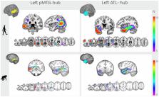
          Communication through language is a great achievement of evolution. In humans, the arcuate fasciculus, white matter that extended dramatically during evolution, is known to subserve language. We investigated whether connections through critical language centers in the temporal lobe are uniquely human. We show that connectivity in the posterior temporal lobe via the arcuate fasciculus expanded bilaterally to frontal and parietal cortices in humans compared with chimpanzees. Concomitantly, the ventral tracts connect more strongly to posterior temporal regions in the chimpanzees than in humans. In the anterior temporal lobe, connections shared between both species and uniquely human expansions are present. Changes to human language streams extend beyond the arcuate fasciculus, including a suite of expansions to connectivity within the temporal lobes.

          Abstract

          The biological foundation for the language-ready brain in the human lineage remains a debated subject. In humans, the arcuate fasciculus (AF) white matter and the posterior portions of the middle temporal gyrus are crucial for language. Compared with other primates, the human AF has been shown to dramatically extend into the posterior temporal lobe, which forms the basis of a number of models of the structural connectivity basis of language. Recent advances in both language research and comparative neuroimaging invite a reassessment of the anatomical differences in language streams between humans and our closest relatives. Here, we show that posterior temporal connectivity via the AF in humans compared with chimpanzees is expanded in terms of its connectivity not just to the ventral frontal cortex but also to the parietal cortex. At the same time, posterior temporal regions connect more strongly to the ventral white matter in chimpanzees as opposed to humans. This pattern is present in both brain hemispheres. Additionally, we show that the anterior temporal lobe harbors a combination of connections present in both species through the inferior fronto-occipital fascicle and human-unique expansions through the uncinate and middle and inferior longitudinal fascicles. These findings elucidate structural changes that are unique to humans and may underlie the anatomical foundations for full-fledged language capacity.

          Related collections

          Most cited references105

          • Record: found
          • Abstract: found
          • Article: not found

          Advances in functional and structural MR image analysis and implementation as FSL.

          The techniques available for the interrogation and analysis of neuroimaging data have a large influence in determining the flexibility, sensitivity, and scope of neuroimaging experiments. The development of such methodologies has allowed investigators to address scientific questions that could not previously be answered and, as such, has become an important research area in its own right. In this paper, we present a review of the research carried out by the Analysis Group at the Oxford Centre for Functional MRI of the Brain (FMRIB). This research has focussed on the development of new methodologies for the analysis of both structural and functional magnetic resonance imaging data. The majority of the research laid out in this paper has been implemented as freely available software tools within FMRIB's Software Library (FSL).
            Bookmark
            • Record: found
            • Abstract: found
            • Article: not found

            Fast robust automated brain extraction.

            An automated method for segmenting magnetic resonance head images into brain and non-brain has been developed. It is very robust and accurate and has been tested on thousands of data sets from a wide variety of scanners and taken with a wide variety of MR sequences. The method, Brain Extraction Tool (BET), uses a deformable model that evolves to fit the brain's surface by the application of a set of locally adaptive model forces. The method is very fast and requires no preregistration or other pre-processing before being applied. We describe the new method and give examples of results and the results of extensive quantitative testing against "gold-standard" hand segmentations, and two other popular automated methods. Copyright 2002 Wiley-Liss, Inc.
              Bookmark
              • Record: found
              • Abstract: found
              • Article: not found

              The minimal preprocessing pipelines for the Human Connectome Project.

              The Human Connectome Project (HCP) faces the challenging task of bringing multiple magnetic resonance imaging (MRI) modalities together in a common automated preprocessing framework across a large cohort of subjects. The MRI data acquired by the HCP differ in many ways from data acquired on conventional 3 Tesla scanners and often require newly developed preprocessing methods. We describe the minimal preprocessing pipelines for structural, functional, and diffusion MRI that were developed by the HCP to accomplish many low level tasks, including spatial artifact/distortion removal, surface generation, cross-modal registration, and alignment to standard space. These pipelines are specially designed to capitalize on the high quality data offered by the HCP. The final standard space makes use of a recently introduced CIFTI file format and the associated grayordinate spatial coordinate system. This allows for combined cortical surface and subcortical volume analyses while reducing the storage and processing requirements for high spatial and temporal resolution data. Here, we provide the minimum image acquisition requirements for the HCP minimal preprocessing pipelines and additional advice for investigators interested in replicating the HCP's acquisition protocols or using these pipelines. Finally, we discuss some potential future improvements to the pipelines. Copyright © 2013 Elsevier Inc. All rights reserved.
                Bookmark

                Author and article information

                Journal
                Proc Natl Acad Sci U S A
                Proc Natl Acad Sci U S A
                pnas
                PNAS
                Proceedings of the National Academy of Sciences of the United States of America
                National Academy of Sciences
                0027-8424
                1091-6490
                5 July 2022
                12 July 2022
                5 January 2023
                : 119
                : 28
                : e2118295119
                Affiliations
                [1] aDonders Institute for Brain, Cognition and Behaviour, Radboud University , 6500 GL Nijmegen, the Netherlands;
                [2] bDonders Centre for Medical Neuroscience, Department of Medical Psychology, Radboud University Medical Center , 6500 HB Nijmegen, the Netherlands;
                [3] cWellcome Centre for Integrative Neuroimaging, Centre for Functional MRI of the Brain, Nuffield Department of Clinical Neurosciences, John Radcliffe Hospital, University of Oxford , Oxford OX3 9DU, United Kingdom;
                [4] dDepartment of Psychiatry, Radboud University Medical Center , 6525 GA Nijmegen, the Netherlands;
                [5] eDepartment of Cognitive Neuroscience, Radboud University Medical Centre , 6500 HB Nijmegen, the Netherlands;
                [6] fInstitute for Science in Society, Radboud University , 6525 AJ Nijmegen, the Netherlands
                Author notes
                2To whom correspondence may be addressed. Email: joanna.sierpowska@ 123456donders.ru.nl .

                Edited by Marcus Raichle, Washington University in St. Louis, St. Louis, MO; received October 7, 2021; accepted May 11, 2022

                Author contributions: J.S., K.L.B., R.B.M., and V.P. designed research; K.L.B. and N.J. performed research; J.S., K.L.B., and G.B.F. contributed new reagents/analytic tools; J.S., K.L.B., N.J., G.B.F., M.R., M.M., and V.P. analyzed data; J.S., K.L.B., N.J., M.M., R.B.M., and V.P. wrote the paper; and G.B.F. and M.R. critically reviewed the manuscript.

                1J.S. and K.L.B. contributed equally to this work.

                Author information
                https://orcid.org/0000-0002-5991-6916
                https://orcid.org/0000-0003-1045-4543
                https://orcid.org/0000-0001-5531-4507
                https://orcid.org/0000-0002-4860-5952
                Article
                202118295
                10.1073/pnas.2118295119
                9282369
                35787056
                94ce4d3f-5c7a-40d2-8f27-8c325d6c9081
                Copyright © 2022 the Author(s). Published by PNAS.

                This article is distributed under Creative Commons Attribution-NonCommercial-NoDerivatives License 4.0 (CC BY-NC-ND).

                History
                : 11 May 2022
                Page count
                Pages: 9
                Funding
                Funded by: Nederlandse Organisatie voor Wetenschappelijk Onderzoek (NWO) 501100003246
                Award ID: 451-17-003
                Award Recipient : Rogier B Mars Award Recipient : Vitoria Piai
                Funded by: Nederlandse Organisatie voor Wetenschappelijk Onderzoek (NWO) 501100003246
                Award ID: 452-13-015
                Award Recipient : Rogier B Mars Award Recipient : Vitoria Piai
                Funded by: Nederlandse Organisatie voor Wetenschappelijk Onderzoek (NWO) 501100003246
                Award ID: 024-001-006
                Award Recipient : Rogier B Mars Award Recipient : Vitoria Piai
                Funded by: European Commission (EC) 501100000780
                Award ID: MSCA-IF 750026
                Award Recipient : Katherine L Bryant
                Funded by: UKRI | Biotechnology and Biological Sciences Research Council (BBSRC) 501100000268
                Award ID: BB/N019814/1
                Award Recipient : Katherine L Bryant Award Recipient : Rogier B Mars
                Funded by: UKRI | Biotechnology and Biological Sciences Research Council (BBSRC) 501100000268
                Award ID: BB/N019814/1
                Award Recipient : Katherine L Bryant Award Recipient : Rogier B Mars
                Funded by: UKRI | Biotechnology and Biological Sciences Research Council (BBSRC) 501100000268
                Award ID: 203129/Z/16/Z
                Award Recipient : Katherine L Bryant Award Recipient : Rogier B Mars
                Categories
                424
                Biological Sciences
                Neuroscience

                white matter,language,evolution,arcuate fasciculus,temporal lobe

                Comments

                Comment on this article