23
views
0
recommends
+1 Recommend
0 collections
    0
    shares
      • Record: found
      • Abstract: found
      • Article: found
      Is Open Access

      Pre-Treatment with Laminarin Protects Hippocampal CA1 Pyramidal Neurons and Attenuates Reactive Gliosis Following Transient Forebrain Ischemia in Gerbils

      research-article

      Read this article at

      Bookmark
          There is no author summary for this article yet. Authors can add summaries to their articles on ScienceOpen to make them more accessible to a non-specialist audience.

          Abstract

          Transient brain ischemia triggers selective neuronal death/loss, especially in vulnerable regions of the brain including the hippocampus. Laminarin, a polysaccharide originating from brown seaweed, has various pharmaceutical properties including an antioxidant function. To the best of our knowledge, few studies have been conducted on the protective effects of laminarin against ischemic injury induced by ischemic insults. In this study, we histopathologically investigated the neuroprotective effects of laminarin in the Cornu Ammonis 1 (CA1) field of the hippocampus, which is very vulnerable to ischemia-reperfusion injury, following transient forebrain ischemia (TFI) for five minutes in gerbils. The neuroprotective effect was examined by cresyl violet staining, Fluoro-Jade B histofluorescence staining and immunohistochemistry for neuronal-specific nuclear protein. Additionally, to study gliosis (glial changes), we performed immunohistochemistry for glial fibrillary acidic protein to examine astrocytes, and ionized calcium-binding adaptor molecule 1 to examine microglia. Furthermore, we examined alterations in pro-inflammatory M1 microglia by using double immunofluorescence. Pretreatment with 10 mg/kg laminarin failed to protect neurons in the hippocampal CA1 field and did not attenuate reactive gliosis in the field following TFI. In contrast, pretreatment with 50 or 100 mg/kg laminarin protected neurons, attenuated reactive gliosis and reduced pro-inflammatory M1 microglia in the CA1 field following TFI. Based on these results, we firmly propose that 50 mg/kg laminarin can be strategically applied to develop a preventative against injuries following cerebral ischemic insults.

          Related collections

          Most cited references41

          • Record: found
          • Abstract: found
          • Article: not found

          Microglia Function in the Central Nervous System During Health and Neurodegeneration.

          Microglia are resident cells of the brain that regulate brain development, maintenance of neuronal networks, and injury repair. Microglia serve as brain macrophages but are distinct from other tissue macrophages owing to their unique homeostatic phenotype and tight regulation by the central nervous system (CNS) microenvironment. They are responsible for the elimination of microbes, dead cells, redundant synapses, protein aggregates, and other particulate and soluble antigens that may endanger the CNS. Furthermore, as the primary source of proinflammatory cytokines, microglia are pivotal mediators of neuroinflammation and can induce or modulate a broad spectrum of cellular responses. Alterations in microglia functionality are implicated in brain development and aging, as well as in neurodegeneration. Recent observations about microglia ontogeny combined with extensive gene expression profiling and novel tools to study microglia biology have allowed us to characterize the spectrum of microglial phenotypes during development, homeostasis, and disease. In this article, we review recent advances in our understanding of the biology of microglia, their contribution to homeostasis, and their involvement in neurodegeneration. Moreover, we highlight the complexity of targeting microglia for therapeutic intervention in neurodegenerative diseases. Expected final online publication date for the Annual Review of Immunology Volume 35 is April 26, 2017. Please see http://www.annualreviews.org/page/journal/pubdates for revised estimates.
            Bookmark
            • Record: found
            • Abstract: found
            • Article: found
            Is Open Access

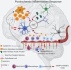
            Neuroinflammation: friend and foe for ischemic stroke

            Stroke, the third leading cause of death and disability worldwide, is undergoing a change in perspective with the emergence of new ideas on neurodegeneration. The concept that stroke is a disorder solely of blood vessels has been expanded to include the effects of a detrimental interaction between glia, neurons, vascular cells, and matrix components, which is collectively referred to as the neurovascular unit. Following the acute stroke, the majority of which are ischemic, there is secondary neuroinflammation that both promotes further injury, resulting in cell death, but conversely plays a beneficial role, by promoting recovery. The proinflammatory signals from immune mediators rapidly activate resident cells and influence infiltration of a wide range of inflammatory cells (neutrophils, monocytes/macrophages, different subtypes of T cells, and other inflammatory cells) into the ischemic region exacerbating brain damage. In this review, we discuss how neuroinflammation has both beneficial as well as detrimental roles and recent therapeutic strategies to combat pathological responses. Here, we also focus on time-dependent entry of immune cells to the ischemic area and the impact of other pathological mediators, including oxidative stress, excitotoxicity, matrix metalloproteinases (MMPs), high-mobility group box 1 (HMGB1), arachidonic acid metabolites, mitogen-activated protein kinase (MAPK), and post-translational modifications that could potentially perpetuate ischemic brain damage after the acute injury. Understanding the time-dependent role of inflammatory factors could help in developing new diagnostic, prognostic, and therapeutic neuroprotective strategies for post-stroke inflammation.
              Bookmark
              • Record: found
              • Abstract: found
              • Article: not found

              Microglial activation and its implications in the brain diseases.

              An inflammatory process in the central nervous system (CNS) is believed to play an important role in the pathway leading to neuronal cell death in a number of neurodegenerative diseases including Parkinson's disease, Alzheimer's disease, prion diseases, multiple sclerosis and HIV-dementia. The inflammatory response is mediated by the activated microglia, the resident immune cells of the CNS, which normally respond to neuronal damage and remove the damaged cells by phagocytosis. Activation of microglia is a hallmark of brain pathology. However, it remains controversial whether microglial cells have beneficial or detrimental functions in various neuropathological conditions. The chronic activation of microglia may in turn cause neuronal damage through the release of potentially cytotoxic molecules such as proinflammatory cytokines, reactive oxygen intermediates, proteinases and complement proteins. Therefore, suppression of microglia-mediated inflammation has been considered as an important strategy in neurodegenerative disease therapy. Several anti-inflammatory drugs of various chemical ingredients have been shown to repress the microglial activation and to exert neuroprotective effects in the CNS following different types of injuries. However, the molecular mechanisms by which these effects occur remain unclear. In recent years, several research groups including ours have attempted to explain the potential mechanisms and signaling pathways for the repressive effect of various drugs, on activation of microglial cells in CNS injury. We provide here a comprehensive review of recent findings of mechanisms and signaling pathways by which microglial cells are activated in CNS inflammatory diseases. This review article further summarizes the role of microglial cells in neurodegenerative diseases and various forms of potential therapeutic options to inhibit the microglial activation which amplifies the inflammation-related neuronal injury in neurodegenerative diseases.
                Bookmark

                Author and article information

                Journal
                Mar Drugs
                Mar Drugs
                marinedrugs
                Marine Drugs
                MDPI
                1660-3397
                12 January 2020
                January 2020
                : 18
                : 1
                : 52
                Affiliations
                [1 ]Department of Neurobiology, School of Medicine, Kangwon National University, Chuncheon 24341, Gangwon, Korea; xorud312@ 123456naver.com (T.-K.L.); nbrkim17@ 123456gmail.com (B.K.); taeparo@ 123456naver.com (Y.E.P.); anajclee@ 123456kangwon.ac.kr (J.-C.L.)
                [2 ]Department of Biomedical Science, Research Institute of Bioscience and Biotechnology, Hallym University, Chuncheon 24252, Gangwon, Korea; jh-ahn@ 123456hallym.ac.kr
                [3 ]Leefarm Co., Ltd., Hongcheon 25117, Gangwon, Korea; flfhflfh@ 123456naver.com
                [4 ]Department of Anatomy, College of Korean Medicine, Dongguk University, Gyeongju 38066, Gyeongbuk, Korea; jh-park@ 123456dongguk.ac.kr
                [5 ]Department of Radiology, Kangwon National University Hospital, Chuncheon 24289, Gangwon, Korea; yangke@ 123456kangwon.ac.kr
                [6 ]Department of Emergency Medicine, School of Medicine, Kangwon National University, Chuncheon 24341, Gangwon, Korea; dr10126@ 123456naver.com (M.C.S.); cjhemd@ 123456kangwon.ac.kr (J.H.C.)
                [7 ]Department of Food Science and Nutrition, Hallym University, Chuncheon 24252, Gangwon, Korea
                Author notes
                [* ]Correspondence: ijkang@ 123456hallym.ac.kr (I.-J.K.); mhwon@ 123456kangwon.ac.kr (M.-H.W.); Tel.: +82-33-250-8891 (I.-J.K.); +82-33-248-2135 (M.-H.W.); Fax: +82-33-256-1614 (I.-J.K.); +82-33-255-4787 (M.-H.W.)
                [†]

                Co-First: Tae-Kyeong Lee and Ji Hyeon Ahn contributed equally to this work.

                Author information
                https://orcid.org/0000-0003-0314-5195
                https://orcid.org/0000-0002-7178-6501
                Article
                marinedrugs-18-00052
                10.3390/md18010052
                7024340
                31940961
                949de62e-ecfa-4ab8-b609-1e36d98a4367
                © 2020 by the authors.

                Licensee MDPI, Basel, Switzerland. This article is an open access article distributed under the terms and conditions of the Creative Commons Attribution (CC BY) license ( http://creativecommons.org/licenses/by/4.0/).

                History
                : 26 November 2019
                : 09 January 2020
                Categories
                Article

                Pharmacology & Pharmaceutical medicine
                gliosis,ischemia-reperfusion,polysaccharide,pyramidal neurons,prevention,rodents

                Comments

                Comment on this article