1
views
0
recommends
+1 Recommend
0 collections
    0
    shares
      • Record: found
      • Abstract: found
      • Article: found
      Is Open Access

      Quantifying differences in fMRI preprocessing pipelines via OGRE (One-step General Registration and Extraction)

      Preprint
      research-article

      Read this article at

      Bookmark
          There is no author summary for this article yet. Authors can add summaries to their articles on ScienceOpen to make them more accessible to a non-specialist audience.

          Abstract

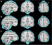
          Volumetric preprocessing methods continue to enjoy great popularity in the analysis of functional MRI (fMRI) data. Among these methods, the software packages FSL (FMRIB, Oxford, UK) and FreeSurfer (LCN, Charlestown, MA) are omnipresent throughout the field. However, it remains unknown what advantages an integrated FSL+FreeSurfer preprocessing approach might provide over FSL alone. Here we developed the One-step General Registration and Extraction (OGRE) pipeline to combine FreeSurfer and FSL tools for brain extraction and registration, for FSL volumetric analysis of fMRI data. We compared preprocessing approaches in a dataset wherein adult human volunteers (N=26) performed a precision drawing task during fMRI scanning. OGRE’s preprocessing, compared to traditional FSL preprocessing, led to lower inter-individual variability across the brain, more precise brain extraction, and greater detected activation in sensorimotor areas contralateral to movement. This demonstrates that the introduction of FreeSurfer tools via OGRE preprocessing can improve fMRI data analysis, in the context of FSL’s volumetric analysis approach. The OGRE pipeline provides a turnkey method to integrate FreeSurfer-based brain extraction and registration with FSL analysis of task fMRI data.

          Related collections

          Most cited references40

          • Record: found
          • Abstract: found
          • Article: not found

          The organization of the human cerebral cortex estimated by intrinsic functional connectivity.

          Information processing in the cerebral cortex involves interactions among distributed areas. Anatomical connectivity suggests that certain areas form local hierarchical relations such as within the visual system. Other connectivity patterns, particularly among association areas, suggest the presence of large-scale circuits without clear hierarchical relations. In this study the organization of networks in the human cerebrum was explored using resting-state functional connectivity MRI. Data from 1,000 subjects were registered using surface-based alignment. A clustering approach was employed to identify and replicate networks of functionally coupled regions across the cerebral cortex. The results revealed local networks confined to sensory and motor cortices as well as distributed networks of association regions. Within the sensory and motor cortices, functional connectivity followed topographic representations across adjacent areas. In association cortex, the connectivity patterns often showed abrupt transitions between network boundaries. Focused analyses were performed to better understand properties of network connectivity. A canonical sensory-motor pathway involving primary visual area, putative middle temporal area complex (MT+), lateral intraparietal area, and frontal eye field was analyzed to explore how interactions might arise within and between networks. Results showed that adjacent regions of the MT+ complex demonstrate differential connectivity consistent with a hierarchical pathway that spans networks. The functional connectivity of parietal and prefrontal association cortices was next explored. Distinct connectivity profiles of neighboring regions suggest they participate in distributed networks that, while showing evidence for interactions, are embedded within largely parallel, interdigitated circuits. We conclude by discussing the organization of these large-scale cerebral networks in relation to monkey anatomy and their potential evolutionary expansion in humans to support cognition.
            Bookmark
            • Record: found
            • Abstract: found
            • Article: not found

            Advances in functional and structural MR image analysis and implementation as FSL.

            The techniques available for the interrogation and analysis of neuroimaging data have a large influence in determining the flexibility, sensitivity, and scope of neuroimaging experiments. The development of such methodologies has allowed investigators to address scientific questions that could not previously be answered and, as such, has become an important research area in its own right. In this paper, we present a review of the research carried out by the Analysis Group at the Oxford Centre for Functional MRI of the Brain (FMRIB). This research has focussed on the development of new methodologies for the analysis of both structural and functional magnetic resonance imaging data. The majority of the research laid out in this paper has been implemented as freely available software tools within FMRIB's Software Library (FSL).
              Bookmark
              • Record: found
              • Abstract: found
              • Article: not found

              Fast robust automated brain extraction.

              An automated method for segmenting magnetic resonance head images into brain and non-brain has been developed. It is very robust and accurate and has been tested on thousands of data sets from a wide variety of scanners and taken with a wide variety of MR sequences. The method, Brain Extraction Tool (BET), uses a deformable model that evolves to fit the brain's surface by the application of a set of locally adaptive model forces. The method is very fast and requires no preregistration or other pre-processing before being applied. We describe the new method and give examples of results and the results of extensive quantitative testing against "gold-standard" hand segmentations, and two other popular automated methods. Copyright 2002 Wiley-Liss, Inc.
                Bookmark

                Author and article information

                Contributors
                Journal
                bioRxiv
                BIORXIV
                bioRxiv
                Cold Spring Harbor Laboratory
                07 October 2023
                : 2023.09.19.558290
                Affiliations
                Program in Occupational Therapy, Washington University School of Medicine, St. Louis, MO, United States.
                Department of Biostatistics, Washington University School of Medicine, St. Louis, MO, United States.
                Department of Biostatistics, Washington University School of Medicine, St. Louis, MO, United States
                Program in Occupational Therapy, Washington University School of Medicine, St. Louis, MO, United States.
                Author notes

                Author contribution statement: MPM was involved in software development (primary) and manuscript writing/editing. BAP was involved in conceptualization, software development, data analysis, funding acquisition, supervision, and manuscript writing/editing. LL and RZ were involved in data analysis and manuscript editing

                [* ]Corresponding author: bphilip@ 123456wustl.edu .
                Article
                10.1101/2023.09.19.558290
                10541115
                37781580
                944c2d6c-abaf-4212-87a8-940300f2d28a

                This work is licensed under a Creative Commons Attribution-NonCommercial-NoDerivatives 4.0 International License, which allows reusers to copy and distribute the material in any medium or format in unadapted form only, for noncommercial purposes only, and only so long as attribution is given to the creator.

                History
                Funding
                Funded by: NIH/NINDS
                Award ID: R01 NS114046
                Categories
                Article

                image analysis pipeline,fmri,brain,software,magnetic resonance imaging,humans,brain mapping

                Comments

                Comment on this article