14
views
0
recommends
+1 Recommend
0 collections
    0
    shares
      • Record: found
      • Abstract: found
      • Article: found
      Is Open Access

      HIF-1α overexpression in mesenchymal stem cell-derived exosomes mediates cardioprotection in myocardial infarction by enhanced angiogenesis

      research-article

      Read this article at

      Bookmark
          There is no author summary for this article yet. Authors can add summaries to their articles on ScienceOpen to make them more accessible to a non-specialist audience.

          Abstract

          Background

          Myocardial infarction (MI) is a severe disease that often associated with dysfunction of angiogenesis. Cell-based therapies for MI using mesenchymal stem cell (MSC)-derived exosomes have been well studied due to their strong proangiogenic effect. Genetic modification is one of the most common methods to enhance exosome therapy. This study investigated the proangiogenic and cardioprotective effect of exosomes derived from hypoxia-inducible factor 1-alpha (HIF-1α)-modified MSCs.

          Methods

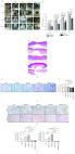
          Lentivirus containing HIF-1α overexpressing vector was packaged and used to infect MSCs. Exosomes were isolated from MSC-conditioned medium by ultracentrifugation. Human umbilical vein endothelial cells (HUVECs) were treated under hypoxia condition for 48 h co-cultured with PBS, control exosomes, or HIF-1α-overexpressed exosomes, respectively. Then the preconditioned HUVECs were subjected to tube formation assay, Transwell assay, and EdU assay to evaluate the protective effect of exosomes. Meanwhile, mRNA and secretion levels of proangiogenic factors were measured by RT-qPCR and ELISA assays. In vivo assays were conducted using the rat myocardial infarction model. PBS, control exosomes, or HIF-1α-overexpressed exosomes were injected through tail vein after MI surgery. Heart function was assessed by echocardiography at days 3, 14, and 28. At day 7, mRNA and protein expression levels of proangiogenic factors in the peri-infarction area and circulation were evaluated, respectively. At day 28, hearts were collected and subjected to H&E staining, Masson’s trichrome staining, and immunofluorescent staining.

          Results

          HIF-1α-overexpressed exosomes rescued the impaired angiogenic ability, migratory function, and proliferation of hypoxia-injured HUVECs. Simultaneously, HIF-1α-overexpressed exosomes preserved heart function by promoting neovessel formation and inhibiting fibrosis in the rat MI model. In addition, both in vitro and in vivo proangiogenic factors mRNA and protein expression levels were elevated after HIF-1α-overexpressed exosome application.

          Conclusion

          HIF-1α-overexpressed exosomes could rescue the impaired angiogenic ability, migration, and proliferation of hypoxia-pretreated HUVECs in vitro and mediate cardioprotection by upregulating proangiogenic factors and enhancing neovessel formation.

          Related collections

          Most cited references28

          • Record: found
          • Abstract: found
          • Article: not found

          Mesenchymal stem cells: biology, pathophysiology, translational findings, and therapeutic implications for cardiac disease.

          Mesenchymal stem cells (MSCs) are a prototypical adult stem cell with capacity for self-renewal and differentiation with a broad tissue distribution. Initially described in bone marrow, MSCs have the capacity to differentiate into mesoderm- and nonmesoderm-derived tissues. The endogenous role for MSCs is maintenance of stem cell niches (classically the hematopoietic), and as such, MSCs participate in organ homeostasis, wound healing, and successful aging. From a therapeutic perspective, and facilitated by the ease of preparation and immunologic privilege, MSCs are emerging as an extremely promising therapeutic agent for tissue regeneration. Studies in animal models of myocardial infarction have demonstrated the ability of transplanted MSCs to engraft and differentiate into cardiomyocytes and vasculature cells, recruit endogenous cardiac stem cells, and secrete a wide array of paracrine factors. Together, these properties can be harnessed to both prevent and reverse remodeling in the ischemically injured ventricle. In proof-of-concept and phase I clinical trials, MSC therapy improved left ventricular function, induced reverse remodeling, and decreased scar size. This article reviews the current understanding of MSC biology, mechanism of action in cardiac repair, translational findings, and early clinical trial data of MSC therapy for cardiac disease.
            Bookmark
            • Record: found
            • Abstract: found
            • Article: not found

            Hypoxia Inducible Factor-1α Potentiates Jagged 1-Mediated Angiogenesis by Mesenchymal Stem Cell-Derived Exosomes

            Insufficient vessel growth associated with ischemia remains an unresolved issue in vascular medicine. Mesenchymal stem cells (MSCs) have been shown to promote angiogenesis via a mechanism that is potentiated by hypoxia. Overexpression of hypoxia inducible factor (HIF)-1α in MSCs improves their therapeutic potential by inducing angiogenesis in transplanted tissues. Here, we studied the contribution of exosomes released by HIF-1α-overexpressing donor MSCs (HIF-MSC) to angiogenesis by endothelial cells. Exosome secretion was enhanced in HIF-MSC. Omics analysis of miRNAs and proteins incorporated into exosomes pointed to the Notch pathway as a candidate mediator of exosome communication. Interestingly, we found that Jagged1 was the sole Notch ligand packaged into MSC exosomes and was more abundant in HIF-MSC than in MSC controls. The addition of Jagged1-containing exosomes from MSC and HIF-MSC cultures to endothelial cells triggered transcriptional changes in Notch target genes and induced angiogenesis in an in vitro model of capillary-like tube formation, and both processes were stimulated by HIF-1α. Finally, subcutaneous injection of Jagged 1-containing exosomes from MSC and HIF-MSC cultures in the Matrigel plug assay induced angiogenesis in vivo, which was more robust when they were derived from HIF-MSC cultures. All Jagged1-mediated effects could be blocked by prior incubation of exosomes with an anti-Jagged 1 antibody. All together, the results indicate that exosomes derived from MSCs stably overexpressing HIF-1α have an increased angiogenic capacity in part via an increase in the packaging of Jagged1, which could have potential applications for the treatment of ischemia-related disease. Stem Cells 2017;35:1747-1759.
              Bookmark
              • Record: found
              • Abstract: found
              • Article: found
              Is Open Access

              MSC-Derived Exosome Promotes M2 Polarization and Enhances Cutaneous Wound Healing

              Mesenchymal stem cell transplantation (MSCT) promotes cutaneous wound healing. Numerous studies have shown that the therapeutic effects of MSCT appear to be mediated by paracrine signaling. However, the cell-cell interaction during MSCT between MSCs and macrophages in the region of cutaneous wound healing is still unknown. In this study, early depletion of macrophages delayed the wound repair with MSC injection, which suggested that MSC-mediated wound healing required macrophages. Moreover, we demonstrated that systemically infused bone marrow MSCs (BMMSCs) and jaw bone marrow MSCs (JMMSCs) could translocate to the wound site, promote macrophages toward M2 polarization, and enhance wound healing. In vitro coculture of MSCs with macrophages enhanced their M2 polarization. Mechanistically, we found that exosomes derived from MSCs induced macrophage polarization and depletion of exosomes of MSCs reduced the M2 phenotype of macrophages. Infusing MSCs without exosomes led to lower number of M2 macrophages at the wound site along with delayed wound repair. We further showed that the miR-223, derived from exosomes of MSCs, regulated macrophage polarization by targeting pknox1. These findings provided the evidence that MSCT elicits M2 polarization of macrophages and may accelerate wound healing by transferring exosome-derived microRNA.
                Bookmark

                Author and article information

                Contributors
                sunjc1993@gmail.com
                shenhan@suda.edu.cn
                shaolianbo1987@126.com
                txm_2020@126.com
                chenyueqiu-216@163.com
                asscenliu@163.com
                skyinger@163.com
                uuzyshen@aliyun.com
                Journal
                Stem Cell Res Ther
                Stem Cell Res Ther
                Stem Cell Research & Therapy
                BioMed Central (London )
                1757-6512
                28 August 2020
                28 August 2020
                2020
                : 11
                : 373
                Affiliations
                GRID grid.263761.7, ISNI 0000 0001 0198 0694, Department of Cardiovascular Surgery of the First Affiliated Hospital & Institute for Cardiovascular Science, , Soochow University, ; No.899, Pinghai Road, Suzhou, 215006 China
                Article
                1881
                10.1186/s13287-020-01881-7
                7455909
                32859268
                941eb72a-611e-48ce-aa4b-34b867cb52e9
                © The Author(s) 2020

                Open AccessThis article is licensed under a Creative Commons Attribution 4.0 International License, which permits use, sharing, adaptation, distribution and reproduction in any medium or format, as long as you give appropriate credit to the original author(s) and the source, provide a link to the Creative Commons licence, and indicate if changes were made. The images or other third party material in this article are included in the article's Creative Commons licence, unless indicated otherwise in a credit line to the material. If material is not included in the article's Creative Commons licence and your intended use is not permitted by statutory regulation or exceeds the permitted use, you will need to obtain permission directly from the copyright holder. To view a copy of this licence, visit http://creativecommons.org/licenses/by/4.0/. The Creative Commons Public Domain Dedication waiver ( http://creativecommons.org/publicdomain/zero/1.0/) applies to the data made available in this article, unless otherwise stated in a credit line to the data.

                History
                : 19 January 2020
                : 26 June 2020
                : 9 August 2020
                Funding
                Funded by: FundRef http://dx.doi.org/10.13039/501100001809, National Natural Science Foundation of China;
                Award ID: 91839101
                Award Recipient :
                Funded by: Introduction Project of Clinical Medicine Expert Team for Suzhou
                Award ID: SZYJTD201704
                Award Recipient :
                Funded by: National Clinical Key Specialty of Cardiovascular Surgery
                Award ID: -
                Award Recipient :
                Funded by: Jiangsu Clinical Research Center for Cardiovascular Surgery
                Award ID: BL2014051
                Award Recipient :
                Categories
                Research
                Custom metadata
                © The Author(s) 2020

                Molecular medicine
                hif-1α,exosome,mesenchymal stem cell,myocardial infarction,angiogenesis
                Molecular medicine
                hif-1α, exosome, mesenchymal stem cell, myocardial infarction, angiogenesis

                Comments

                Comment on this article