2
views
0
recommends
+1 Recommend
0 collections
    0
    shares
      • Record: found
      • Abstract: found
      • Article: found
      Is Open Access

      Neural correlates of lateral modulation and perceptual filling-in in center-surround radial sinusoidal gratings: an fMRI study

      research-article

      Read this article at

      Bookmark
          There is no author summary for this article yet. Authors can add summaries to their articles on ScienceOpen to make them more accessible to a non-specialist audience.

          Abstract

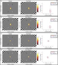
          We investigated lateral modulation effects with functional magnetic resonance imaging. We presented radial sinusoidal gratings in random sequence: a scotoma grating with two arc-shaped blank regions (scotomata) in the periphery, one in the left and one in the right visual field, a center grating containing pattern only in the scotoma regions, and a full-field grating where the pattern occupied the whole screen. On each trial, one of the three gratings flickered in counterphase for 10 s, followed by a blank period. Observers were instructed to perform a fixation task and report whether filling-in was experienced during the scotoma condition. The results showed that the blood-oxygen-level-dependent signal was reduced in areas corresponding to the scotoma regions in the full-field compared to the center condition in V1 to V3 areas, indicating a lateral inhibition effect when the surround was added to the center pattern. The univariate analysis results showed no difference between the filling-in and no-filling-in trials. However, multivariate pattern analysis results showed that classifiers trained on activation pattern in V1 to V3 could differentiate between filling-in and no-filling-in trials, suggesting that the neural activation pattern in visual cortex correlated with the subjective percept.

          Related collections

          Most cited references81

          • Record: found
          • Abstract: found
          • Article: not found

          LIBSVM: A library for support vector machines

          LIBSVM is a library for Support Vector Machines (SVMs). We have been actively developing this package since the year 2000. The goal is to help users to easily apply SVM to their applications. LIBSVM has gained wide popularity in machine learning and many other areas. In this article, we present all implementation details of LIBSVM. Issues such as solving SVM optimization problems theoretical convergence multiclass classification probability estimates and parameter selection are discussed in detail.
            Bookmark
            • Record: found
            • Abstract: found
            • Article: not found

            FreeSurfer.

            FreeSurfer is a suite of tools for the analysis of neuroimaging data that provides an array of algorithms to quantify the functional, connectional and structural properties of the human brain. It has evolved from a package primarily aimed at generating surface representations of the cerebral cortex into one that automatically creates models of most macroscopically visible structures in the human brain given any reasonable T1-weighted input image. It is freely available, runs on a wide variety of hardware and software platforms, and is open source. Copyright © 2012 Elsevier Inc. All rights reserved.
              Bookmark
              • Record: found
              • Abstract: found
              • Article: not found

              Cortical surface-based analysis. I. Segmentation and surface reconstruction.

              Several properties of the cerebral cortex, including its columnar and laminar organization, as well as the topographic organization of cortical areas, can only be properly understood in the context of the intrinsic two-dimensional structure of the cortical surface. In order to study such cortical properties in humans, it is necessary to obtain an accurate and explicit representation of the cortical surface in individual subjects. Here we describe a set of automated procedures for obtaining accurate reconstructions of the cortical surface, which have been applied to data from more than 100 subjects, requiring little or no manual intervention. Automated routines for unfolding and flattening the cortical surface are described in a companion paper. These procedures allow for the routine use of cortical surface-based analysis and visualization methods in functional brain imaging. Copyright 1999 Academic Press.
                Bookmark

                Author and article information

                Contributors
                mark.greenlee@ur.de
                Journal
                Sci Rep
                Sci Rep
                Scientific Reports
                Nature Publishing Group UK (London )
                2045-2322
                27 September 2022
                27 September 2022
                2022
                : 12
                : 16143
                Affiliations
                [1 ]GRID grid.7727.5, ISNI 0000 0001 2190 5763, Institute of Experimental Psychology, , University of Regensburg, ; Universitätsstraße 31, 93053 Regensburg, Germany
                [2 ]GRID grid.19188.39, ISNI 0000 0004 0546 0241, Department of Psychology, , National Taiwan University, ; Taipei, Taiwan
                [3 ]GRID grid.19188.39, ISNI 0000 0004 0546 0241, Neurobiology and Cognitive Science Center, , National Taiwan University, ; Taipei, Taiwan
                Article
                20592
                10.1038/s41598-022-20592-y
                9515077
                36167763
                92b1b84e-c08c-426f-9810-f56e1a033539
                © The Author(s) 2022

                Open Access This article is licensed under a Creative Commons Attribution 4.0 International License, which permits use, sharing, adaptation, distribution and reproduction in any medium or format, as long as you give appropriate credit to the original author(s) and the source, provide a link to the Creative Commons licence, and indicate if changes were made. The images or other third party material in this article are included in the article's Creative Commons licence, unless indicated otherwise in a credit line to the material. If material is not included in the article's Creative Commons licence and your intended use is not permitted by statutory regulation or exceeds the permitted use, you will need to obtain permission directly from the copyright holder. To view a copy of this licence, visit http://creativecommons.org/licenses/by/4.0/.

                History
                : 18 May 2022
                : 15 September 2022
                Funding
                Funded by: FundRef http://dx.doi.org/10.13039/501100001655, Deutscher Akademischer Austauschdienst;
                Award ID: Research Grants – Doctoral Programmes in Germany, 2017/18
                Award Recipient :
                Funded by: FundRef http://dx.doi.org/10.13039/501100004663, Ministry of Science and Technology, Taiwan;
                Award ID: TOP 2021
                Award ID: 109- 603 2410-H-002-086-MY4
                Award Recipient :
                Funded by: FundRef http://dx.doi.org/10.13039/501100001659, Deutsche Forschungsgemeinschaft;
                Award ID: GR988-27-1
                Award Recipient :
                Funded by: Universität Regensburg (3161)
                Categories
                Article
                Custom metadata
                © The Author(s) 2022

                Uncategorized
                pattern vision,human behaviour,striate cortex
                Uncategorized
                pattern vision, human behaviour, striate cortex

                Comments

                Comment on this article