13
views
0
recommends
+1 Recommend
0 collections
    0
    shares
      • Record: found
      • Abstract: found
      • Article: found
      Is Open Access

      A simple and reproducible method for quantification of human tear lipids with ultrahigh-performance liquid chromatography-mass spectrometry

      research-article

      Read this article at

      Bookmark
          There is no author summary for this article yet. Authors can add summaries to their articles on ScienceOpen to make them more accessible to a non-specialist audience.

          Abstract

          Purpose

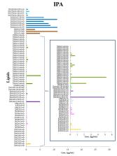
          The purpose was to select a simple and reproducible method for lipid measurements of human tears with ultrahigh-performance liquid chromatography-mass spectrometry (UHPLC-MS). Two sample preparation procedures were evaluated and compared: the Bligh and Dyer (BD) liquid-liquid extraction method with chloroform and methanol and protein precipitation with isopropanol (IPA).

          Methods

          Reproducibility and recovery efficiencies of 20 non-endogenous internal lipid standards were tested in 10-µl tear samples from healthy subjects. The lipid coverage and the simplicity of execution were also assessed. Lipid profiles of the tear extracts were acquired with UHPLC-MS, uhpland the lipids were identified using SimLipid software.

          Results

          Both methods were robust producing good lipid coverage and reproducibility and high recovery efficiencies. The two protocols identified a 69-feature tear lipidome that covered 11 lipid classes from six different lipid categories. The main differences in recovery were due to the intrinsic lipid selectivity of each solvent. Although both methods were similarly efficient in recovering O-acyl-ω-hydroxy fatty acid (OAHFAs) and non-polar lipids, polar lipids were more efficiently recovered with IPA precipitation, which, in turn, exhibited higher reproducibility. In addition, IPA precipitation is automatable and simpler than the BD approach.

          Conclusions

          IPA precipitation is an excellent procedure for extracting lipids from small tear volumes for quantitative large-scale, untargeted lipid profiling, which may be useful for identifying lipid biomarkers in tears from patients with different ocular surface pathologies, allowing personalized therapies to be designed.

          Related collections

          Most cited references27

          • Record: found
          • Abstract: not found
          • Article: not found

          A rapid method of total lipid extraction and purification.

            Bookmark
            • Record: found
            • Abstract: found
            • Article: not found

            Extensive characterization of human tear fluid collected using different techniques unravels the presence of novel lipid amphiphiles.

            The tear film covers the anterior eye and the precise balance of its various constituting components is critical for maintaining ocular health. The composition of the tear film amphiphilic lipid sublayer, in particular, has largely remained a matter of contention due to the limiting concentrations of these lipid amphiphiles in tears that render their detection and accurate quantitation tedious. Using systematic and sensitive lipidomic approaches, we validated different tear collection techniques and report the most comprehensive human tear lipidome to date; comprising more than 600 lipid species from 17 major lipid classes. Our study confers novel insights to the compositional details of the existent tear film model, in particular the disputable amphiphilic lipid sublayer constituents, by demonstrating the presence of cholesteryl sulfate, O-acyl-ω-hydroxyfatty acids, and various sphingolipids and phospholipids in tears. The discovery and quantitation of the relative abundance of various tear lipid amphiphiles reported herein are expected to have a profound impact on the current understanding of the existent human tear film model.
              Bookmark
              • Record: found
              • Abstract: found
              • Article: not found

              Tear analysis in ocular surface diseases.

              The thin layer of tears covering the ocular surface are a complex body fluid containing thousands of molecules of varied form and function of several origins. In this review, we have discussed some key issues in the analysis of tears in the context of understanding and diagnosing eye disease using current technologies of proteomics and metabolomics, and for their potential for clinical application. In the last several years, advances in proteomics/metabolomics/lipidomics technologies have greatly expanded our knowledge of the chemical composition of tear fluid. The quickened pace of studies has shown that tears as a complex extra-cellular fluid of the ocular surface contains a great deal of molecular information useful for the diagnosis, prognosis, and treatment of ocular surface diseases that has the ability to addresses the emphasis on personalized medicine and biomarkers of disease. Future research directions will likely include (1) standardize tear collection, storage, extraction, and sample preparation; (2) quantitative proteomic analysis of tear proteins using multiple reaction monitoring (MRM)-based mass spectrometry; (3) population based studies of human tear proteomics/metabolomics; (4) tear proteomics/metabolomics for systemic diseases; and (5) functional studies of tear proteins. Copyright © 2012 Elsevier Ltd. All rights reserved.
                Bookmark

                Author and article information

                Journal
                Mol Vis
                Mol. Vis
                MV
                Molecular Vision
                Molecular Vision
                1090-0535
                2019
                31 December 2019
                : 25
                : 934-948
                Affiliations
                [1 ]Department of Cell Biology and Histology, Faculty of Medicine and Nursing, University of the Basque Country UPV/EHU, Leioa, Spain.
                [2 ]Servicio Central de Análisis, Facultad de Ciencia y Tecnología, University of the Basque Country UPV/EHU, Leioa, Spain
                [3 ]Department of Physiology, Faculty of Medicine and Nursing, University of the Basque Country UPV/EHU, Leioa, Spain.
                [4 ]Department of Organic Chemistry II, Faculty of Science and Technology, University of the Basque Country UPV/EHU, Leioa, Spain
                [5 ]Instituto Clínico Quirurgico de Oftalmología and University of the Basque Country, UPV/EHU, Bizkaia, Spain
                Author notes
                Correspondence to: E. Vecino, Department of Cell Biology and Histology, University of the Basque Country UPV/EHU, Leioa, Spain; +34946012820; FAX: +34946012881; email: elena.vecino@ehu.eus
                Article
                85 2019MOLVIS0168
                6992877
                32038094
                927dc1ed-d5cd-4293-950a-5a4c6d881119
                Copyright © 2019 Molecular Vision.

                This is an open-access article distributed under the terms of the Creative Commons Attribution License, which permits unrestricted use, distribution, and reproduction in any medium, provided the original work is properly cited, used for non-commercial purposes, and is not altered or transformed.

                History
                : 17 June 2019
                : 31 December 2019
                Categories
                Research Article
                Custom metadata
                Export to XML

                Vision sciences
                Vision sciences

                Comments

                Comment on this article

                Similar content104

                Cited by7

                Most referenced authors503