6
views
0
recommends
+1 Recommend
0 collections
    0
    shares
      • Record: found
      • Abstract: found
      • Article: found
      Is Open Access

      Surgical management of scalp arterio-venous malformation and scalp venous malformation: An experience of eleven cases

      research-article

      Read this article at

      Bookmark
          There is no author summary for this article yet. Authors can add summaries to their articles on ScienceOpen to make them more accessible to a non-specialist audience.

          Abstract

          Aims:

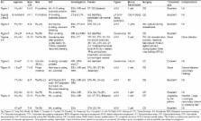
          Scalp arterio-venous malformation (AVM) and scalp venous malformation (SVM) are rare conditions that usually need surgical treatment. Here, we have reported our experience of the surgical management of such lesions with a short review of the literature.

          Materials and Methods:

          In this prospective study, 11 patients with scalp AVM and SVM, who underwent surgical excision of lesion in our hospital from 2006 to 2012, were included. All suspected high-flow AVM were investigated with the selective internal and external carotid digital subtraction angiogram (DSA) ± computed tomography (CT) scan of brain with CT angiogram or magnetic resonance imaging (MRI) of brain with MR angiogram, and all suspected low-flow vascular malformation (VM) was investigated with MRI of brain + MR angiogram. Eight were high-flow and three were low-flow VM.

          Results:

          All lesions were successfully excised. Scalp cosmetic aspects were acceptable in all cases. There was no major post-operative complication or recurrence till last follow-up.

          Conclusions:

          With preoperative appropriate surgical planning, scalp AVM and SVM can be excised without major complication.

          Related collections

          Most cited references24

          • Record: found
          • Abstract: found
          • Article: not found

          Arteriovenous malformations of the scalp.

          We report twenty-four patients with cirsoid aneurysms of the scalp. For nine patients (38%), the lesions were related to trauma. Each of the patients presented with a pulsatile scalp swelling with a bruit. No focal neurological deficits were noted in any of the patients. Scalp malformations in all patients were confirmed by selective internal and external carotid angiography, with no intracerebral component revealed in any of the patients. Twenty-one patients had the lesions surgically excised, with good results. The remaining three refused surgical intervention. Meticulous surgical technique, which includes removal of the pericranial component of the malformation, was paramount.
            Bookmark
            • Record: found
            • Abstract: found
            • Article: not found

            Traumatic arteriovenous fistula of the superficial temporal artery.

            Arteriovenous fistula of the scalp is relatively rare. The superficial temporal artery is particularly vulnerable to trauma due to its long and relatively exposed course in the scalp. We report a traumatic arteriovenous fistula of the superficial temporal artery treated by complete surgical excision and review the literature with regard to etiology, clinical manifestations, pathogenesis, diagnosis and management of these unusual lesions.
              Bookmark
              • Record: found
              • Abstract: found
              • Article: not found

              Scalp arteriovenous malformations.

              We discuss our experience with the surgical management of scalp vascular malformation and review the literature on the subject. A prospective case-control study of eight patients with scalp vascular malformations admitted to our hospital between 1997 and 2002. All the patients were investigated with selective internal and external carotid angiography. Depending upon the origin of feeding arteries, the scalp vascular malformations were classified into two categories: Group I: the primary scalp arteriovenous malformations and Group II: secondary venous dilatations. Six patients belonged to Group I and two patients were in Group II. Five patients belonging to Group I underwent successful excision of the arteriovenous malformation. There was no recurrence in this group. Of the two patients in Group II, one patient who had scalp vascular dilatation simulating a primary scalp vascular malformation underwent excision of the lesion. This patient developed severe postoperative brain edema and died. Primary scalp vascular malformation can be excised safely. However, excision of secondary scalp venous dilatation without treatment of the intracranial component can be dangerous.
                Bookmark

                Author and article information

                Journal
                Indian J Plast Surg
                Indian J Plast Surg
                IJPS
                Indian Journal of Plastic Surgery : Official Publication of the Association of Plastic Surgeons of India
                Medknow Publication & Media Pvt Ltd (India )
                0970-0358
                1998-376X
                Jan-Apr 2013
                : 46
                : 1
                : 98-107
                Affiliations
                [1]Department of Neurosurgery, Dhaka Medical College Hospital, Dhaka, Bangladesh
                Author notes
                Address for correspondence: Dr. Forhad Hossain Chowdhury, Department of Neurosurgery, Dhaka Medical College Hospital, Dhaka, Bangladesh. E-mail: forhadchowdhury74@ 123456yahoo.com
                Article
                IJPS-46-98
                10.4103/0970-0358.113723
                3745130
                23960313
                90c000a7-a5af-49c9-9084-ce6a6acbf149
                Copyright: © Indian Journal of Plastic Surgery

                This is an open-access article distributed under the terms of the Creative Commons Attribution-Noncommercial-Share Alike 3.0 Unported, which permits unrestricted use, distribution, and reproduction in any medium, provided the original work is properly cited.

                History
                Categories
                Original Article

                Surgery
                arterio-venous malformation,cavernous angioma,cirsoid aneurysm,scalp avm,scalp venous malformatiom,sinus pericranii

                Comments

                Comment on this article