34
views
0
recommends
+1 Recommend
0 collections
    0
    shares
      • Record: found
      • Abstract: found
      • Article: found
      Is Open Access

      Structures of membrane proteins

      review-article
      * , *
      Quarterly Reviews of Biophysics
      Cambridge University Press

      Read this article at

      Bookmark
          There is no author summary for this article yet. Authors can add summaries to their articles on ScienceOpen to make them more accessible to a non-specialist audience.

          Abstract

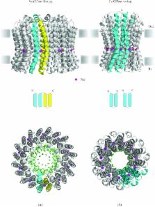
          In reviewing the structures of membrane proteins determined up to the end of 2009, we present in words and pictures the most informative examples from each family. We group the structures together according to their function and architecture to provide an overview of the major principles and variations on the most common themes. The first structures, determined 20 years ago, were those of naturally abundant proteins with limited conformational variability, and each membrane protein structure determined was a major landmark. With the advent of complete genome sequences and efficient expression systems, there has been an explosion in the rate of membrane protein structure determination, with many classes represented. New structures are published every month and more than 150 unique membrane protein structures have been determined. This review analyses the reasons for this success, discusses the challenges that still lie ahead, and presents a concise summary of the key achievements with illustrated examples selected from each class.

          Related collections

          Most cited references325

          • Record: found
          • Abstract: found
          • Article: not found

          High-resolution crystal structure of an engineered human beta2-adrenergic G protein-coupled receptor.

          Heterotrimeric guanine nucleotide-binding protein (G protein)-coupled receptors constitute the largest family of eukaryotic signal transduction proteins that communicate across the membrane. We report the crystal structure of a human beta2-adrenergic receptor-T4 lysozyme fusion protein bound to the partial inverse agonist carazolol at 2.4 angstrom resolution. The structure provides a high-resolution view of a human G protein-coupled receptor bound to a diffusible ligand. Ligand-binding site accessibility is enabled by the second extracellular loop, which is held out of the binding cavity by a pair of closely spaced disulfide bridges and a short helical segment within the loop. Cholesterol, a necessary component for crystallization, mediates an intriguing parallel association of receptor molecules in the crystal lattice. Although the location of carazolol in the beta2-adrenergic receptor is very similar to that of retinal in rhodopsin, structural differences in the ligand-binding site and other regions highlight the challenges in using rhodopsin as a template model for this large receptor family.
            Bookmark
            • Record: found
            • Abstract: found
            • Article: not found

            Crystal structure of rhodopsin: A G protein-coupled receptor.

            Heterotrimeric guanine nucleotide-binding protein (G protein)-coupled receptors (GPCRs) respond to a variety of different external stimuli and activate G proteins. GPCRs share many structural features, including a bundle of seven transmembrane alpha helices connected by six loops of varying lengths. We determined the structure of rhodopsin from diffraction data extending to 2.8 angstroms resolution. The highly organized structure in the extracellular region, including a conserved disulfide bridge, forms a basis for the arrangement of the seven-helix transmembrane motif. The ground-state chromophore, 11-cis-retinal, holds the transmembrane region of the protein in the inactive conformation. Interactions of the chromophore with a cluster of key residues determine the wavelength of the maximum absorption. Changes in these interactions among rhodopsins facilitate color discrimination. Identification of a set of residues that mediate interactions between the transmembrane helices and the cytoplasmic surface, where G-protein activation occurs, also suggests a possible structural change upon photoactivation.
              Bookmark
              • Record: found
              • Abstract: found
              • Article: not found

              Molecular basis of bacterial outer membrane permeability revisited.

              Gram-negative bacteria characteristically are surrounded by an additional membrane layer, the outer membrane. Although outer membrane components often play important roles in the interaction of symbiotic or pathogenic bacteria with their host organisms, the major role of this membrane must usually be to serve as a permeability barrier to prevent the entry of noxious compounds and at the same time to allow the influx of nutrient molecules. This review summarizes the development in the field since our previous review (H. Nikaido and M. Vaara, Microbiol. Rev. 49:1-32, 1985) was published. With the discovery of protein channels, structural knowledge enables us to understand in molecular detail how porins, specific channels, TonB-linked receptors, and other proteins function. We are now beginning to see how the export of large proteins occurs across the outer membrane. With our knowledge of the lipopolysaccharide-phospholipid asymmetric bilayer of the outer membrane, we are finally beginning to understand how this bilayer can retard the entry of lipophilic compounds, owing to our increasing knowledge about the chemistry of lipopolysaccharide from diverse organisms and the way in which lipopolysaccharide structure is modified by environmental conditions.
                Bookmark

                Author and article information

                Journal
                Q Rev Biophys
                Q. Rev. Biophys
                QRB
                Quarterly Reviews of Biophysics
                Cambridge University Press (New York, USA )
                0033-5835
                1469-8994
                February 2010
                February 2010
                : 43
                : 1
                : 65-158
                Affiliations
                [001]MRC Laboratory of Molecular Biology, Cambridge, UK
                Author notes
                [* ]Authors for correspondence: K. R. Vinothkumar and R. Henderson, MRC Laboratory of Molecular Biology, Hill Road, Cambridge CB2 0QH, UK. Tel.: 44-1223-402405 and 44-1223-402215; E-mail: vkumar@ 123456mrc-lmb.cam.ac.uk and rh15@ 123456mrc-lmb.cam.ac.uk
                Article
                S0033583510000041 00004
                10.1017/S0033583510000041
                3604715
                20667175
                90bea40b-020b-48be-88f9-5cd608ae61d5
                Copyright © Cambridge University Press 2010 The online version of this article is published within an Open Access environment subject to the conditions of the Creative Commons Attribution-NonCommercial-ShareAlike licence <http://creativecommons.org/licenses/by-nc-sa/2.5/>. The written permission of Cambridge University Press must be obtained for commercial re-use.

                The online version of this article is published within an Open Access environment subject to the conditions of the Creative Commons Attribution-NonCommercial-ShareAlike licence < http://creativecommons.org/licenses/by-nc-sa/2.5/>. The written permission of Cambridge University Press must be obtained for commercial re-use.

                History
                Page count
                Pages: 94
                Categories
                Review Article

                Comments

                Comment on this article