104
views
0
recommends
+1 Recommend
0 collections
    0
    shares
      • Record: found
      • Abstract: found
      • Article: found
      Is Open Access

      Alteration of Basal Ganglia and Right Frontoparietal Network in Early Drug-Naïve Parkinson’s Disease during Heat Pain Stimuli and Resting State

      research-article

      Read this article at

      Bookmark
          There is no author summary for this article yet. Authors can add summaries to their articles on ScienceOpen to make them more accessible to a non-specialist audience.

          Abstract

          Background

          The symptoms and pathogenesis of Parkinson’s disease (PD) are complicated and an accurate diagnosis of PD is difficult, particularly in early-stage. Because functional magnetic resonance imaging (fMRI) is non-invasive and is characterized by the integration of different brain areas in terms of functional connectivity (FC), fMRI has been widely used in PD research. Non-motor symptom (NMS) features are also frequently present in PD before the onset of classical motor symptoms with pain as the primary NMS. Considering that PD could affect the pain process at multiple levels, we hypothesized that pain is one of the earliest symptoms in PD and investigated whether FC of the pain network was disrupted in PD without pain. To better understand the pathogenesis of pain in PD, we combined resting state and pain-stimuli-induced task state fMRI to identify alterations in FC related to pain in PD.

          Methods

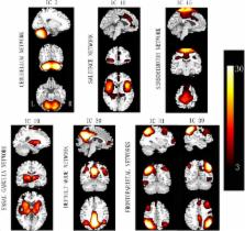
          Fourteen early drug-naïve PD without pain and 17 age- and sex-matched healthy controls (HC) participated in our testing task. We used independent component analysis to select seven functional networks related to PD and pain. We focused on abnormalities in FC and in functional network connectivity (FNC) in PD compared with HC during the task (51°C heat pain stimuli) and at rest.

          Results

          Compared with HC, PD showed decreased FC in putamen within basal ganglia network (BGN) in task state and decreased FC in putamen of salience network (SN) and mid-cingulate cortex of sensorimotor network in rest state. FNC between the BGN and the SN are reduced during both states in PD compared with HC. In addition, right frontoparietal network (RFPN), which is considered as a bridge between the SN and default-mode network, was significantly disturbed during the task.

          Conclusion

          These findings suggest that BGN plays a role in the pathological mechanisms of pain underlying PD, and RFPN likely contributes greatly to harmonization between intrinsic brain activity and external stimuli.

          Related collections

          Most cited references32

          • Record: found
          • Abstract: found
          • Article: not found

          A method for functional network connectivity among spatially independent resting-state components in schizophrenia.

          Functional connectivity of the brain has been studied by analyzing correlation differences in time courses among seed voxels or regions with other voxels of the brain in healthy individuals as well as in patients with brain disorders. The spatial extent of strongly temporally coherent brain regions co-activated during rest has also been examined using independent component analysis (ICA). However, the weaker temporal relationships among ICA component time courses, which we operationally define as a measure of functional network connectivity (FNC), have not yet been studied. In this study, we propose an approach for evaluating FNC and apply it to functional magnetic resonance imaging (fMRI) data collected from persons with schizophrenia and healthy controls. We examined the connectivity and latency among ICA component time courses to test the hypothesis that patients with schizophrenia would show increased functional connectivity and increased lag among resting state networks compared to controls. Resting state fMRI data were collected and the inter-relationships among seven selected resting state networks (identified using group ICA) were evaluated by correlating each subject's ICA time courses with one another. Patients showed higher correlation than controls among most of the dominant resting state networks. Patients also had slightly more variability in functional connectivity than controls. We present a novel approach for quantifying functional connectivity among brain networks identified with spatial ICA. Significant differences between patient and control connectivity in different networks were revealed possibly reflecting deficiencies in cortical processing in patients.
            Bookmark
            • Record: found
            • Abstract: found
            • Article: not found

            Causal interactions between fronto-parietal central executive and default-mode networks in humans.

            Information processing during human cognitive and emotional operations is thought to involve the dynamic interplay of several large-scale neural networks, including the fronto-parietal central executive network (CEN), cingulo-opercular salience network (SN), and the medial prefrontal-medial parietal default mode networks (DMN). It has been theorized that there is a causal neural mechanism by which the CEN/SN negatively regulate the DMN. Support for this idea has come from correlational neuroimaging studies; however, direct evidence for this neural mechanism is lacking. Here we undertook a direct test of this mechanism by combining transcranial magnetic stimulation (TMS) with functional MRI to causally excite or inhibit TMS-accessible prefrontal nodes within the CEN or SN and determine consequent effects on the DMN. Single-pulse excitatory stimulations delivered to only the CEN node induced negative DMN connectivity with the CEN and SN, consistent with the CEN/SN's hypothesized negative regulation of the DMN. Conversely, low-frequency inhibitory repetitive TMS to the CEN node resulted in a shift of DMN signal from its normally low-frequency range to a higher frequency, suggesting disinhibition of DMN activity. Moreover, the CEN node exhibited this causal regulatory relationship primarily with the medial prefrontal portion of the DMN. These findings significantly advance our understanding of the causal mechanisms by which major brain networks normally coordinate information processing. Given that poorly regulated information processing is a hallmark of most neuropsychiatric disorders, these findings provide a foundation for ways to study network dysregulation and develop brain stimulation treatments for these disorders.
              Bookmark
              • Record: found
              • Abstract: found
              • Article: not found

              Cognitive impairment and resting-state network connectivity in Parkinson's disease.

              The purpose of this work was to evaluate changes in the connectivity patterns of a set of cognitively relevant, dynamically interrelated brain networks in association with cognitive deficits in Parkinson's disease (PD) using resting-state functional MRI. Sixty-five nondemented PD patients and 36 matched healthy controls were included. Thirty-four percent of PD patients were classified as having mild cognitive impairment (MCI) based on performance in attention/executive, visuospatial/visuoperceptual (VS/VP) and memory functions. A data-driven approach using independent component analysis (ICA) was used to identify the default-mode network (DMN), the dorsal attention network (DAN) and the bilateral frontoparietal networks (FPN), which were compared between groups using a dual-regression approach controlling for gray matter atrophy. Additional seed-based analyses using a priori defined regions of interest were used to characterize local changes in intranetwork and internetwork connectivity. Structural group comparisons through voxel-based morphometry and cortical thickness were additionally performed to assess associated gray matter atrophy. ICA results revealed reduced connectivity between the DAN and right frontoinsular regions in MCI patients, associated with worse performance in attention/executive functions. The DMN displayed increased connectivity with medial and lateral occipito-parietal regions in MCI patients, associated with worse VS/VP performance, and with occipital reductions in cortical thickness. In line with data-driven results, seed-based analyses mainly revealed reduced within-DAN, within-DMN and DAN-FPN connectivity, as well as loss of normal DAN-DMN anticorrelation in MCI patients. Our findings demonstrate differential connectivity changes affecting the networks evaluated, which we hypothesize to be related to the pathophysiological bases of different types of cognitive impairment in PD. © 2014 Wiley Periodicals, Inc.
                Bookmark

                Author and article information

                Contributors
                Journal
                Front Hum Neurosci
                Front Hum Neurosci
                Front. Hum. Neurosci.
                Frontiers in Human Neuroscience
                Frontiers Media S.A.
                1662-5161
                24 August 2015
                2015
                : 9
                : 467
                Affiliations
                [1] 1Key Laboratory for NeuroInformation of Ministry of Education, High-Field Magnetic Resonance Brain Imaging Key Laboratory of Sichuan Province, Center for Information in Medicine, University of Electronic Science and Technology of China , Chengdu, China
                [2] 2School of Computer Science and Technology, Southwest University for Nationalities , Chengdu, China
                [3] 3Neurology Department, Affiliated Hospital of North Sichuan Medical College, North Sichuan Medical College , Nanchong, China
                [4] 4School of Life and Science Engineering, Southwest Jiaotong University , Chengdu, China
                Author notes

                Edited by: Rachael D. Seidler, University of Michigan, USA

                Reviewed by: Mingrui Xia, Beijing Normal University, China; Manousos A. Klados, Max Planck Institute for Human Cognitive and Brain Sciences, Germany

                *Correspondence: Xiaoming Wang and Cheng Luo, University of Electronic Science and Technology of China, No. 2006, Xiyuan Avenue, Chengdu 611731, China, wangxm238@ 123456163.com ; chengluo@ 123456uestc.edu.cn

                Ying Tan and Juan Tan have contributed equally to this work.

                Article
                10.3389/fnhum.2015.00467
                4547030
                26379530
                9094e70f-9db6-4d24-b454-3de3a9ce915b
                Copyright © 2015 Tan, Tan, Deng, Cui, He, Yang, Deng, Xiao, Huang, Zhang, Tan, Shen, Liu, Wang, Yao and Luo.

                This is an open-access article distributed under the terms of the Creative Commons Attribution License (CC BY). The use, distribution or reproduction in other forums is permitted, provided the original author(s) or licensor are credited and that the original publication in this journal is cited, in accordance with accepted academic practice. No use, distribution or reproduction is permitted which does not comply with these terms.

                History
                : 31 March 2015
                : 10 August 2015
                Page count
                Figures: 5, Tables: 2, Equations: 0, References: 49, Pages: 9, Words: 6265
                Categories
                Neuroscience
                Original Research

                Neurosciences
                parkinson’s disease,fmri,functional connectivity,functional network connectivity,basal ganglia network,right frontoparietal network

                Comments

                Comment on this article