439
views
0
recommends
+1 Recommend
0 collections
    0
    shares
      • Record: found
      • Abstract: found
      • Article: found
      Is Open Access

      Characterization of Discrete Subpopulations of Progenitor Cells in Traumatic Human Extremity Wounds

      research-article

      Read this article at

      Bookmark
          There is no author summary for this article yet. Authors can add summaries to their articles on ScienceOpen to make them more accessible to a non-specialist audience.

          Abstract

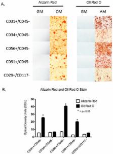
          Here we show that distinct subpopulations of cells exist within traumatic human extremity wounds, each having the ability to differentiate into multiple cells types in vitro. A crude cell suspension derived from traumatized muscle was positively sorted for CD29, CD31, CD34, CD56 or CD91. The cell suspension was also simultaneously negatively sorted for either CD45 or CD117 to exclude hematopoietic stem cells. These subpopulations varied in terms their total numbers and their abilities to grow, migrate, differentiate and secrete cytokines. While all five subpopulations demonstrated equal abilities to undergo osteogenesis, they were distinct in their ability to undergo adipogenesis and vascular endotheliogenesis. The most abundant subpopulations were CD29+ and CD34+, which overlapped significantly. The CD29+ and CD34+ cells had the greatest proliferative and migratory capacity while the CD56+ subpopulation produced the highest amounts of TGFß1 and TGFß2. When cultured under endothelial differentiation conditions the CD29+ and CD34+ cells expressed VE-cadherin, Tie2 and CD31, all markers of endothelial cells. These data indicate that while there are multiple cell types within traumatized muscle that have osteogenic differentiation capacity and may contribute to bone formation in post-traumatic heterotopic ossification (HO), the major contributory cell types are CD29+ and CD34+, which demonstrate endothelial progenitor cell characteristics.

          Related collections

          Most cited references47

          • Record: found
          • Abstract: found
          • Article: not found

          Conversion of vascular endothelial cells into multipotent stem-like cells.

          Mesenchymal stem cells can give rise to several cell types, but varying results depending on isolation methods and tissue source have led to controversies about their usefulness in clinical medicine. Here we show that vascular endothelial cells can transform into multipotent stem-like cells by an activin-like kinase-2 (ALK2) receptor-dependent mechanism. In lesions from individuals with fibrodysplasia ossificans progressiva (FOP), a disease in which heterotopic ossification occurs as a result of activating ALK2 mutations, or from transgenic mice expressing constitutively active ALK2, chondrocytes and osteoblasts expressed endothelial markers. Lineage tracing of heterotopic ossification in mice using a Tie2-Cre construct also suggested an endothelial origin of these cell types. Expression of constitutively active ALK2 in endothelial cells caused endothelial-to-mesenchymal transition and acquisition of a stem cell-like phenotype. Similar results were obtained by treatment of untransfected endothelial cells with the ligands transforming growth factor-β2 (TGF-β2) or bone morphogenetic protein-4 (BMP4) in an ALK2-dependent manner. These stem-like cells could be triggered to differentiate into osteoblasts, chondrocytes or adipocytes. We suggest that conversion of endothelial cells to stem-like cells may provide a new approach to tissue engineering.
            Bookmark
            • Record: found
            • Abstract: found
            • Article: not found

            Expression of Cd34 and Myf5 Defines the Majority of Quiescent Adult Skeletal Muscle Satellite Cells

            Skeletal muscle is one of a several adult post-mitotic tissues that retain the capacity to regenerate. This relies on a population of quiescent precursors, termed satellite cells. Here we describe two novel markers of quiescent satellite cells: CD34, an established marker of hematopoietic stem cells, and Myf5, the earliest marker of myogenic commitment. CD34+ve myoblasts can be detected in proliferating C2C12 cultures. In differentiating cultures, CD34+ve cells do not fuse into myotubes, nor express MyoD. Using isolated myofibers as a model of synchronous precursor cell activation, we show that quiescent satellite cells express CD34. An early feature of their activation is alternate splicing followed by complete transcriptional shutdown of CD34. This data implicates CD34 in the maintenance of satellite cell quiescence. In heterozygous Myf5 nlacZ/+ mice, all CD34+ve satellite cells also express β-galactosidase, a marker of activation of Myf5, showing that quiescent satellite cells are committed to myogenesis. All such cells are positive for the accepted satellite cell marker, M-cadherin. We also show that satellite cells can be identified on isolated myofibers of the myosin light chain 3F-nlacZ-2E mouse as those that do not express the transgene. The numbers of satellite cells detected in this way are significantly greater than those identified by the other three markers. We conclude that the expression of CD34, Myf5, and M-cadherin defines quiescent, committed precursors and speculate that the CD34−ve, Myf5−ve minority may be involved in maintaining the lineage-committed majority.
              Bookmark
              • Record: found
              • Abstract: found
              • Article: not found

              LRP: role in vascular wall integrity and protection from atherosclerosis.

              Vascular smooth muscle cell (SMC) proliferation and migration are important events in the development of atherosclerosis. The low-density lipoprotein receptor-related protein (LRP1) mediates suppression of SMC migration induced by platelet-derived growth factor (PDGF). Here we show that LRP1 forms a complex with the PDGF receptor (PDGFR). Inactivation of LRP1 in vascular SMCs of mice causes PDGFR overexpression and abnormal activation of PDGFR signaling, resulting in disruption of the elastic layer, SMC proliferation, aneurysm formation, and marked susceptibility to cholesterol-induced atherosclerosis. The development of these abnormalities was reduced by treatment with Gleevec, an inhibitor of PDGF signaling. Thus, LRP1 has a pivotal role in protecting vascular wall integrity and preventing atherosclerosis by controlling PDGFR activation.
                Bookmark

                Author and article information

                Contributors
                Role: Editor
                Journal
                PLoS One
                PLoS ONE
                plos
                plosone
                PLoS ONE
                Public Library of Science (San Francisco, USA )
                1932-6203
                2014
                9 December 2014
                : 9
                : 12
                : e114318
                Affiliations
                [1 ]Department of Surgery, Uniformed Services University of Health Sciences, Bethesda, MD, United States of America
                [2 ]Clinical and Experimental Orthopaedics, National Institute of Arthritis and Musculoskeletal and Skin Diseases, National Institutes of Health, Bethesda, MD, United States of America
                [3 ]Laboratory of Genome Integrity, Nation Cancer Institute, National Institutes of Health, Bethesda, MD, United States of America
                [4 ]Department of Orthopaedic Surgery, Walter Reed National Military Medical Center, Bethesda, MD, United States of America
                University of California, San Diego, United States of America
                Author notes

                Competing Interests: The authors have declared that no competing interests exist.

                Conceived and designed the experiments: LJN WMJ GEW DJH. Performed the experiments: GEW DJH GTC YJ. Analyzed the data: GEW DJH YJ LJN WMJ KMW. Contributed reagents/materials/analysis tools: GEW YJ GTC DJH KMW. Wrote the paper: GEW DJH WMJ LJN.

                Article
                PONE-D-14-26277
                10.1371/journal.pone.0114318
                4260839
                25490403
                9071dcd9-84d3-4af6-8446-b69e2bf743a7
                Copyright @ 2014

                This is an open-access article, free of all copyright, and may be freely reproduced, distributed, transmitted, modified, built upon, or otherwise used by anyone for any lawful purpose. The work is made available under the Creative Commons CC0 public domain dedication.

                History
                : 23 June 2014
                : 5 November 2014
                Page count
                Pages: 20
                Funding
                Support was provided by the Military Amputee Research Program at Walter Reed Army Medical Center (P05-A011), the Comprehensive Neurosciences Program (CNP-2008-CR01), the Defense Medical Research and Development Program (D10_1_AR_J5_920) and the NIH Intramural Research Program (Z01 AR41131). The funders had no role in study design, data collection and analysis, decision to publish, or preparation of the manuscript.
                Categories
                Research Article
                Biology and Life Sciences
                Biotechnology
                Bioengineering
                Tissue Engineering
                Cell Biology
                Cellular Types
                Animal Cells
                Stem Cells
                Adult Stem Cells
                Mesenchymal Stem Cells
                Stem Cell Niche
                Physiology
                Physiological Processes
                Tissue Repair
                Wound Healing
                Engineering and Technology
                Medicine and Health Sciences
                Clinical Genetics
                Stem Cell Therapy
                Surgical and Invasive Medical Procedures
                Transplantation
                Cell Transplantation
                Custom metadata
                The authors confirm that all data underlying the findings are fully available without restriction. All relevant data are within the paper.

                Uncategorized
                Uncategorized

                Comments

                Comment on this article