13
views
0
recommends
+1 Recommend
0 collections
    0
    shares
      • Record: found
      • Abstract: found
      • Article: found
      Is Open Access

      Improved Diagnostics Help to Identify Clinical Features and Biomarkers That Predict Mycoplasma pneumoniae Community-acquired Pneumonia in Children

      research-article

      Read this article at

      Bookmark
          There is no author summary for this article yet. Authors can add summaries to their articles on ScienceOpen to make them more accessible to a non-specialist audience.

          Abstract

          Background

          There are no reliable signs or symptoms that differentiate Mycoplasma pneumoniae ( Mp) infection in community-acquired pneumonia (CAP) from other etiologies. Additionally, current diagnostic tests do not reliably distinguish between Mp infection and carriage. We previously determined that the measurement of Mp-specific immunoglobulin M antibody-secreting cells (ASCs) by enzyme-linked immunospot assay allowed for differentiation between infection and carriage. Using this new diagnostic test, we aimed to identify clinical and laboratory features associated with Mp infection.

          Methods

          This is a prospective cohort study of children, 3–18 years of age, with CAP from 2016 to 2017. Clinical features and biomarkers were compared between Mp-positive and -negative groups by Mann-Whitney U test or Fisher exact test, as appropriate. Area under the receiver operating characteristic curve (AUC) differences and optimal thresholds were determined by using the DeLong test and Youden J statistic, respectively.

          Results

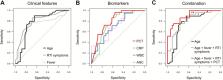
          Of 63 CAP patients, 29 were Mp-positive (46%). Mp positivity was statistically associated with older age (median, 8.6 vs 4.7 years), no underlying disease, family with respiratory symptoms, prior antibiotic treatment, prolonged prodromal respiratory symptoms and fever, and extrapulmonary (skin) manifestations. Lower levels of C-reactive protein, white blood cell count, absolute neutrophil count, and procalcitonin (PCT), specifically PCT <0.25 μg/L, were statistically associated with Mp infection. A combination of age >5 years (AUC = 0.77), prodromal fever and respiratory symptoms >6 days (AUC = 0.79), and PCT <0.25 μg/L (AUC = 0.81) improved diagnostic performance (AUC = 0.90) ( P = .05).

          Conclusions

          A combination of clinical features and biomarkers may aid physicians in identifying patients at high risk for Mp CAP.

          Abstract

          Community-acquired pneumonia (CAP) caused by Mycoplasma pneumoniae ( Mp) is difficult to diagnose. A combination of clinical features and biomarkers may aid physicians in identifying patients at high risk for Mp CAP.

          Related collections

          Most cited references40

          • Record: found
          • Abstract: found
          • Article: not found

          Community-acquired pneumonia requiring hospitalization among U.S. children.

          Incidence estimates of hospitalizations for community-acquired pneumonia among children in the United States that are based on prospective data collection are limited. Updated estimates of pneumonia that has been confirmed radiographically and with the use of current laboratory diagnostic tests are needed.
            Bookmark
            • Record: found
            • Abstract: found
            • Article: found
            Is Open Access

            Causes of severe pneumonia requiring hospital admission in children without HIV infection from Africa and Asia: the PERCH multi-country case-control study

            Summary Background Pneumonia is the leading cause of death among children younger than 5 years. In this study, we estimated causes of pneumonia in young African and Asian children, using novel analytical methods applied to clinical and microbiological findings. Methods We did a multi-site, international case-control study in nine study sites in seven countries: Bangladesh, The Gambia, Kenya, Mali, South Africa, Thailand, and Zambia. All sites enrolled in the study for 24 months. Cases were children aged 1–59 months admitted to hospital with severe pneumonia. Controls were age-group-matched children randomly selected from communities surrounding study sites. Nasopharyngeal and oropharyngeal (NP-OP), urine, blood, induced sputum, lung aspirate, pleural fluid, and gastric aspirates were tested with cultures, multiplex PCR, or both. Primary analyses were restricted to cases without HIV infection and with abnormal chest x-rays and to controls without HIV infection. We applied a Bayesian, partial latent class analysis to estimate probabilities of aetiological agents at the individual and population level, incorporating case and control data. Findings Between Aug 15, 2011, and Jan 30, 2014, we enrolled 4232 cases and 5119 community controls. The primary analysis group was comprised of 1769 (41·8% of 4232) cases without HIV infection and with positive chest x-rays and 5102 (99·7% of 5119) community controls without HIV infection. Wheezing was present in 555 (31·7%) of 1752 cases (range by site 10·6–97·3%). 30-day case-fatality ratio was 6·4% (114 of 1769 cases). Blood cultures were positive in 56 (3·2%) of 1749 cases, and Streptococcus pneumoniae was the most common bacteria isolated (19 [33·9%] of 56). Almost all cases (98·9%) and controls (98·0%) had at least one pathogen detected by PCR in the NP-OP specimen. The detection of respiratory syncytial virus (RSV), parainfluenza virus, human metapneumovirus, influenza virus, S pneumoniae, Haemophilus influenzae type b (Hib), H influenzae non-type b, and Pneumocystis jirovecii in NP-OP specimens was associated with case status. The aetiology analysis estimated that viruses accounted for 61·4% (95% credible interval [CrI] 57·3–65·6) of causes, whereas bacteria accounted for 27·3% (23·3–31·6) and Mycobacterium tuberculosis for 5·9% (3·9–8·3). Viruses were less common (54·5%, 95% CrI 47·4–61·5 vs 68·0%, 62·7–72·7) and bacteria more common (33·7%, 27·2–40·8 vs 22·8%, 18·3–27·6) in very severe pneumonia cases than in severe cases. RSV had the greatest aetiological fraction (31·1%, 95% CrI 28·4–34·2) of all pathogens. Human rhinovirus, human metapneumovirus A or B, human parainfluenza virus, S pneumoniae, M tuberculosis, and H influenzae each accounted for 5% or more of the aetiological distribution. We observed differences in aetiological fraction by age for Bordetella pertussis, parainfluenza types 1 and 3, parechovirus–enterovirus, P jirovecii, RSV, rhinovirus, Staphylococcus aureus, and S pneumoniae, and differences by severity for RSV, S aureus, S pneumoniae, and parainfluenza type 3. The leading ten pathogens of each site accounted for 79% or more of the site's aetiological fraction. Interpretation In our study, a small set of pathogens accounted for most cases of pneumonia requiring hospital admission. Preventing and treating a subset of pathogens could substantially affect childhood pneumonia outcomes. Funding Bill & Melinda Gates Foundation.
              Bookmark
              • Record: found
              • Abstract: found
              • Article: not found

              British Thoracic Society guidelines for the management of community acquired pneumonia in children: update 2011.

              The British Thoracic Society first published management guidelines for community acquired pneumonia in children in 2002 and covered available evidence to early 2000. These updated guidelines represent a review of new evidence since then and consensus clinical opinion where evidence was not found. This document incorporates material from the 2002 guidelines and supersedes the previous guideline document.
                Bookmark

                Author and article information

                Journal
                Clin Infect Dis
                Clin. Infect. Dis
                cid
                Clinical Infectious Diseases: An Official Publication of the Infectious Diseases Society of America
                Oxford University Press (US )
                1058-4838
                1537-6591
                26 October 2019
                26 October 2019
                : ciz1059
                Affiliations
                [1 ] Division of Infectious Diseases and Hospital Epidemiology, University Children’s Hospital Zurich , Zurich, Switzerland
                [2 ] Emergency Medicine and Hospital Medicine, Children’s Hospital Colorado , Denver, Colorado, USA
                [3 ] Emergency Department, University Children’s Hospital Zurich , Zurich, Switzerland
                [4 ] Institute of Medical Virology, University of Zurich , Zurich, Switzerland
                [5 ] Division of Diagnostic Imaging, University Children’s Hospital Zurich , Zurich, Switzerland
                [6 ] Division of Anesthesiology, University Children’s Hospital Zurich , Zurich, Switzerland
                [7 ] Division of Otolaryngology, University Children’s Hospital Zurich , Zurich, Switzerland
                [8 ] Medignition Inc Research Consultants , Zurich, Switzerland
                [9 ] Department of Pediatrics, Division of Pediatric Infectious Diseases and Immunology, Erasmus MC University Medical Center–Sophia Children’s Hospital , Rotterdam, The Netherlands
                Author notes
                Correspondence: P. M. Meyer Sauteur, Division of Infectious Diseases and Hospital Epidemiology, University Children’s Hospital Zurich, Steinwiesstrasse 75, CH-8032 Zurich, Switzerland ( patrick.meyer@ 123456kispi.uzh.ch ).
                Author information
                http://orcid.org/0000-0002-4312-9803
                http://orcid.org/0000-0002-6242-607X
                http://orcid.org/0000-0002-1263-5818
                http://orcid.org/0000-0002-3904-1606
                http://orcid.org/0000-0003-2737-0354
                http://orcid.org/0000-0001-9122-2431
                http://orcid.org/0000-0002-9520-8693
                http://orcid.org/0000-0003-1405-2250
                http://orcid.org/0000-0002-9868-154X
                http://orcid.org/0000-0002-1259-477X
                http://orcid.org/0000-0002-2373-8804
                Article
                ciz1059
                10.1093/cid/ciz1059
                7108170
                31665253
                90687ccc-cdee-49e7-b1d6-fa6e8ab5d3a4
                © The Author(s) 2019. Published by Oxford University Press for the Infectious Diseases Society of America.

                This is an Open Access article distributed under the terms of the Creative Commons Attribution-NonCommercial-NoDerivs licence ( http://creativecommons.org/licenses/by-nc-nd/4.0/), which permits non-commercial reproduction and distribution of the work, in any medium, provided the original work is not altered or transformed in any way, and that the work is properly cited. For commercial re-use, please contact journals.permissions@oup.com

                History
                : 25 July 2019
                : 23 October 2019
                : 14 February 2020
                Page count
                Pages: 13
                Funding
                Funded by: Promedica Foundation
                Funded by: Starr International Foundation
                Categories
                Major Article
                Custom metadata
                PAP
                corrected-proof

                Infectious disease & Microbiology
                antibiotics,c-reactive protein,diagnosis,procalcitonin,treatment
                Infectious disease & Microbiology
                antibiotics, c-reactive protein, diagnosis, procalcitonin, treatment

                Comments

                Comment on this article