6
views
0
recommends
+1 Recommend
0 collections
    0
    shares
      • Record: found
      • Abstract: not found
      • Article: not found

      A nomogram to predict pathologic complete response (pCR) and the value of tumor-infiltrating lymphocytes (TILs) for prediction of response to neoadjuvant chemotherapy (NAC) in breast cancer patients

      Read this article at

      ScienceOpenPublisherPubMed
      Bookmark
          There is no author summary for this article yet. Authors can add summaries to their articles on ScienceOpen to make them more accessible to a non-specialist audience.

          Related collections

          Most cited references16

          • Record: found
          • Abstract: found
          • Article: not found

          Pathological complete response and long-term clinical benefit in breast cancer: the CTNeoBC pooled analysis.

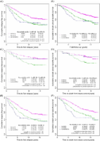
          Pathological complete response has been proposed as a surrogate endpoint for prediction of long-term clinical benefit, such as disease-free survival, event-free survival (EFS), and overall survival (OS). We had four key objectives: to establish the association between pathological complete response and EFS and OS, to establish the definition of pathological complete response that correlates best with long-term outcome, to identify the breast cancer subtypes in which pathological complete response is best correlated with long-term outcome, and to assess whether an increase in frequency of pathological complete response between treatment groups predicts improved EFS and OS. We searched PubMed, Embase, and Medline for clinical trials of neoadjuvant treatment of breast cancer. To be eligible, studies had to meet three inclusion criteria: include at least 200 patients with primary breast cancer treated with preoperative chemotherapy followed by surgery; have available data for pathological complete response, EFS, and OS; and have a median follow-up of at least 3 years. We compared the three most commonly used definitions of pathological complete response--ypT0 ypN0, ypT0/is ypN0, and ypT0/is--for their association with EFS and OS in a responder analysis. We assessed the association between pathological complete response and EFS and OS in various subgroups. Finally, we did a trial-level analysis to assess whether pathological complete response could be used as a surrogate endpoint for EFS or OS. We obtained data from 12 identified international trials and 11 955 patients were included in our responder analysis. Eradication of tumour from both breast and lymph nodes (ypT0 ypN0 or ypT0/is ypN0) was better associated with improved EFS (ypT0 ypN0: hazard ratio [HR] 0·44, 95% CI 0·39-0·51; ypT0/is ypN0: 0·48, 0·43-0·54) and OS (0·36, 0·30-0·44; 0·36, 0·31-0·42) than was tumour eradication from the breast alone (ypT0/is; EFS: HR 0·60, 95% CI 0·55-0·66; OS 0·51, 0·45-0·58). We used the ypT0/is ypN0 definition for all subsequent analyses. The association between pathological complete response and long-term outcomes was strongest in patients with triple-negative breast cancer (EFS: HR 0·24, 95% CI 0·18-0·33; OS: 0·16, 0·11-0·25) and in those with HER2-positive, hormone-receptor-negative tumours who received trastuzumab (EFS: 0·15, 0·09-0·27; OS: 0·08, 0·03, 0·22). In the trial-level analysis, we recorded little association between increases in frequency of pathological complete response and EFS (R(2)=0·03, 95% CI 0·00-0·25) and OS (R(2)=0·24, 0·00-0·70). Patients who attain pathological complete response defined as ypT0 ypN0 or ypT0/is ypN0 have improved survival. The prognostic value is greatest in aggressive tumour subtypes. Our pooled analysis could not validate pathological complete response as a surrogate endpoint for improved EFS and OS. US Food and Drug Administration. Copyright © 2014 Elsevier Ltd. All rights reserved.
            Bookmark
            • Record: found
            • Abstract: found
            • Article: found
            Is Open Access

            Ki67 Index, HER2 Status, and Prognosis of Patients With Luminal B Breast Cancer

            Background Gene expression profiling of breast cancer has identified two biologically distinct estrogen receptor (ER)-positive subtypes of breast cancer: luminal A and luminal B. Luminal B tumors have higher proliferation and poorer prognosis than luminal A tumors. In this study, we developed a clinically practical immunohistochemistry assay to distinguish luminal B from luminal A tumors and investigated its ability to separate tumors according to breast cancer recurrence-free and disease-specific survival. Methods Tumors from a cohort of 357 patients with invasive breast carcinomas were subtyped by gene expression profile. Hormone receptor status, HER2 status, and the Ki67 index (percentage of Ki67-positive cancer nuclei) were determined immunohistochemically. Receiver operating characteristic curves were used to determine the Ki67 cut point to distinguish luminal B from luminal A tumors. The prognostic value of the immunohistochemical assignment for breast cancer recurrence-free and disease-specific survival was investigated with an independent tissue microarray series of 4046 breast cancers by use of Kaplan–Meier curves and multivariable Cox regression. Results Gene expression profiling classified 101 (28%) of the 357 tumors as luminal A and 69 (19%) as luminal B. The best Ki67 index cut point to distinguish luminal B from luminal A tumors was 13.25%. In an independent cohort of 4046 patients with breast cancer, 2847 had hormone receptor–positive tumors. When HER2 immunohistochemistry and the Ki67 index were used to subtype these 2847 tumors, we classified 1530 (59%, 95% confidence interval [CI] = 57% to 61%) as luminal A, 846 (33%, 95% CI = 31% to 34%) as luminal B, and 222 (9%, 95% CI = 7% to 10%) as luminal–HER2 positive. Luminal B and luminal–HER2-positive breast cancers were statistically significantly associated with poor breast cancer recurrence-free and disease-specific survival in all adjuvant systemic treatment categories. Of particular relevance are women who received tamoxifen as their sole adjuvant systemic therapy, among whom the 10-year breast cancer–specific survival was 79% (95% CI = 76% to 83%) for luminal A, 64% (95% CI = 59% to 70%) for luminal B, and 57% (95% CI = 47% to 69%) for luminal–HER2 subtypes. Conclusion Expression of ER, progesterone receptor, and HER2 proteins and the Ki67 index appear to distinguish luminal A from luminal B breast cancer subtypes.
              Bookmark
              • Record: found
              • Abstract: found
              • Article: not found

              Tumor-associated lymphocytes as an independent predictor of response to neoadjuvant chemotherapy in breast cancer.

              PURPOSE Preclinical data suggest a contribution of the immune system to chemotherapy response. In this study, we investigated the prespecified hypothesis that the presence of a lymphocytic infiltrate in cancer tissue predicts the response to neoadjuvant chemotherapy. METHODS We investigated intratumoral and stromal lymphocytes in a total of 1,058 pretherapeutic breast cancer core biopsies from two neoadjuvant anthracycline/taxane-based studies (GeparDuo, n = 218, training cohort; and GeparTrio, n = 840, validation cohort). Molecular parameters of lymphocyte recruitment and activation were evaluated by kinetic polymerase chain reaction in 134 formalin-fixed, paraffin-embedded tumor samples. Results In a multivariate regression analysis including all known predictive clinicopathologic factors, the percentage of intratumoral lymphocytes was a significant independent parameter for pathologic complete response (pCR) in both cohorts (training cohort: P = .012; validation cohort: P = .001). Lymphocyte-predominant breast cancer responded, with pCR rates of 42% (training cohort) and 40% (validation cohort). In contrast, those tumors without any infiltrating lymphocytes had pCR rates of 3% (training cohort) and 7% (validation cohort). The expression of inflammatory marker genes and proteins was linked to the histopathologic infiltrate, and logistic regression showed a significant association of the T-cell-related markers CD3D and CXCL9 with pCR. CONCLUSION The presence of tumor-associated lymphocytes in breast cancer is a new independent predictor of response to anthracycline/taxane neoadjuvant chemotherapy and provides useful information for oncologists to identify a subgroup of patients with a high benefit from this type of chemotherapy.
                Bookmark

                Author and article information

                Journal
                Breast Cancer Research and Treatment
                Breast Cancer Res Treat
                Springer Science and Business Media LLC
                0167-6806
                1573-7217
                January 2019
                October 15 2018
                January 2019
                : 173
                : 2
                : 255-266
                Article
                10.1007/s10549-018-4981-x
                30324273
                90189bad-c00f-4d67-bb7b-1be145cbd2ee
                © 2019

                http://www.springer.com/tdm

                History

                Comments

                Comment on this article