8
views
0
recommends
+1 Recommend
1 collections
    0
    shares
      • Record: found
      • Abstract: found
      • Article: found
      Is Open Access

      Region-specific blood–brain barrier transporter changes leads to increased sensitivity to amisulpride in Alzheimer’s disease

      research-article

      Read this article at

      ScienceOpenPublisherPMC
      Bookmark
          There is no author summary for this article yet. Authors can add summaries to their articles on ScienceOpen to make them more accessible to a non-specialist audience.

          Abstract

          Background

          Research into amisulpride use in Alzheimer’s disease (AD) implicates blood–brain barrier (BBB) dysfunction in antipsychotic sensitivity. Research into BBB transporters has been mainly directed towards the ABC superfamily, however, solute carrier (SLC) function in AD has not been widely studied. This study tests the hypothesis that transporters for organic cations contribute to the BBB delivery of the antipsychotics (amisulpride and haloperidol) and is disrupted in AD.

          Methods

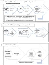
          The accumulation of [ 3H]amisulpride (3.7–7.7 nM) and [ 3H]haloperidol (10 nM) in human (hCMEC/D3) and mouse (bEnd.3) brain endothelial cell lines was explored. Computational approaches examined molecular level interactions of both drugs with the SLC transporters [organic cation transporter 1 (OCT1), plasma membrane monoamine transporter (PMAT) and multi-drug and toxic compound extrusion proteins (MATE1)] and amisulpride with the ABC transporter (P-glycoprotein). The distribution of [ 3H]amisulpride in wildtype and 3×transgenic AD mice was examined using in situ brain perfusion experiments. Western blots determined transporter expression in mouse and human brain capillaries .

          Results

          In vitro BBB and in silico transporter studies indicated that [ 3H]amisulpride and [ 3H]haloperidol were transported by the influx transporter, OCT1, and efflux transporters MATE1 and PMAT. Amisulpride did not have a strong interaction with OCTN1, OCTN2, P-gp, BCRP or MRP and could not be described as a substrate for these transporters. Amisulpride brain uptake was increased in AD mice compared to wildtype mice, but vascular space was unaffected. There were no measurable changes in the expression of MATE1, MATE2, PMAT OCT1, OCT2, OCT3, OCTN1, OCTN2 and P-gp in capillaries isolated from whole brain homogenates from the AD mice compared to wildtype mice. Although, PMAT and MATE1 expression was reduced in capillaries obtained from specific human brain regions (i.e. putamen and caudate) from AD cases (Braak stage V–VI) compared to age matched controls (Braak stage 0–II).

          Conclusions

          Together our research indicates that the increased sensitivity of individuals with Alzheimer’s to amisulpride is related to previously unreported changes in function and expression of SLC transporters at the BBB (in particular PMAT and MATE1). Dose adjustments may be required for drugs that are substrates of these transporters when prescribing for individuals with AD.

          Related collections

          Most cited references50

          • Record: found
          • Abstract: found
          • Article: not found

          P-glycoprotein in the blood-brain barrier of mice influences the brain penetration and pharmacological activity of many drugs.

          The mouse mdr1a (also called mdr3) P-GP is abundant in the blood-brain barrier, and its absence in mdr1a (-/-) mice leads to highly increased levels of the drugs ivermectin, vinblastine, digoxin, and cyclosporin A in the brain. We show here that the drugs loperamide, domperidone, and ondansetron are transported substrates for the mouse mdr1a P-GP and its human homologue MDR1. Phenytoin is a relatively weaker substrate for each, and the drugs haloperidol, clozapine, and flunitrazepam are transported hardly or not at all. Tissue distribution studies demonstrated that the relative brain penetration of radiolabeled ondansetron and loperamide (and their metabolites) is increased four- and sevenfold, respectively, in mdr1a (-/-) mice. A pilot toxicity study with oral loperamide showed that this peripherally acting antidiarrheal agent gains potent opiatelike activity in the central nervous system of mdr1a (-/-) mice. mdr1a (-/-) mice also showed increased sensitivity to neurolepticlike side effects of oral domperidone. These results point to the possible role that the drug-transporting P-GP(s) may play in the clinical use of many drugs, especially those with potential targets in the central nervous system.
            Bookmark
            • Record: found
            • Abstract: found
            • Article: not found

            Females exhibit more extensive amyloid, but not tau, pathology in an Alzheimer transgenic model.

            Epidemiological studies indicate that women have a higher risk of Alzheimer's disease (AD) even after adjustment for age. Though transgenic mouse models of AD develop AD-related amyloid beta (Abeta) and/or tau pathology, gender differences have not been well documented in these models. In this study, we found that female 3xTg-AD transgenic mice expressing mutant APP, presenilin-1 and tau have significantly more aggressive Abeta pathology. We also found an increase in beta-secretase activity and a reduction of neprilysin in female mice compared to males; this suggests that a combination of increased Abeta production and decreased Abeta degradation may contribute to higher risk of AD in females. In contrast to significantly more aggressive Abeta pathology in females, gender did not affect the levels of phosphorylated tau in 3xTg-AD mice. These results point to the involvement of Abeta pathways in the higher risk of AD in women. In addition to comparison of pathology between genders at 9, 16 and 23 months of age, we examined the progression of Abeta pathology at additional age points; i.e., brain Abeta load, intraneuronal oligomeric Abeta distribution and plaque load, in male 3xTg-AD mice at 3, 6, 9, 12, 16, 20 and 23 months of age. These findings confirm progressive Abeta pathology in 3xTg-AD transgenic mice, and provide guidance for their use in therapeutic research.
              Bookmark
              • Record: found
              • Abstract: found
              • Article: not found

              The human brain endothelial cell line hCMEC/D3 as a human blood-brain barrier model for drug transport studies.

              The human brain endothelial capillary cell line hCMEC/D3 has been developed recently as a model for the human blood-brain barrier. In this study a further characterization of this model was performed with special emphasis on permeability properties and active drug transport. Para- or transcellular permeabilities (P(e)) of inulin (0.74 x 10(-3) cm/min), sucrose (1.60 x 10(-3) cm/min), lucifer yellow (1.33 x 10(-3) cm/min), morphine (5.36 x 10(-3) cm/min), propranolol (4.49 x 10(-3) cm/min) and midazolam (5.13 x 10(-3) cm/min) were measured. By addition of human serum the passive permeability of sucrose could be reduced significantly by up to 39%. Furthermore, the expression of a variety of drug transporters (ABCB1, ABCG2, ABCC1-5) as well as the human transferrin receptor was demonstrated on the mRNA level. ABCB1, ABCG2 and transferrin receptor proteins were detected and functional activity of ABCB1, ABCG2 and the ABCC family was quantified in efflux experiments. Furthermore, ABCB1-mediated bidirectional transport of rhodamine 123 was studied. The transport rate from the apical to the basolateral compartment was significantly lower than that in the inverse direction, indicating directed p-glycoprotein transport. The results of this study demonstrate the usefulness of the hCMEC/D3 cell line as an in vitro model to study drug transport at the level of the human blood-brain barrier.
                Bookmark

                Author and article information

                Contributors
                sarah.thomas@kcl.ac.uk
                Journal
                Fluids Barriers CNS
                Fluids Barriers CNS
                Fluids and Barriers of the CNS
                BioMed Central (London )
                2045-8118
                17 December 2019
                17 December 2019
                2019
                : 16
                : 38
                Affiliations
                [1 ]ISNI 0000 0001 2322 6764, GRID grid.13097.3c, Faculty of Life Sciences and Medicine, School of Cancer and Pharmaceutical Sciences, , King’s College London, ; Franklin-Wilkins Building, 150 Stamford Street, Waterloo, London, SE1 9NH UK
                [2 ]ISNI 0000 0004 0621 7673, GRID grid.411810.d, Faculty of Pharmacy, , Misr International University, ; Cairo, 11431 Egypt
                [3 ]ISNI 0000 0001 2322 6764, GRID grid.13097.3c, Wolfson Centre for Age-Related Diseases, , King’s College London, ; Guy’s Campus, London, SE1 1UL UK
                [4 ]ISNI 0000 0001 2322 6764, GRID grid.13097.3c, Maurice Wohl Clinical Neuroscience Institute, , King’s College London, ; 125 Coldharbour Lane, Camberwell, London, SE5 9N UK
                [5 ]ISNI 0000000121901201, GRID grid.83440.3b, Division of Psychiatry, Faculty of Brain Sciences, , University College London, ; 149 Tottenham Court Road, London, W1T 7NF UK
                Author information
                http://orcid.org/0000-0002-0053-0154
                Article
                158
                10.1186/s12987-019-0158-1
                6915870
                31842924
                8fb64518-7c84-45fd-8cc9-276d191afcef
                © The Author(s) 2019

                Open AccessThis article is licensed under a Creative Commons Attribution 4.0 International License, which permits use, sharing, adaptation, distribution and reproduction in any medium or format, as long as you give appropriate credit to the original author(s) and the source, provide a link to the Creative Commons licence, and indicate if changes were made. The images or other third party material in this article are included in the article’s Creative Commons licence, unless indicated otherwise in a credit line to the material. If material is not included in the article’s Creative Commons licence and your intended use is not permitted by statutory regulation or exceeds the permitted use, you will need to obtain permission directly from the copyright holder. To view a copy of this licence, visit http://creativecommons.org/licenses/by/4.0/. The Creative Commons Public Domain Dedication waiver ( http://creativecommons.org/publicdomain/zero/1.0/) applies to the data made available in this article, unless otherwise stated in a credit line to the data.

                History
                : 10 October 2019
                : 2 December 2019
                Funding
                Funded by: Medical Research Council
                Award ID: MR/K500811/1
                Funded by: FundRef http://dx.doi.org/10.13039/100004440, Wellcome Trust;
                Award ID: 080268
                Award Recipient :
                Funded by: FundRef http://dx.doi.org/10.13039/501100000268, Biotechnology and Biological Sciences Research Council;
                Award ID: BB/L01534X/1
                Funded by: Medical Research Council DTP
                Award ID: MR/N013700/1
                Funded by: FundRef http://dx.doi.org/10.13039/501100012317, UCLH Biomedical Research Centre;
                Funded by: KCL ARUK network fund
                Funded by: GSTT MRes-PhD Studentship
                Funded by: The Edmond and Lily Safra Research Foundation
                Categories
                Research
                Custom metadata
                © The Author(s) 2019

                Neurology
                amisulpride,mate1,pmat,oct1,blood–brain barrier,alzheimer’s
                Neurology
                amisulpride, mate1, pmat, oct1, blood–brain barrier, alzheimer’s

                Comments

                Comment on this article