68
views
0
recommends
+1 Recommend
0 collections
    0
    shares
      • Record: found
      • Abstract: found
      • Article: found
      Is Open Access

      Extensive migration of young neurons into the infant human frontal lobe

      research-article

      Read this article at

      Bookmark
          There is no author summary for this article yet. Authors can add summaries to their articles on ScienceOpen to make them more accessible to a non-specialist audience.

          Abstract

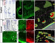
          The first few months after birth, when a child begins to interact with the environment, are critical to human brain development. The human frontal lobe is important for social behavior and executive function; it has increased in size and complexity relative to other species, but the processes that have contributed to this expansion are unknown. Our studies of postmortem infant human brains revealed a collection of neurons that migrate and integrate widely into the frontal lobe during infancy. Chains of young neurons move tangentially close to the walls of the lateral ventricles and along blood vessels. These cells then individually disperse long distances to reach cortical tissue, where they differentiate and contribute to inhibitory circuits. Late-arriving interneurons could contribute to developmental plasticity, and the disruption of their postnatal migration or differentiation may underlie neurodevelopmental disorders.

          Related collections

          Most cited references37

          • Record: found
          • Abstract: found
          • Article: not found

          Interneuron dysfunction in psychiatric disorders.

          Schizophrenia, autism and intellectual disabilities are best understood as spectrums of diseases that have broad sets of causes. However, it is becoming evident that these conditions also have overlapping phenotypes and genetics, which is suggestive of common deficits. In this context, the idea that the disruption of inhibitory circuits might be responsible for some of the clinical features of these disorders is gaining support. Recent studies in animal models demonstrate that the molecular basis of such disruption is linked to specific defects in the development and function of interneurons - the cells that are responsible for establishing inhibitory circuits in the brain. These insights are leading to a better understanding of the causes of schizophrenia, autism and intellectual disabilities, and may contribute to the development of more-effective therapeutic interventions.
            Bookmark
            • Record: found
            • Abstract: found
            • Article: not found

            Learning enhances adult neurogenesis in the hippocampal formation.

            Thousands of hippocampal neurons are born in adulthood, suggesting that new cells could be important for hippocampal function. To determine whether hippocampus-dependent learning affects adult-generated neurons, we examined the fate of new cells labeled with the thymidine analog bromodeoxyuridine following specific behavioral tasks. Here we report that the number of adult-generated neurons doubles in the rat dentate gyrus in response to training on associative learning tasks that require the hippocampus. In contrast, training on associative learning tasks that do not require the hippocampus did not alter the number of new cells. These findings indicate that adult-generated hippocampal neurons are specifically affected by, and potentially involved in, associative memory formation.
              Bookmark
              • Record: found
              • Abstract: found
              • Article: not found

              Corridors of Migrating Neurons in Human Brain and Their Decline during Infancy

              The subventricular zone (SVZ) of many adult non-human mammals generates large numbers of new neurons destined for the olfactory bulb (OB) 1–6 . Along the walls of the lateral ventricles, immature neuronal progeny migrate in tangentially-oriented chains that coalesce into a rostral migratory stream (RMS) connecting the SVZ to the OB. The adult human SVZ, in contrast, contains a hypocellular gap layer separating the ependymal lining from a periventricular ribbon of astrocytes 7 . Some of these SVZ astrocytes can function as neural stem cells in vitro, but their function in vivo remains controversial. An initial report finds few SVZ proliferating cells and rare migrating immature neurons in the RMS of adult humans 7 . In contrast, a subsequent study indicates robust proliferation and migration in the human SVZ and RMS 8,9 . Here, we find that the infant human SVZ and RMS contain an extensive corridor of migrating immature neurons before 18 months of age, but, contrary to previous reports 8 , this germinal activity subsides in older children and is nearly extinct by adulthood. Surprisingly, during this limited window of neurogenesis, not all new neurons in the human SVZ are destined for the OB – we describe a major migratory pathway that targets the prefrontal cortex in humans. Together, these findings reveal robust streams of tangentially migrating immature neurons in human early postnatal SVZ and cortex. These pathways represent potential targets of neurological injuries affecting neonates.
                Bookmark

                Author and article information

                Journal
                0404511
                7473
                Science
                Science
                Science (New York, N.Y.)
                0036-8075
                1095-9203
                23 March 2017
                07 October 2016
                18 May 2017
                : 354
                : 6308
                : aaf7073
                Affiliations
                [1 ]Edythe Broad Institute for Stem Cell Research and Regeneration Medicine University of California San Francisco CA 94143 USA
                [2 ]Department of Neurology, University of California San Francisco CA 94143 USA
                [3 ]Department of Neurological Surgery University of California San Francisco CA 94143 USA
                [4 ]Laboratory of Comparative Neurobiology Instituto Cavanilles, Universidad de Valencia CIBERNED Valencia Spain
                [5 ]Multiple Sclerosis and Neural Regeneration Unit Department of Neurology Hospital Universitario y Politecnico La Fe 46026 Valencia Spain
                [6 ]Department of Radiology and Biomedical Imaging University of California San Francisco CA 94143 USA
                [7 ]Department of Pathology University of California San Francisco CA 94143 USA
                [8 ]Department of Pediatrics University of California, San Francisco CA 94143 USA
                [9 ]Department of Paediatrics University of Cambridge Cambridge CB2 0QQ, UK
                Article
                NIHMS856057
                10.1126/science.aaf7073
                5436574
                27846470
                8fb1c113-4791-4ee9-869f-797ad4362dbc

                Permissions: Obtain information about reproducing this article: http://www.sciencemag.org/about/permissions.dtl

                History
                Categories
                Article

                Uncategorized
                Uncategorized

                Comments

                Comment on this article