
- Record: found
- Abstract: found
- Article: found
Update of the Brazilian Guideline on Nuclear Cardiology - 2020
research-article
Luiz Eduardo Mastrocola
1 ,
Barbara Juarez Amorim
2
,
3 ,
João Vicente Vitola
4 ,
Simone Cristina Soares Brandão
5 ,
Gabriel Blacher Grossman
6
,
7 ,
Ronaldo de Souza Leão Lima
8
,
9
,
10 ,
Rafael Willain Lopes
1 ,
William Azem Chalela
11 ,
Lara Cristiane Terra Ferreira Carreira
12 ,
José Roberto Nolasco de Araújo
13 ,
Cláudio Tinoco Mesquita
14 ,
José Claudio Meneghetti
11
February 2020
There is no author summary for this article yet. Authors can add summaries to their articles on ScienceOpen to make them more accessible to a non-specialist audience.
Abstract
Declaration of potential conflict of interest of authors/collaborators of Update of
the Brazilian Guidelines on Nuclear Cardiology - 2020If the last three years the author/developer
of the Update:
Names Members of the Update
Participated in clinical studies and/or experimental trials supported by pharmaceutical
or equipment related to the guideline in question
Has spoken at events or activities sponsored by industry related to the guideline
in question
It was (is) advisory board member or director of a pharmaceutical or equipment
Committees participated in completion of research sponsored by industry
Personal or institutional aid received from industry
Produced scientific papers in journals sponsored by industry
It shares the industry
Barbara Juarez Amorim
No
No
No
No
No
No
No
Claudio Tinoco Mesquita
NIH
Bayer
No
No
Pfizer
No
No
Gabriel Blacher Grossman
No
No
No
No
No
No
No
João Vicente Vitola
No
No
No
No
No
No
No
José Claudio Meneghetti
No
No
No
No
No
No
No
José Roberto Nolasco de Araújo
No
No
No
No
No
No
No
Lara Cristiane Terra Ferreira Carreira
No
No
No
No
No
No
No
Luiz Eduardo Mastrocola
No
No
No
No
No
No
No
Rafael Willain Lopes
No
No
No
No
No
No
No
Ronaldo de Souza Leao Lima
No
No
No
No
No
No
No
Simone Cristina Soares Brandão
No
No
No
No
No
No
No
William Azem Chalela
No
No
No
No
No
No
No
List of Abbreviations and Acronyms
HED - 11C - meta-hydroxyephedrine labeled with Carbon-11
PIB-11C - PET - pittsburgh B compound labeled with carbon-11 by PET imaging
MIBG-123I - metaiodobenzylguanidine labeled with iodine 123
13NH3 - ammonia labeled with Nitrogen-13
H2O-15O - water labeled with Oxygen-15
FDG-18F - fluorodeoxyglucose labeled with Fluorine-18
FDG-18F - PET/TC - fluorodeoxyglucose labeled with fluorine-18 by hybrid imaging (positron
emission tomography coupled with computerized tomography)
Sodium fluoride-18F - fluorine-18 labeled Sodium Fluoride for PET Amyloid Imaging
201Hg - mercury-201
201Tl - thallium-201
82Rb - rubidium-82
82Sr - strontium-82
99mTc - technetium-99m
MIBI-99mTc - technetium-99m-labeled SESTAMIBI or MIBI
Pyrophosphate-99mTc - technetium-99m-labeled pyrophosphate
ACEI - angiotensin converting enzyme inhibitors
ACS - acute coronary syndrome
Aden - adenosine
ADMIRE-HF - AdreView Myocardial Imaging for Risk Evaluation in HF
AF - atrial fibrillation
AHA - American Heart Association
AL - light chain immunoglobulin
ALARA - as low as reasonably achievable
AMI - acute myocardial infarction
angio-CT - angiotomography of coronary arteries
ARB - angiotensin receptor blockers
ATP III - Adult Treatment Panel, from the Program for Detection, Evaluation, and Treatment
of High Cholesterol in Adults
AUC - area under the curve
AVB - atrioventricular blockage
BMI - body mass index
BNP - B-natriuretic peptide
CA - cardiac amyloidosis
CABG - coronary artery bypass graft
CC - coronary calcium
CAD - coronary artery disease
CCA - coronary cineangiography
CFR - coronary flow reserve
CHF - congestive heart failure
CIED - cardiac implantable electronic devices
CMR - cardiac magnetic resonance
CONFIRM - Coronary CT Angiography Evaluation for Clinical Outcomes: an International
Multicenter Registry
COURAGE - Clinical Outcomes Utilizing Revascularization and Aggressive druG Evaluation
Trial
CPU - chest pain unit
CRP - C reactive protein
CRT - cardiac resynchronization therapy
CS - calcium score
CTX - cardiotoxicity
CV - cardiovascular
Cx - circumflex coronary artery
CZT - cadmium zinc telluride semiconductors
DDD - artificial pacemaker stimulation mode
DG1 - diagonal 1 coronary artery
Dipy. - dipyridamole
DM - diabetes mellitus
Dobut. - dobutamine
DS - duke score
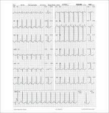
ECG - 12-lead electrocardiogram
ECHO - echocardiogram
EDV - end diastolic volume
ERASE Chest Pain -The Emergency Room Assessment of Sestamibi for Evaluation of Chest
Pain Trial
ESV - end systolic volume
ET - exercise testing
FAME - Fractional Flow Reserve versus Angiography for Guidance of PCI in Patients
with Multivessel Coronary Artery Disease
FBP - filtered back-projection
FDA - food and drug administration
FDG-6-P - fluorodeoxyglucose - 6 - phosphate
FFA - free fatty acids
FFR - fractional flow reserve
FRS - Framingham risk score
Gated-SPECT - myocardial perfusion imaging by single photon emission computed tomography
technique synchronized with electrocardiogram
HBP - high blood pressure
HF - heart failure
HFpEF - heart failure with preserved ejection fraction
HFrEF - heart failure with reduced ejection fraction
HMR - heart to mediastinum ratio
HR - heart rate
IAEA - International Atomic Energy Agency
ICD - implantable cardioverter defibrillator
ICNC - International Conference of Nuclear Cardiology
IE - infectious endocarditis
IFR - instantaneous flow reserve/instantaneous wave-free ratio
INCAPS - IAEA Nuclear Cardiology Protocols Cross-Sectional Study
ISCHEMIA - International Study of Comparative Health Effectiveness with Medical and
Invasive Approaches
IV - intravenously/intravenous
keV - kilo-electron volts
LAD - left anterior descending coronary artery
LAFB - left anterior fascicular block
LBBB - left bundle branch block
LV - left ventricle
LVAD / VAD - left ventricular assist device / ventricular assist devices
LVEF - left ventricular ejection fraction
MBF - myocardial blood flow
MBFR - myocardial blood flow reserve
MBq - megabequerel
mCi - milicurie
MET - metabolic equivalent.
MFR - myocardial flow reserve
MIBI / SESTAMIBI - 2-methoxy-isobutyl-isonitrile
MPS - myocardial perfusion scintigraphy
MR - magnetic resonance
MRS - myocardial revascularization surgery
mSv - millisieverts
MVO2 - myocardial oxygen consumption
NaI - sodium iodine
NE - norepinephrine
NPV - negative predictive value
NSTEMI - non-ST segment elevation myocardial infarction
NSVT - nonsustained ventricular tachycardia
NYHA HF - New York Heart Association Heart Failure Class
OMT - optimized medical therapy
OR - odds ratio
OSEM - ordered subset expectation maximization
PAREPET - prediction of arrhythmic events with positron emission tomography
PARR-2 - PET and Recovery after Revascularization study
PCI - percutaneous coronary intervention
PET - positron emission tomography
PET/CT - positron emission tomography coupled with computed tomography (hybrid imaging)
PET/MR - positron emission tomography coupled with magnetic resonance (hybrid imaging)
PM - pacemaker
PREMIER - Performance of Rest Myocardial Perfusion Imaging in the Management of Acute
Chest Pain in the Emergency Room in Developing Nations
PROCAM - PROSpective CArdiovascular Munster Study
PROMISE - Prospective Multicenter Imaging Study for Evaluation of Chest Pain
RCA - right coronary artery
Regad. - regadenoson
RESCUE - Randomized Evaluation of patients with Stable angina Comparing diagnostic
Examinations
ROC - receiver operating characteristics
ROI - regions of interest
ROMICAT II - rule out myocardial infarction by cardiac computed tomography
RV - right ventricle/ventricular
SBC - Brazilian Society of Cardiology
SBMN - Brazilian Society of Nuclear Medicine
SBP - systolic blood pressure
SCORE - Systematic Coronary Risk Evaluation Study
SDS - summed difference score
Shining / Shine Through - residual activity effect
SPECT - myocardial perfusion imaging by single photon emission computed tomography
SRS - summed rest/redistribution score
SSS - summed stress score
STEMI - ST segment elevation myocardial infarction
STICH - Surgical Treatment for Ischemic Heart Failure study
SUS - Brazil’s public Single Health System (acronym in Portuguese)
SUV - standard uptake value
TIA - transient ischemic attack
TID - transient ischemic dilatation
TOF - time of flight
TTR - transthyretin
TTR CA - transthyretin cardiac amyloidosis
UA - unstable angina
USA - United States of America
VAD - ventricular assist devices
VF - ventricular fibrillation
VT - ventricular tachycardia
WR - myocardial washout rate
1. Introduction
Nuclear cardiology is a non-anatomical, physiological imaging method. The use of radioactive
or radiopharmaceutical substances makes it possible to study several physiopathological
mechanisms of cardiovascular disease in vivo. Via this imaging technique, it is also
possible to visualize and accompany an instituted therapy’s physiological effects
on cardiac function, on the cellular and biochemical level. Of all the applications
of nuclear medicine in cardiology, scintigraphy or myocardial perfusion imaging with
technetium-99m-labeled radiopharmaceuticals synchronized with electrocardiogram (Gated-SPECT),
is the most common exam in clinical practice. For this reason, this technique will
be the most discussed in these Guidelines.
Recent years have, however, seen a growing concern among the scientific community
regarding rational and optimized use of ionizing radiation in medicine. Cardiovascular
imaging, moreover, encompasses all functional and anatomical imaging techniques and
should, in this context, be used rationally and cost-effectively. Other applications
of nuclear medicine in cardiology have also emerged and gained prominence during the
past decades, especially positron emission tomography (PET) for the study of coronary
flow reserve, cardiac sympathetic activity, and inflammatory/infectious processes,
and cardiac amyloidosis (CA). All of these aspects have been taken into consideration
and will be covered in detail in the chapters developed herein.
Guidelines recommendations are highly valuable tools for medical activity of the highest
quality. The objective is to support and aid doctors in making decisions regarding
their patients, by elaborating orientations which may be useful as part of the decision-making
process. No Guidelines, however, should be replaced by the abilities, experience,
and clinical judgments of specialized professionals who are have the final say in
their decisions concerning each individual patient.
In general, whenever possible and applicable, classifications of recommendation have
been adopted for indicating cardiac scintigraphy, supported by levels of evidence,
in accordance with the recommendations established by classical cardiology guidelines
(Table 1).
Table 1
Classes of recommendation and levels of evidence
Classes of recommendation
Class I - Conditions for which there is conclusive evidence or, in the absence of
conclusive evidence, general consensus that the procedure is safe and useful/effective
Class II - Conditions for which there are conflicting evidence and/or divergent opinions
regarding the procedure's safety and usefulness/effectiveness
Class IIA - Weight or evidence/opinion in favor of the procedure. The majority of
studies/experts approve.
Class IIB - Safety and usefulness/effectiveness less well established, with no prevailing
opinions in favor
Class III - Conditions for which there is evidence and/or consensus that the procedure
is not useful/effective and could, in some cases, be harmful
Levels of Evidence
Level A - Data obtained from multiple concordant large randomized trials and/or robust
meta-analysis of randomized clinical trials
Level B - Data obtained from less robust meta-analysis, from a single randomized trial,
or from non-randomized (observational) trials
Level C - Data obtained through consensus of expert opinion
Based on current evidence, this document, which does not function as a substitute,
practically and objectively adds important data to and updates the Brazilian Cardiology
Society’s (SBC) First Guidelines and Update on Nuclear Cardiology, both of which were
published by the Brazilian Archives of Cardiology (Arquivos Brasileiros de Cardiologia),
in 2002 and 2005, respectively.
As in the previously mentioned documents, those who participated in the elaboration
of these Guidelines are considered specialists in their respective areas and were,
for this reason, chosen to develop the chapters thereon. The committed involvement
of all colleagues representing the SBC and the Brazilian Society of Nuclear Medicine
(SBMN) have made the elaboration of these new update of Brazilian Guidelines on Nuclear
Cardiology possible. It is our hope that they will be of great use, especially to
Cardiologists and Nuclear Medicine and Clinical Physicians in Brazil. The Organizing
Committee appreciates the collaboration of all those involved.
2. Addendum to the ISCHEMIA Study*
At the time of publication of this guideline, the International Study of Comparative
Health Effectiveness with Medical and Invasive Approaches (ISCHEMIA) had not been
published yet, although the main findings were presented on November 16, 2019 at the
American Heart Association (AHA) annual congress in Philadelphia, USA, available on
the study’s website. Considering its importance for medical decision-making and the
potential implications for nuclear cardiology, a few relevant concerns should be highlighted
on the findings available so far:
The main objective of the ISCHEMIA study was to assess whether patients (P) with at
least moderate ischemia on a functional examination would benefit from myocardial
revascularization (coronary artery bypass grafting or percutaneous coronary intervention)
added to optimal medical therapy). Were randomized 5,179 patients with stable CAD
and myocardial ischemia documented by one of many different methods (myocardial perfusion
scintigraphy, stress echocardiography, cardiac magnetic resonance imaging, exercise
testing not associated with cardiac imaging). These noninvasive methods were used
to define the etiology of chest pain and for cardiovascular risk stratification, a
management approach established in clinical practice that is not invalidated by the
findings of the study. Prior knowledge indicates that patients with lower ischemic
burden have a better prognosis than individuals with larger and more intense ischemia;
The ISCHEMIA trial demonstrated no benefit of myocardial revascularization (Invasive
Group - IG) versus optimal medical therapy (OMT) to reduce the major outcomes of “death”
and “acute myocardial infarction.” Despite the methodological differences, these results
were somewhat similar to those of the COURAGE study. It is noteworthy that the mortality
curves began to separate after two years of medical follow-up, apparently benefiting
the IG and potential long-term implications, which justified the increased clinical
follow-up of P, underway at the moment. Note that the IG had an improved quality of
life assessment, reduced frequency of angina and lower use of specific medication
compared to the OMT group;
The ISCHEMIA trial is one of the most relevant studies on stable CAD, with important
messages for clinical practice. The validity of the results is emphasized for the
population sample evaluated in the study and for the definitions of ischemia and its
severity levels employed. However, for exclusion situations, such as P with left main
disease, recent acute coronary syndrome, angioplasty in the previous 12 months, ejection
fraction < 35% and progressive or unstable symptoms, prior knowledge remains unchanged.
Both CAD and ischemic heart disease represent a broad spectrum of patients, with inherent
heterogeneity and important prognostic implications (extensive evidence base in the
literature and described in detail in the current guideline). Were excluded from the
trial an impressive number of P that had at least moderate angina and ischemia in
the absence of coronary obstructions, showing the diversity of the disease and the
value of functional assessment;
The main question is whether the ISCHEMIA study has properly evaluated a significant
number of P with moderate/severe ischemia, aiming to determine whether myocardial
revascularization adds prognostic benefit to these patients, as documented by scintigraphy,
which was not the exclusive method of documentation. There was the inclusion (randomization)
of cases with nonexistent or mild ischemia (12% of the total randomized), which is
surprising for a study that was initially intended to include only patients with moderate
to severe ischemia. There was also a change in the criteria for inclusion of P with
severe ischemia in the study, with a significant number based on the results of exercise
testing, without imaging, a decision made after the study was in progress. From this
change, the percentage of these P that would effectively have severe myocardial ischemia
on scintigraphy is questioned;
Therefore, the Editorial Board of this guideline believes that the definitive analysis
of the results will only be possible after the formal publication of the trial results.
3. The Application of Nuclear Medicine Techniques to Justify Financial Resources Available
for Attending Cardiology Patients in Brazil
3.1. Introduction
Cardiovascular diseases are the main cause of death in Brazil, and they are responsible
for 30% of deaths worldwide every year.
1
They are responsible for approximately 8% of total healthcare costs in Brazil, a figure
which has been increasingly annually, in parallel with population aging.
1
Teich and Araújo estimated that in 2011, approximately 200,000 events associated with
acute coronary syndromes occurred in Brazil, entailing a massive impact of 3.88 billion
Brazilian reals, considering only hospital and indirect costs, associated with loss
of productivity.
2
Considering these findings, it has been demonstrated that (preventive) measures play
a crucial role in reducing morbidity and mortality, and they should be a priority
in national healthcare policy design, as they have profound additional impacts on
reducing costs and maintaining productivity. Another significant point, however, which
has contributed to reducing the outcome of “cardiovascular death” and to justifying
expenses, involves the use of tools which make accurate diagnosis of a determined
condition possible (?) and which aid and guide the conduct of physicians, based on
these results. Myocardial perfusion scintigraphy (MPS) plays a significant role in
justifying financial resources for attending patients with established or potential
cardiovascular disease.
3.2. Cost-Effectiveness in Comparison with Cardiac Catheterization
One of the main fundaments of MPS is its good ability to identify low-risk patients,
who do not require invasive intervention, in spite of established coronary disease,
such as anatomical lesions on coronary angiography.
3
Observational studies in the 1990’s have demonstrated that MPS was able to identify
high- and low-risk groups, resulting in reduced costs for patients with coronary artery
disease (CAD) and avoiding procedures that are not associated with improved patient
health outcomes. A major prospective study carried out in the United States of America
(USA) recruited 11,372 patients with stable angina, who were referred to either MPS
or cardiac catheterization. Patients were adjusted by clinical risk, and the costs
of direct cardiac catheterization (aggressive strategy) were compared to initial scintigraphy
followed by selective catheterization in high-risk patients (conservative strategy).
Although both strategies had similar adverse outcomes, such as cardiac death and non-fatal
myocardial infarction, revascularization rates were higher (between 13% and 50%) in
patients who underwent catheterization directly.
4
This reflex of revascularizing anatomical lesions which do not determine ischemia
led to unnecessary associated medical costs of around 5,000 dollars per patient in
this study.
4
Currently, the use of medical resources for conditions that do not have consequences
for patients or that could be managed conservatively is known as “overtreatment.”
5
The study of the impact of MPS on reducing costs has shown that its main function
is to prevent patients who have low or moderate risks on single photon emission computed
tomography (SPECT) from being treated with unnecessary catheterizations and revascularizations.
Similarly to this North American study, Underwood et al.
6
have demonstrated that strategies which incorporate myocardial scintigraphy to evaluate
patients with stable coronary diseases are both cheaper than and as effective as strategies
involving invasive anatomical assessment.
6
Cerci et al.
7
evaluated the impact of diagnostic exams on patients with CAD in different scenarios
within Brazil’s public Single Health System (SUS, acronym in Portuguese). The study’s
most relevant finding is that, although non-invasive functional tests are the most
frequently solicited exams for evaluating patients with suspected or known CAD, the
majority of healthcare costs for these patients are related to procedures/invasive
treatment. In other words, in the Brazilian context, the costs of diagnostic exams
continue to be significantly lower than those of invasive and therapeutic procedures.
In this manner, it seems logical to affirm that, if scintigraphy exams are made available
to patients attended by the SUS, there will be a similar impact on the reduction of
healthcare costs, which has been the case in the USA and some countries in Europe.
Another relevant piece of data from this study refers to the fact that the majority
of patients who were revascularized had not undergone tests to document ischemic burden;
only anatomical diagnostic techniques had been applied.
7
3.3. Cost-Effectiveness of Myocardial Perfusion Scintigraphy in Relation to Coronary
Angiotomography
Angiotomography (angio-CT) of coronary arteries offers very accurate, non-invasive
anatomical assessment, and it has proved to be an excellent technique for ruling out
obstructive coronary disease in low- to intermediate-risk patients. Angio-CT, however,
has presented results similar to those of cardiac catheterization in relation to triggering
a higher number of myocardial revascularizations, which do not necessarily (means)
reduced cardiovascular outcomes. In a recent meta-analysis comparing angio-CT to functional
methods, no differences were observed regarding the outcomes of death or cardiac hospitalization,
but there was a 29% reduction in the number of non-fatal infarctions. On the other
hand, the use of this method was associated with 33% and 86% higher rates of invasive
coronary angiography and myocardial revascularization, respectively. It is not known
whether the reduction in non-fatal infarctions may be attributed to the higher number
of revascularizations, which is (unlikely) considering in light of other studies on
stable CAD, or to the higher use of statins and aspirin associated with the recognition
of anatomical coronary lesions.
8
With the objective of elucidating the role of angio-CT on cost-effectiveness of approaches
to stable CAD in comparison with myocardial scintigraphy, the Randomized Evaluation
of Patients With Stable Angina Comparing Diagnostic Examinations (RESCUE) study, which
is being developed, is expected to compare these strategies in a prospective, randomized
manner.
9
The authors of a recent meta-analysis published by the American Heart Association
(AHA)/Circulation, have reinforced 2 important aspects of cost-effectiveness:
10
The importance of performing appropriate exams as a way of (ensuring) their cost-effectiveness,
especially techniques like MPS.
The results of appropriate exams should effectively lead to appropriate decision making
in clinical conduct and patient management.
4. Indications for Myocardial Perfusion Scintigraphy
Over the past years, different medical societies have published criteria for defining
scenarios in which myocardial scintigraphy may be adequately utilized. In addition
to traditional classification of recommendation and levels of evidence, more recent
criteria on appropriate MPS exam referral have been suggested, dividing indications
into appropriate, possibly appropriate, and rarely appropriate, resulting from the
application of scores constructed based on clinical scenarios and specific methodologies.
11
In this classification, indications with scores from 1 to 3 are considering rarely
appropriate; 4 to 6, possibly appropriate; and 7 to 9, appropriate. Published documents
are based on evidence from American and European Guidelines, as well as the recently
published Brazilian Guidelines on stable coronary disease.
12-15
Regardless of classification type, there is consensus that symptomatic patients with
intermediate risks of ischemic heart disease are the ones who most benefit from MPS
in terms of diagnostic and prognostic evaluation. The exam should preferably be performed
in association with physical exercise in patients with sufficient physical and clinical
conditions (estimated ability for activities of daily living with metabolic expenditure
greater than 5 METs), in order to measure their functional capacity, hemodynamic responses
(heart rate and blood pressure behavior), stress-induced arrhythmias, and other responses.
It is recommended that patients with complete left bundle branch block, regardless
of functional ability, undergo MPS under pharmacological stress (dipyridamole or adenosine).
In the same manner, regardless of pretest probability of ischemic heart disease, patients
with low functional ability or uninterpretable electrocardiogram (ECG) are indicated
to undergo MPS. On the other hand, patients with low probability of ischemic heart
disease, higher functional ability, and interpretable ECG are not indicated for MPS
(Table 2).
Table 2
Indication criteria for myocardial perfusion scintigraphy in symptomatic patients
Assessment of patients with non-acute chest pain or ischemic equivalent
Score
Low pretest probability of CAD, with interpretable resting ECG and ability to exercise
3
Low pretest probability of CAD, with uninterpretable resting ECG or inability to exercise
7
Intermediate pretest probability of CAD, with interpretable resting ECG and ability
to exercise
7
Intermediate pretest probability of CAD, with uninterpretable resting ECG or inability
to exercise
9
High pretest probability of CAD, regardless of interpretable resting ECG and ability
to exercise
8
ACS: acute coronary syndrome; CAD: coronary artery disease; ECG: 12-lead electrocardiogram.
In patients with heart failure (HF) and left ventricular systolic dysfunction or recent-onset
atrial fibrillation (AF), ventricular tachycardia (VT) or syncope, the indication
for MPS is appropriate or possibly appropriate, unless the patient in question is
low risk or has low pretest probability. Asymptomatic patients with no history of
ischemic heart disease and without abnormal exercise testing (ET) generally do not
benefit from undergoing MPS. In specific situations, in patients with high calcium
scores (greater than or equal to 400), diabetes, chronic renal insufficiency, or a
prevalent family history of ischemic heart disease, performing MPS may aggregate value
to the medical decision-making process, with satisfactory cost-effectiveness. Asymptomatic
patients with abnormal stress ECG who are re-stratified with the use of prognostic
scores, such as the Duke score, may also benefit from complementary investigation
via MPS, especially if their risk scores are intermediate or high (Table 3). Diverse
examples of clinical situations cited in Table 3 may also be found in the section
on integration of diagnostic modalities.
Table 3
Indication criteria for myocardial perfusion scintigraphy in asymptomatic patients
and/or patients with prior exams
Asymptomatic patients - detection of CAD/risk stratification
Score
Low risk (ATP III criteria)
1
Intermediate risk (ATP III criteria) - interpretable ECG
3
Intermediate risk (ATP III criteria) - uninterpretable ECG
5
High risk (ATP III criteria)
7
High risk and calcium score (Agatston) between 100 and 400
7
Calcium score (Agatston) > 400
7
Low-risk Duke score (> +5)
2
Intermediate-risk Duke score (between -11 and + 5)
7
High-risk Duke score (< -11)
8
Agatston: score that defines the presence and quantity of calcium in coronary arteries,
characterizing atherosclerosis; ATP III: Adult Treatment Panel, from the program for
detection, evaluation, and treatment of high cholesterol in adults; CAD: coronary
artery disease.
When patients have established ischemic heart disease and are asymptomatic, early
myocardial perfusion studies with radiopharmaceuticals should be avoided following
percutaneous coronary intervention and/or myocardial revascularization surgery procedures.
In the event of percutaneous coronary intervention and myocardial revascularization
surgery, the application of MPS has been observed to have a favorable cost-benefit
ratio for follow up after more than 2 and 5 years, respectively, even in asymptomatic
patients. Symptomatic patients with specific clinical conditions (or equivalent manifestations)
may benefit from the exam before this period (Table 4).
Table 4
Indication criteria for myocardial perfusion scintigraphy in patients who have undergone
revascularization procedures (CABG or PCI)
Previous percutaneous revascularization or surgical procedures
Score
Symptomatic
8
Asymptomatic, CABG less than 5 years prior
5
Asymptomatic, CABG 5 or more years prior
7
Asymptomatic, percutaneous revascularization less than 2 years prior
3
Asymptomatic, percutaneous revascularization 2 or more years prior
6
CABG: myocardial revascularization surgery; PCI percutaneous coronary intervention.
For patients with previous exams who manifest new symptoms or who require assessment
of the repercussion of diagnosed intermediate lesions and characterization of arteries
with obstructive lesions “responsible” for a larger myocardial area at risk, as well
as patients with multivascular diseases, the indication for MPS is classified as appropriate
or possibly appropriate. In patients with established coronary disease and worsening
symptoms, MPS may aid in quantifying ischemic burden (extent and intensity of defects)
and in determining medical management. In clinically stable patients with previous
exams performed more than 2 years prior, MPS may be appropriate (Table 5).
Table 5
Indication criteria for myocardial perfusion scintigraphy for risk stratification
and prognostic assessment of patients with proven stable coronary artery disease and/or
prior exams
Asymptomatic patients or patients with stable symptoms - previously "normal" stress
imaging exams
Score
Intermediate/high risk (ATP III) - stress imaging exam ≥ 2 years prior
6
Asymptomatic patients or patients with stable symptoms - CCA or abnormal imaging exams,
without prior CABG
CAD on CCA or "abnormal" stress imaging exam (exam performed > 2 years prior)
5
CAD on CCA or "abnormal" stress imaging exam (exam performed < 2 years prior)
3
Previously "unclear," "contradictory," or "borderline" non-invasive assessment - obstructive
CAD as initial concern
8
New, recent, or progressive symptoms
Abnormal CCA or abnormal stress imaging exam
9
Normal CCA or normal stress imaging exam
6
Coronary cineangiography (invasive or non-invasive)
Coronary stenosis or anatomical abnormality whose significance is unclear
9
ACS: acute coronary syndrome; ATP III: Adult Treatment Panel, from the program for
detection, evaluation, and treatment of high cholesterol in adults; CAD: coronary
artery disease; CCA: coronary cineangiography; CABG: myocardial revascularization
surgery.
In patients who present acute chest pain, with clinical suspicion of acute coronary
syndrome (ACS), normal or uninterpretable ECG (old left bundle branch block or pacemaker)
and normal biomarkers, resting myocardial scintigraphy may exclude acute cardiovascular
events with a high degree of safety (high negative predictive value [NPV]), allowing
patients to be discharged from the emergency room. If the exam is normal, investigation
may continue with outpatient tests involving physical or pharmacological stress, whether
associated or non-associated with non-invasive imaging, and even anatomical assessment
via coronary angio-CT, in specific conditions. For patients with ACS who are clinically
stable, with neither recurring chest pain nor HF, and who have not undergone any invasive
exam, MPS is useful for detecting presence and extent of myocardial ischemia (Table
6).
Table 6
Indication criteria for myocardial perfusion scintigraphy in patients with acute chest
pain or post-acute coronary syndrome
Assessment of patients with acute chest pain
Score
Resting image only
Possible ACS - ECG without ischemic alterations or LBBB or pacemaker; low-risk TIMI
score; borderline, minimally elevated, or negative troponin
8
Possible ACS - ECG without ischemic alterations or LBBB or pacemaker; high-risk TIMI
score; borderline, minimally elevated, or negative troponin
7 / 8
Possible ACS - ECG without ischemic alterations or LBBB or pacemaker; negative initial
troponin. Recent (up to 2 hours) or evolving chest pain
7
Assessment of post-ACS patients (infarction with or without elevated ST segment)
Stable, post-AMI patients, with ST segment elevation, for assessment of ischemia;
cardiac catheterization not performed
8
Stable, post-AMI patients, without ST segment elevation, for assessment of ischemia;
cardiac catheterization not performed
9
ACS: acute coronary syndrome; AMI: acute myocardial infarction; CAD: coronary artery
disease; ECG: 12-lead electrocardiogram; LBBB: left bundle branch block.
Indications for MPS to assess pre-operative risk of non-cardiac surgeries and vascular
surgeries have also been recently revised.
16
Patients who will undergo low-risk surgeries do not need to undergo MPS. If the surgery
is not low-risk, functional capacity is the factor that determines whether MPS will
be necessary. In patients with functional capacity estimated at greater than or equal
to 4 METs, without cardiac symptoms, regardless of clinical or surgical risk, non-invasive
assessment of myocardial ischemia is generally not recommended. However, for patients
with low functional capacity and elevated clinical/surgical risks, there is an indication
to perform MPS under pharmacological stress. The following are considered clinical
risks: history of ischemic heart disease, congestive heart failure (CHF), cerebrovascular
disease, diabetes mellitus (DM), and renal insufficiency (creatinine > 2.0 mg/dl).
In the absence of these risk factors, regardless of functional capacity, surgery may
be performed without complementary functional exams (Table 7).
Table 7
Indication criteria for myocardial perfusion scintigraphy for pre-operative assessment
of non-cardiac surgeries
Pre-operative assessment of non-cardiac surgeries
Score
Low-risk surgery
1
Intermediate-risk surgery or vascular surgeryFunctional capacity greater than or equal
to 4 METs
1
Intermediate-risk surgery or vascular surgeryFunctional capacity unknown or less than
4 METsNo clinical risk factors
1
Intermediate-risk surgeryFunctional capacity unknown or less than 4 METsOne or more
clinical risk factors
7
Vascular surgeryFunctional capacity unknown or less than 4 METsOne or more clinical
risk factors
8
MET: metabolic equivalent.
In patients with accentuated left ventricular dysfunction who are eligible for myocardial
revascularization, assessment of myocardial viability may aid selection of patients
who will benefit from this treatment (Table 8).
Table 8
Indication criteria for myocardial perfusion scintigraphy for assessment of myocardial
viability
Assessment of myocardial viability
Score
Accentuated left ventricular dysfunction
9
Eligible for myocardial revascularization
9
MPS is, therefore, an appropriate indication in diverse clinical manifestations of
ischemic heart disease, from acute manifestations in the emergency room to diagnostic
investigation of stable patients, aiding in therapeutic decision making through various
tools which make it possible to define disease severity, as well as in pre-operative
assessment in specific situations and in defining the benefits of revascularization
for patients with significant myocardial viability. It is worth noting that, for diagnostic
investigation, patients with intermediate probability of ischemic heart disease are
those who most benefit from MPS and that it is rarely appropriate in patients with
low probability.
5. Myocardial Perfusion Scintigraphy Methods - types of Cardiovascular Stress
5.1. Radiopharmaceuticals Used to Perform Myocardial Perfusion Scintigraphy
In Brazil, the main radiopharmaceuticals available for obtaining images of the myocardium
are thallium-201 (201Tl) and those labeled with technetium-99m (99mTc), which mainly
include 2-methoxy-isobutyl-isonitrile, known as Sestamibi (or MIBI), and tetrofosmin.
Given that these are the most widely used, the specific methods used for acquiring
images with them will be presented.
Thallium-201 or 201Tl
17
is a monovalent cation with biological properties analogous to those of potassium.
It is both intracellular and absent in scar tissue, and it is thus designated for
differentiating ischemic myocardium from fibrosis. It has a physical half-life of
73 hours, and it decays by electron capture to mercury-201 (201Hg), and the photons
emitted for imaging are primarily x-rays (of 201Hg itself) between 68 and 80 kilo-electron
volts (keV), in addition to lower quantities of gamma radiation in the energy range
of 135 keV and 166 KeV. Upon intravenous injection, initial myocardial uptake is proportional
to regional blood flow, depending on the integrity of the cellular membrane. It penetrates
the cellular membrane via active transport, involving energy expenditure (Na+/K+ATPase
system), with a high first-pass extraction fraction in the myocardium (the proportion
of 201Tl which is extracted from blood and absorbed by myocytes), of around 70% to
85%.
Maximum concentration of thallium-201 in the myocardium occurs approximately 5 minutes
after injection, which is generally administered during peak exercise or clinical
and/or electrocardiographic alterations triggered during an ET or a pharmacological
test. It presents rapid disappearance or clearance from the intravascular compartment.
Following initial distribution of the radioisotope throughout the myocardium, related
to blood flow, the phenomenon of redistribution begins 10 to 15 minutes after injection.
This is dependent on clearance or washout of thallium-201 from the myocardium, which
no longer depends on blood flow but rather on the concentration gradient between myocytes
and blood levels. Redistribution of thallium-201 is quicker in normal myocardium than
in ischemic myocardium, resulting in different activities in these tissues (differential
“washout”).
Due to the characteristics described and the ability to evaluate the integrity of
the cellular membrane, thallium-201 has the additional property of studying myocardial
viability, predominantly related to hibernating myocardium (Figure 1).
18-20
This represents the condition of resting left ventricular dysfunction, resulting from
chronic hypoperfusion in myocardial regions where, although the myocytes have remained
viable (alive), they have chronically depressed contractile function. Hibernation
may also be seen as a “flow-contraction” agreement process, where metabolism remains
dependent on residual myocardial flow in a manner sufficient for minimum substrate
supply and inhibitory substance removal. Therefore, the condition of hibernation,
notwithstanding reduced resting coronary flow, is not necessarily associated with
the presence of chronic ischemia, given that oxygen supply and consumption ratio may
be preserved.
21,22
Figure 1
Hibernation represented as persistent decrease of blood flow and contractile function.
Recovery of function is immediate following restoration of coronary flow. %: percent
values; NLS: normals.
Source: Adapted from Dilszian.
277
Technetium-99m-labeled SESTAMIBI or MIBI (MIBI- 99mTc):
23,24
The most frequently used marker for myocardial perfusion studies, is 2-methoxy-isobutyl-isonitrile,
a stable, lipophilic, cationic compound belonging to the isonitrile family, which
has the property of crossing cellular (sarcolemmal) membranes and binding to myocyte
mitochondria through the mechanism of passive diffusion, depending on the electrochemical
transmembrane gradient. It therefore involves no energy expenditure. It has a lower
first-pass extraction fraction in the myocardium than thallium-201, of approximately
60%.
25
It does not expressively present the phenomenon of redistribution, largely remaining
retained within mitochondria. This property makes it necessary to deliver 2 separate
injections of the radiopharmaceutical, 1 during the resting and 1 during the stress
phase. This may be done either on the same day or on different days. As MIBI is not
radioactive, it must be labeled with technetium-99m (99mTc), which has a physical
half-life of 6 hours and emits gamma photons in the energy range of 140 keV (photopeak).
Similarly to thallium-201, initial myocardial uptake is proportional to regional blood
flow, depending on the integrity of the cellular membrane. In this manner, a linear
relationship is observed between the intravenous dose per gram of myocardium and blood
flow per minute (Figure 2), starting at minimal flow ranges of approximately 2.0 to
2.5 milliliters per gram.minute-1, values normally found in maximum exercise testings.
When very high coronary flow are reached, generally over 3.0 mililiters per gram.minute
-1, the linear relationship between this variable and myocardial uptake is lost, with
decreased blood extraction of the radiopharmaceutical, in a phenomenon known as “roll
off”.
26-28
Nonetheless, owing to higher energy emission (higher photopeak), measured in keV,
it presents higher quality images, in comparison with thallium-201. Finally, the elimination
of MIBI-99mTc takes place through the hepatobiliary system, whereas elimination of
thallium-201 is mainly achieved through the renal system. Regarding other isonitriles
approved by the FDA for assessment of obstructive CAD, only tetrofosmin, whose properties
are similar to those of MIBI-99mTc, has been made available for clinical use.
Figure 2
Linear association between intravenous dose per gram of myocardium and blood flow
per minute, using the radiopharmaceuticals 201Tl and MIBI-99mTc. Once coronary flow
exceeds 2.5 ml.min.g-1, a loss of linear relationship is observed (phenomenon of “roll
off”).
Source: Adapted from Berman DS.
116
5.2. Myocardial Perfusion Scintigraphy with Tomography Imaging (SPECT)
Technological evolution of computerized systems has made it possible to divide the
myocardium of the left ventricle (LV) into tomographic slices measuring only a few
millimeters. In conventional gamma cameras (with iodide sodium crystals) the size
of a pixel (the smallest component of a digital image) is 6.4 mm, and in CZT (cadmium
zinc telluride semiconductors) technology it is 4 mm, representing related cross sections
and, consequently, the method’s spatial resolution.
29-31
The resulting images facilitate the separation of nearby regions, improving contrast
resolution and allowing for better detection of differences in concentrations of radioactivity
in the myocardium. The SPECT technique also allows for detection of ischemic regions,
even those that are small in size, i.e., approximately 2% of LV mass, in tissue with
relatively normal tracer concentration.
Protocols: The preferred means of obtaining perfusion images of the myocardium and
LV function with tracers labeled with technetium-99m (99mTc) is known as the “1-day
protocol” (Figure 3A), made up of 2 stages, (resting-stress or stress-resting). During
the first step, the injected dose of MIBI-99mTc, measured in millicuries (mCi) or
megabecquerels (mBq), is three times lower than the dose administered during the second
phase, thus avoiding the residual activity effect or “shining through” phenomenon.
Another option is the “2-day protocol” (Figure 3B), where in each phase is performed
on a separate day. In this case, similar doses and acquisition parameters are used.
It is important to emphasize that, in situations where stress images are taken before
resting ones, even if the perfusion images are normal, it is nevertheless important
to obtain resting images, except in specific cases, given that analysis of LV function
in both situations may provide relevant information, including the possibility of
detecting patients with homogenous tracer distribution due to balanced severe coronary
diseases. Furthermore, the detection of transient LV dilatation may also be useful
in this case, and this requires that both phases be performed. However, in asymptomatic
patients who have intermediate/low risks and no clinical evidence of CAD, who have
undergone the stress phase as the initial MPS stage and whose perfusion images are
normal, it is possible to dispense with the resting phase, in what is known as the
“stress only protocol.” In this situation, recent studies have provided evidence that
the test’s prognostic value is maintained and that diagnostic ability is similar to
the costs of high sensitivity. Furthermore, the patient receives a lower dose of radioactive
activity, and total exam time is reduced.
32,33
Figure 3
Perfusion image acquisition and myocardial function with the radiopharmaceuticals
sestamibi (MIBI) or tetrofosmin labeled with technetium-99m or 99mTc: “one-day” protocol
(A) and “two-day” protocol (B). The legends “99mTc Injection/Resting” and “99mTc Injection/Stress”
represent administration of the radiopharmaceutical MIBI-99mTc during both stages,
with dosage measured in millicuries (mCi), established in accordance with equipment
and acquisition model used, as well as patient weight. In Protocol A, the stress dose
is 3 times higher than the resting dose; in Protocol B, the resting and stress doses
are similar, considering an interval of 24 hours between image acquisition.
5.3. Myocardial Perfusion Scintigraphy with Tomographic Images Synchronized with Electrocardiogram
(Gated-SPECT)
34-41
Cardiac images should be acquired synchronized with patient ECG, allowing for additional
analysis of ventricular function, simultaneous with myocardial perfusion evaluation.
This information adds data to the medical decision-making process within known incremental
prognostic values, and it improves test accuracy, especially regarding specificity
values. Considering this aspect, in situations where there are doubts between persistent
perfusion defects and/or artifacts (due to breast or diaphragmatic attenuation), analysis
of ventricular wall motility and thickness may contribute to differentiating these
two causes. When apparent reduced relative uptake of a radiopharmaceutical is due
to an artifact, the motility and systolic thickness of this wall are normal.
The estimated results of left ventricular ejection fraction (LVEF) that are conventionally
considered normal vary according to technique and methodology employed. With the Gated-SPECT
technique, this value is ≥ 50% for both sexes; there are few references with differentiated
values for men and women, in addition to different established limits of normality.
Due to specific aspects related to methodologies used to calculate LVEF, values found
in individuals who are shorter and individuals with smaller ventricular cavities and/or
hypertrophic ventricles, especially in women, may be overestimated, at times exceeding
values of 75% to 80%.
Calculations of LVEF and ventricular volumes obtained by Gated-SPECT may be utilized
for prognostic stratification. LVEF < 45% and end systolic volume (ESV) > 70 ml are
associated with increased risks of cardiac death.
42,43
This analysis may be carried out either while resting or under stress; it should preferably
be done during both steps, however, considering the possibility of detecting transient
LV dysfunctions induced by physical exercise or pharmacological stress.
Cardiac arrhythmias pose difficulties to the acquisition of ECG-synchronized images
and may significantly influence the results obtained for ejection fraction and produce
artifacts in myocardial perfusion images. There is a technically defined time window
for RR interval variation, generally around 20%, after which point heartbeats are
rejected. This situation means that if there is an arrhythmia which produces variations
between RR intervals above these established limits, such as persistent AF, the corresponding
data from that specific cardiac cycle will be rejected, and there will consequently
be lower counting statistics. In these cases, images should be acquired without ECG
synchronization in order to avoid the occurrence of artifacts.
5.4. Cardiovascular Stress
The basic principle of using cardiovascular stress associated with myocardial perfusion
images consists of creating heterogeneity in blood flow between vascular territories
irrigated by normal coronary arteries with significant obstructive stenoses.
44,45
The use of myocardial perfusion agents makes it possible to visualize this heterogeneity
in regional blood flow. In practice, of all existing cardiovascular stressors, only
ET and pharmacological tests have been used.
Both stress modalities, physical exercise and pharmacological vasodilation, have similar
sensitivity and specificity for the detection of CAD via analysis of perfusion images.
46-48
Physical stress: ET is the associated method of choice for diagnostic and prognostic
values, which have already been established in conformity with clinical, hemodynamic,
and electrocardiographic variables obtained during exercise, which add incremental
data to myocardial perfusion study. Stress tests have a higher chance of revealing
abnormalities in patients with more severe and extensive obstructive arterial disease.
Chest pain and/or decreased systolic blood pressure (SBP) during low levels of exercise
are highly important findings that are associated with adverse prognoses and multivessel
coronary disease. Other markers of unfavorable prognosis include high-magnitude ST
segment depression, with a horizontal or downsloping aspect, which may appear early
during low workloads or be characterized by late recovery after stress has ceased,
present in multiple leads, among others (Table 9).
Table 9
Exercise testing parameters associated with unfavorable prognosis and multivessel
coronary disease.
• ECG:
- ST-segment depression ≥ 2 mm, with descending morphology and early appearance
(metabolic load < 5 - 6 METs), involving multiple leads, usually lasting for ≥ 5 minutes
of recovery
- Exercise-induced ST-segment elevations
- Reproducible, symptomatic, or sustained ventricular tachycardia (> 30 s)
• Metabolic load < 5 - 6 METs*
• Chronotropic incompetence
• Systolic blood pressure: inability to reach values ≥ 120 mmHg, or sustained decrease
≥ 10 mmHg, or fall below resting values during progressive exercise
• Symptoms: angina pectoris when performing a lower workload , generally during the
beginning of exercise, when conventional protocols are applied
ECG: electrocardiogram; MET: metabolic equivalent. (*1 MET = oxygen consumption in
supine resting conditions, equivalent to 3.5 mL.kg-1.min-1)
Some studies have incorporated stress test variables into diagnostic and prognostic
scores.
49
The most widely used in our context is the Duke prognostic score. Using Cox’s regression
analysis, Mark DB et al. proposed
50
and validated
51
this score for use with the exercise treadmill test and the Bruce protocol. It is
calculated by the following formula:
DS
=
T
min
−
5
xST
−
4
xAI
or
Duke
Score
=
exercise
time
in
min
utes
−
5
×
ST
deviation
in
mil
lim
eters
−
4
×
angina
index
The angina index has a value of 0 (zero) if there are no symptoms during exercise,
1 (one) if non-limiting chest pain occurs, and 2 (two) if the pain is impeditive (growing
intensity) as exercise proceeds. In accordance with the results of the regression
equation, patients are classified as follows:
High-risk group: patients with scores ≤ -11, with an annual cardiovascular mortality
rate ≥ 5%.
Low-risk group: patients with scores ≥ 5, with an annual cardiovascular mortality
rate < 1%. In clinical practice, when patients are considered high-risk, this reinforces
a priori the indication for invasive study with the aim of managing and directing
medical treatment, be it interventional or not, while always taking the possibility
of improving morbimortality and quality of life into account. In patients with intermediates
results, i.e., scores between > -11 and < +5, in order to reclassify risk, complementary
exams associated with imaging, such as the following, may be required:
- Myocardial perfusion scintigraphy (MPS) with ET or vasodilators.
- Vasodilator stress cardiac magnetic resonance (technique associated with inability
to exercise).
- Doppler echocardiogram under stress or specific conditions.
- Computerized angiotomography of coronary arteries.
Finally, in patients considered low-risk, medical management is related to prevention
measures. On the other hand, based on a growing base of evidence, these methods,
52
especially MPS, have become of paramount importance for quantifying ischemic area,
even in patients who are considered high-risk, with the aim of assisting and directing
the medical approach to be adopted,
53-58
notwithstanding the unavailability of information from randomized clinical trials
such as the “Ischemia Study,” which will be able to assist in better management of
patients with extensive areas of the myocardium at risk.
59
Furthermore, emphasis given to exercise as the primary stress-producing agent of choice
within the cardiovascular system has become clear, given that it is the most physiological
method for triggering myocardial ischemia, based on sympathetic stimulation and the
increase in the main determinants of myocardial oxygen consumption (MVO2), such as
HR, blood pressure, and myocardial contractility. Likewise, exercise leads to coronary
vasodilation through biochemical mechanisms, resulting in increased blood flow to
the myocardium and greater oxygen supply, thus meeting the necessary demands imposed
during the application of extreme effort. This ability to increase coronary blood
flow, which reaches three to four times baseline values during peak exercise, in the
absence of significant obstructive coronary lesions, conceptually represents the phenomenon
known as “coronary reserve,” considered the main characteristic of MPS with radiopharmaceuticals.
Moreover, with respect to the limitations and contraindications of this methodology,
60
joint analysis of both stress test and cardiac imaging exams will play a fundamental
role in the medical decision-making process, albeit in view of previous clinical information
or pretest probability of obstructive CAD.
With relation to the main methodological aspects, the following stand out:
Prior venous access in an arm, in a “Y” shape (separate routes), for radiopharmaceutical
injection during peak exercise and subsequent flush with saline solution, respectively.
Safety criteria for administering and interrupting stress should be in accordance
with established guidelines, reinforcing the need for a maximum test.
61
Following intravenous administration of the radiopharmaceutical, stimulate continuation
of stress for 1 more minute.
When using MIBI-99mTc (absolute preference in Brazil), image acquisition follows conventional
protocols (30 to 60 minutes after stopping stress). Variations in initial acquisition
time depend on patient type (obesity, prior abdominal surgery, prominent extracardiac
activity in the resting images phase.
When using thallium-201, considering the phenomenon of redistribution, images should
be taken 10 to 15 minutes after stopping stress.
Pharmacological tests: Represent excellent alternatives for evaluating patients with
physical limitations or clinical impediments to undergoing efficacious exercise testing
. The most frequent conditions are found in Table 10. They represent around 20% to
30% of all cases of scintigraphy referral and approximately 50% of elderly patients.
62
The drugs used in these circumstances are dipyridamole, adenosine or regadenoson,
and dobutamine. These drugs induce maximum vasodilation and increase coronary flow,
allowing for assessment of coronary reserve, with diagnostic and prognostic power
similar to that of exercise,
63,64
which has recently been extended to elderly patients and women.
65,66
Table 10
Main indications for use of pharmacological stress in patients with contraindications
or limitations to undergoing exercise stress
24,46
• Motor sequelae from cerebral vascular insufficiency and degenerative or inflammatory
musculoskeletal pathologies
• Compensated congestive heart failure
• Chronic pulmonary obstructive disease with important functional restriction, but
without recent hyperresponsiveness
• Low functional capacity
• Other non-cardiac conditions that result in an inability to exercise efficiently
• Severe arterial hypertension
• Complex ventricular arrhythmias triggered by effort
• Pre-operative cardiological assessment for major abdominal vascular surgery
• Presence of left bundle branch intraventricular conduction disorders
• Risk stratification for recent evolution of myocardial infarction
• Use of drugs that interfere with oxygen consumption elevation
• Presence of artificial electric stimulation
In cases of left His bundle branch block or artificial pacemaker with ventricular
stimulation, the first option is a pharmacological test with dipyridamole or adenosine,
with the aim of avoiding what are known as false-positive results (alterations in
relative radiopharmaceutical uptake, in the absence of obstructive lesions). These
are caused by atypical movement of the interventricular septum, which occurs in these
situations and is accentuated when myocardial scintigraphy is performed with ET. Reduced
radiopharmaceutical uptake is often observed in these patients and is most frequently
related to the septal region, which may be exacerbated by the stress test, as increased
HR increases paradoxical septal motion and, consequently, reduces perfusion in this
wall.
67,68
Primary vasodilators: Dipyridamole, adenosine, and regadenoson (not available for
routine clinical practice in Brazil) provoke a significant increase in coronary flow
in normal arteries and a small or nonexistent increase in arteries with functionally
significant stenosis, thus resulting in relative heterogeneity of flow between LV
walls. During maximum vasodilatation, when the radioisotope is injected, the difference
in relative radiopharmaceutical uptake in LV walls will also be observed, making it
possible to diagnose coronary disease:
Dipyridamole: the total dose of dipyridamole is 0.56 mg.kg-1 up to a maximum dose
of 60 mg or 6 vials (a 2-ml vial = 10 mg), administered intravenously (IV), preferably
with a 4-minute infusion pump, diluted in 50 ml of saline solution (SS). It may, alternatively,
be injected manually (with a 20-ml syringe), using the same dilution. Alternatively,
a more elevated dose of 0.84 mg.kg-1 may be used in select cases. The radiopharmaceutical
is administered IV during hyperemia or maximum vasodilation, 2 to 4 minutes after
the end of dipyridamole infusion (Figure 4). Dipyridamole inhibits the action of the
enzyme adenosine deaminase, wich degrades endogenous adenosine, in addition to blocking
reuptake of adenosine into the cellular membrane, with a consequent increase in extracellular
concentration and resulting coronary vasodilation. Its biological half-life is approximately
45 minutes.
Figure 4
Myocardial perfusion scintigraphy associated with injection of dipyridamole. The moment
of maximum vasodilation or coronary hyperemia occurs between 2 and 4 minutes after
completing intravenous dipyridamole administration (blue arrow, 4 minutes), at which
point the radiopharmaceutical (99mTc-tetrofosmin or MIBI-99mTc, orange arrow) is injected.
Clinical observation should be continuous throughout the exam, registering blood pressure,
heart rate, and electrocardiogram every 2 minutes or in accordance with medical decision,
with a typical total exam time of 9 to 10 minutes.
24,46
Adenosine: The usual dose is 140 µg.kg-1.min-1, and it must mandatorily be administered
via a 6-minute continuous infusion pump, diluted in 50 ml of SS, with the injection
of the radiopharmaceutical administered during the third minute via a different intravenous
access (Figure 5). It is, also, possible to inject the solution for 4 minutes, in
which case the radiopharmaceutical is administered during the second minute.
69
Because xanthines block the vasodilation effect, patients should be instructed to
suspend them for 24 hours before a scheduled exam with dipyridamole or 12 hours before
a scheduled exam with adenosine, in addition to any other drug or product, food, or
drink that contains methylxanthines or theophyllines, including coffee, tea, soft
drinks, chocolate, energy drinks, compound analgesics containing caffeine, especially
for treatment of muscular pain or migraines, et al. Reference lists are available
for consultation.
70
Adenosine induces coronary vasodilation via specific activation of A2A receptors in
the cellular membrane, resulting in increased coronary flow up to 4- or 5-fold resting
values.
Figure 5
Myocardial perfusion scintigraphy associated with injection of adenosine. The need
for continuous intravenous administration is due to the drug’s ultrashort plasma half-life
(2 to 10 seconds), with the aim of maintaining coronary hyperemia, which reaches its
peak close to the third minute. At this moment, the radiopharmaceutical (MIBI-99mTc)
is injected. After completing the solution at 6 minutes, frequent monitoring of blood
pressure, heart rate, and electrocardiographic registers is maintained for a variable
time of 4 to 6 minutes
Accuracy for detecting CAD with the use of MPS is comparable between both drugs. It
is worth reiterating that, in exams using dipyridamole and adenosine, modifications
in the ST segment occur relatively infrequently, even in patients with obstructive
CAD (lower sensibility). In some instances, only the relative difference in flow observed
in patients with different degrees of luminal obstruction and coronary reserve will
determine perfusion defects, and ischemia will not necessarily be present. For this
condition, collateral circulation is necessary, which causes coronary steal, with
consequent alterations in contractility. Nevertheless, the sensitivity of scintigraphy
images associated with the use of pharmacological agents or stress tests is similar.
Adverse effects or “paraeffects” of using these drugs
23,71
occur in approximately 50% of patients with dipyridamole and in up to 80% of patients
with adenosine. Common side effects include headache, dizziness, flushed face, feeling
hot, chest pain, ST alterations and others (Tables 11 and 12).
72
These manifestations generally do not last long, and in most cases they may be reversed
by administering intravenous aminophylline at 1 to 2 mg.kg-1 or 72 mg (3 ml) to 240
mg (10 ml or 1 vial) 2 minutes after injecting the radiotracer, when MPS is associated
with dipyridamole. When adenosine is used, there is no need to inject an antagonist,
given its ultrashort half-life, from 2 - 10 seconds, the recommendation being simply
to interrupt the infusion. When it is not medically possible to perform either the
physical stress or the pharmacological dilation modality with dipyridamole or adenosine,
intravenous administration of dobutamine solution may be the best option for assessing
coronary reserve flow, with regards to increased MVO2. Contraindications to dipyridamole
and adenosine use are listed in Table 13.
Table 11
Adverse effects or "paraeffects" related to intravenous administration of dipyridamole
for performance of myocardial perfusion scintigraphy
24,46
Adverse effects or paraeffects
%
Chest pain
20
Headache
12
Dizziness
12
Alterations in ST
8
Ventricular extrasystoles
5
Nausea
5
Arterial hypotension
5
Facial flushing
3
Atrioventricular blockage
2
Fatal or non-fatal myocardial infarction
Extremely rare
Any minor event
50
Table 12
Adverse effects or "paraeffects" related to intravenous adenosine administration via
infusion pump for performance of myocardial perfusion scintigraphy
24,46
Adverse effects or paraeffects
%
Facial flushing
35 to 40
Chest pain
25 to 30
Shortness of breath
20
Dizziness
7
Nausea
5
Symptoms of hypotension
5
Atrioventricular blockage
8
Alterations in ST
5 - 7
Atrial fibrillation
Case reports
Convulsions
Case reports
Hemorrhagic/ischemic stroke
Case reports
Any minor event
80
Table 13
Contraindications to use of adenosine and dipyridamole
24,46
Absolute
• Bronchospastic disease during activity, recent hyperreactivity (< 3 months),
status asthmaticus
• Second- or third-degree atrioventricular blockage, in the absence of a pacemaker
• Arterial hypotension (systolic blood pressure less than 90 mmHg)
• Recent transient ischemic attack or cerebrovascular accident (< 2 months)
• Recent use (less than 24 hours) of dipyridamole in patients who are to receive
adenosine
Relative
• History of reactive pulmonary disease, with no recent crises (> 3 months)
• Sinus node disease
• Severe sinus bradycardia
• Severe bilateral carotid disease
It is, finally, important to stress that, with both dipyridamole and adenosine, no
significant increases are observed in MVO2, which, in clinical practice, is translated
as the product of heart rate (HR) × systolic blood pressure (SBP), or the double product.
During pharmacological stimulation, SBP values generally drop by around 10% while
HR increases by approximately the same proportion, with no consequent increase in
MVO2.
Drugs that promote elevated myocardial oxygen consumption: These drugs represent an
alternative for patients who cannot undergo ET or pharmacological stress with dipyridamole
or adenosine. Examples include patients who have contraindications or limitations
for stress test, as well as pulmonary obstructive disease with recent crises of bronchial
hyperreactivity, arterial hypotension (SBP < 90 mmHg), and significant obstructive
carotid artery lesions on both sides. This is also an alternative modality in patients
indicated for dipyridamole or adenosine who have ingested substances derived from
caffeine or methylxanthines (competitive antagonists) over the past 24 and 12 hours,
respectively. The most commonly used is dobutamine, which acts on beta-1 (β-1) adrenergic
receptors, with chronotropic and inotropic stimulation, depending on the infused dose,
in addition to direct effects on beta-2 (β-2) receptors, with peripheral vasodilation
response. This results in an increase in cardiac output, HR, and SBP, leading to an
increase in MVO2 and, consequently, in coronary vasodilation. Protocol: The protocol
begins with venous administration of the solution (250 mg of dobutamine diluted in
250 ml of saline solution - 1 mg per 1 ml) via infusion pump at a dose of 10 ug.kg-1.min-1
for 3 minutes (first step), followed by 20 µg.kg-1.min-1 for 3 minutes (second step),
adding 10 µg.kg-1.min-1 every 3 minutes (third and fourth steps) until the maximum
dose of 40 µg.kg-1.min-1 has been reached (Figure 6).
73,74
In patients who have not reached submaximal HR and who do not have evidence of ischemia,
it is possible to associate intravenous atropine (0.25 to 2 mg) and perform isometric
stress with hand grip maneuvers (e.g., compressing a tennis ball). A Brazilian study
has demonstrated that early use of atropine (following the first phase of dobutamine
infusion) is safe and that it reduces infusion time and complaints during stress,
without affecting diagnostic precision.
75
Furthermore, the presence of perfusion defects induced by pharmacological vasodilatation
and motility abnormalities triggered by stress aggregate incremental prognostic value
to the test, which has recently been validated with the use of ultrarapid cameras
(CZT technology).
76
Contraindications to dobutamine use may be found in Table 14. Patients on betablockers
should stop taking these medications for 48 to 72 hours before the test. Special attention
should be given to patients with bronchospasm undergoing MPS with dobutamine, whose
plasma half-life is around 2 to 3 minutes, considering that its antagonist is metoprolol
at an intravenous dose of 5 mg and that it is contraindicated in the presence of pulmonary
obstructive disease. The most frequent adverse events or paraeffects associated with
administration of dobutamine solution are listed in Table 15. To reverse them, in
addition to metoprolol, other intravenous short-acting betablockers, such as esmolol
(0.5 mg.kg), which is available, should be injected after the first minute of radiotracer
injection.
Figure 6
Myocardial perfusion scintigraphy associated with intravenous administration of dobutamine
solution (250 mg or 1 vial diluted in 250 ml of saline solution). It may begin with
an alternative initial dose of 5 mcg.kg-1.min-1, for 3 minutes, with sequentially
increasing doses every 3 minutes, up to 40 mcg.kg-1.min-1 or until 85% of maximum
heart rate has been reached (explained in the figure and the text), at which point
the radiopharmaceutical (MIBI-99mTc or 99mTc-tetrofosmin) is injected. In the event
of inadequate increase in heart rate and in the absence of contraindications (glaucoma,
prostatic hypertrophy), atropine is additionally recommended, either early on or starting
at the third step.
Table 14
Contraindications to dobutamine use
24,46
Absolute
• Cardiac arrhythmias including atrial fibrillation and ventricular tachycardia
(sustained or non-sustained)
• Severe aortic stenosis and hypertrophic obstructive cardiomyopathy
• Systolic arterial hypotension (< 90 mmHg), uncontrolled systolic arterial hypertension
(systolic > 200 mmHg), severe or stage III hypertension
• Unstable angina or recent myocardial infarction
• Aneurysms or aortic dissection
• Symptomatic vascular cerebral insufficiency
• Presence of implanted cardiac defibrillator
• Alterations in metabolism of potassium
Relative
• Abdominal aortic aneurysm (> 5 cm in diameter)
• Presence of thrombi in left ventricle
• Left ventricular ejection fraction < 25% (due to increased risk of ventricular
arrhythmias)
Table 15
Adverse effects related to dobutamine infusion for myocardial perfusion scintigraphy
24,46
Adverse effects
%
ST alterations
33
Precordial pain
31
Palpitation
29
Headache
14
Facial flushing
14
Dyspnea
14
Significant arrhythmias (supraventricular and ventricular)
8 to 10
Combined stress: The association of dynamic stress with low workloads (e.g., until
the second stage of the Bruce protocol or until feeling light fatigue, equivalent
to the number 13 on the subjective Borg stress scale) and vasodilators has been shown
to reduce subdiaphragmatic (hepatic) activity and improve the ratio of radiation activity
emitted between the target organ and the viscera (background), with consequent improvements
in image quality.
77
It has similarly shown a decrease in the occurrence of adverse effects resulting from
the infusion of dipyridamole or adenosine, as well as the incidence of atrioventricular
blockage. This protocol is ideal for patients who are able to exercise but who are
using medications that limit increases in HR (betablockers, antiarrhythmic drugs,
et al.).
New drugs: There are 3 types of adenosine receptors (Table 16). The use of specific
selective antagonists to A2 receptors has shown evidence of adequate coronary hyperemia
and lower intensity of systemic effects, especially chest pain and atrioventricular
blockage. A double-blind, randomized (regadenoson or adenosine), multicenter study
78
involving 784 patients has shown that diagnostic information is similar and that there
were no serious adverse effects; regadenoson, however, was tolerated better than adenosine.
Second-degree atrioventricular blockage occurred in 3 patients with adenosine and
in no patients with regadenoson. Regadenoson’s short biological half-life minimizes
and limits the duration of adverse effects, diminishing monitoring time. It is administered
via bolus, and it is not necessary to adjust dose to body weight (Figure 7). Its use
is promising in patients with chronic obstructive pulmonary disease. The incidence
of serious complications
79
with the performance of cardiovascular stress is related in Table 17.
Table 16
Types of existing receptors in the cellular membrane and responses to stimuli
Type
Resulting effects
A1
Atrioventricular blockage
A2a
Coronary artery vasodilation
A2b
Peripheral vasodilation, bronchospasm
A3
Bronchospasm
Table 17
Serious adverse events related to cardiovascular stress methods (rate of events observed
per 1,000 individuals)
79
Serious events
ET
Dobut
Dipy
Aden
Regad
Any event
0.1 - 3.46
2.988
0.714 - 2.6
0.97
CR
Death
0 to 0.25
CR
0.5
CR
CR
VF/VT
0 to 25.7
0.6 - 1.35
NR
NR
NR
AMI
0.038
0.3 - 3
1
0.108
CR
Cardiac rupture
Unk
CR
NR
NR
NR
High-grade AVB / ASY
Unk
NR
CR
CR
CR
Bronchospasm
Unk
NR
1.5
0.76
CR
Stroke/TIA
Unk
CR
NR
NR
CR
AF
Unk
5 - 40
NR
NR
CR
Seizure
Unk
CR
NR
1.5
CR
Aden: adenosine; AVB: atrioventricular blockage; AF: atrial fibrillation; AMI: acute
myocardial infarction; ASY: asystole; CR: case report; Dipy: dipyridamole; Dobut:
dobutamine; ET: exercise testing; NR: not reported; Regad: regadenoson; TIA: transient
ischemic attack; Unk: Unknown; VF/VT: ventricular fibrillation/ventricular tachycardia.
Figure 7
Myocardial perfusion scintigraphy associated with intravenous administration of regadenoson,
a specific agonist of adenosine A2A receptors in the cellular membrane. Activation
of the receptor produces coronary vasodilation with a consequent increase in flow,
similar to dipyridamole and adenosine. Maximum plasma concentration is reached 1 to
4 minutes after injection, with a biological half-life of 2 to 4 minutes during the
first phase. The intermediate and the late phases follow, with approximate duration
of 30 minutes (loss of pharmocodynamic effect) and 2 hours (decline in plasma concentration).
The radiopharmaceutical, MIBI-99mTc or Tetrofosmin- 99mTc, is injected at the moment
of maximum hyperemia, close to 30 seconds after injection of regadenoson.
5.5. Image Generation and Perfusion Defects in Myocardial Scintigraphy with Radioisotopes
Resting coronary flow is 1 ml.g.min-1, increased 3- to 5-fold during maximal vasodilation
or hyperemia, under physical or pharmacological stress (Figure 8).
28
In the presence of obstructive coronary lesions, resting coronary flow decreases when
luminal narrowing is greater than 80%, due to exhaustion of the coronary reserve.
When physical or pharmacological stress are applied, early exhaustion of the coronary
reserve is observed, and it then exhibits a drop, generally beginning with lesions
with luminal narrowing of 50%.
80
This information has currently been validated based on invasive measures of coronary
flow reserve (CFR), fractional flow reserve (FFR), and instantaneous flow reserve
(IFR), considered “standard” for characterizing myocardial ischemia; some have also
been reproduced by non-invasive PET methods.
81-86
Tests with pharmacological stimulation using dipyridamole or adenosine associated
with MPS are considered frequently to result in coronary flows in the range of 4 ml
per gram of myocardium per minute,
87-89
generating homogenous relative uptake patterns of the radioisotopes in the myocardium,
and scintigraphy images are considered normal when the coronary arteries are free
of atherosclerotic processes. There are, however, specific situations in which patients
with balanced multivessel disease (lesions in 3 arteries with similar coronary reserve)
in which perfusion images appear with apparently homogeneous radiopharmaceutical distribution.
90
Figure 8
Effects of different types of stress methods on coronary flow elevation and values
reached during maximum hyperemia. Baseline: resting coronary flow, 1 ml.min.g-1; Exercise:
reaching values 2.5 to 3.5 times baseline coronary flow value; Dobut (dobutamine):
reaching values around 2.0 to 2.5 times baseline coronary flow value; Dipy (dipyridamole)
and Aden (adenosine): reaching values as high as 5.0 times baseline coronary flow
value.
28
From the conceptual point of view, it is necessary to comprehend that the generation
of scintigraphy images is based on relative radiopharmaceutical uptake, which is injected
intravenously during physical exercise or pharmacological test, predominantly in the
LV myocardium. Comparison of radiopharmaceutical uptake between ventricular walls
is expressed in images based on a scale of colors, created by specific computer programs,
which, in addition to allowing for subjective analysis of perfusion, make semi-quantitative
and quantitative evaluation of affected myocardial area possible.
5.6. Possible Scintigraphy Imaging Results, Using Qualitative, Semi-quantitative,
and Quantitative Analyses
Visual or qualitative analysis: By simply inspecting images resulting from perfusion
tomography and ventricular function exams (Gated-SPECT technique), it is possible
to assess blood flow and regional contractility of the LV myocardium indirectly. Tomography
images are reconstructed as multiple slices along the anatomical axis of the LV, defined
as corresponding regions and respective relations with coronary territory. The slices
are taken on the short, long vertical, and long horizontal axes (Figure 9). Characterization
of uptake of the radiopharmaceutical MIBI-99mTc or Tetrofosmin-99mTc during both exam
stages (resting and stress, 1 day protocol) and thallium-201 during the stress and
redistribution phases focuses on the anterior, septal, inferior , lateral, and apical
regions of the LV (Figure 9). The short-axis projection uses transverse tomographic
slices of the LV, sweeping from the apex or distal portion, through the middle of
the cavity, to the basal portion. All regions and subdivisions are numerically identified,
in accordance with the established scoring system, with the aim of standardizing segmentary
analysis of the LV myocardium for perfusion study. Division into 17 segments has consensually
been accepted, resulting in less interpretation subjectivity (Figure 10 and Table
18). Different radiopharmaceutical uptake and retention patterns allow for differentiation
of normal, ischemic, and fibrotic tissues. The normal myocardium has similar uptake
during both the stress and resting/redistribution phases, whereas the ischemic myocardium
shows reduced relative uptake in stress images and normal uptake during resting/redistribution.
Fibrotic tissue, on the other hand, shows reduced relative uptake during both study
phases. If fibrotic tissue coexists with an ischemic yet viable myocardium, reduced
relative uptake will be observed during the stress phase, with partial improvement
during the resting/redistribution phase. Hibernating myocardium will also show persistent
reduced uptake, or be it, reduced uptake that is similar in both the stress and the
resting phases. To differentiate it from fibrotic tissue, it is possible to perform
assessment of viable myocardium with thallium-201, in which case it is sometimes necessary
to add another phase or stage, namely that of late redistribution or reinjection,
interpreted in the same manner.
Figure 9
Two-dimensional reconstruction of scintigraphy images representing normal perfusion
patterns (lower images), in line with minor axis (1), vertical long axis (2), and
horizontal long axis (3) cross sections and their respective corresponding anatomical
cross sections (upper images). A: anterior; AP: apical; I: inferior; L: lateral; S:
septal. Adapted from Mastrocola LE.
195
Figure 10
Numerical segmentation model of the left ventricular myocardium in 17 parts, considering
tomographic slices of the minor and long vertical axes (distal or apical, middle,
and basal or proximal portions), representing the myocardial regions; furthermore,
correspondence of segments may be seen as presented in the polar map, which represents
radiopharmaceutical distribution throughout the left ventricular myocardium in the
form of a polar map, whose center corresponds to the apex and whose peripheries correspond
to the basal portions. The correspondence between the numerical classifications and
their respective segments is described in Table 18.
Table 18
Numerical classification of segmentary division of the left ventricular myocardium
into 17 parts, in cross sections (slices ?) of the minor and long-vertical axes
Regions/walls
Distal/apical portion
Middle portion
Proximal/basal portion
Anterior
13
7
1
Anteroseptal
-
8
2
Inferoseptal
-
9
3
Septal
14
-
-
Inferior
15
10
4
Inferolateral
-
11
5
Anterolateral
-
12
6
Lateral
16
-
-
Apex
17
-
-
Obs: apical region includes the apex and distal or apical portion of all walls (13,14,15,16).
Semiquantitative analysis: With the aim of numerically assessing the intensity of
radiopharmaceutical uptake (perfusion), within the established standards (17-segment
model), specific scores have been developed: a) perfusion - considers the following
numerical scale: 0 = normal; 1 = mildly reduced radiopharmaceutical uptake; 2 = moderately
reduced uptake; 3 = severely reduced uptake; 4 = absence of radiopharmaceutical uptake.
Scores of 3 or 4 are normally associated with coronary stenosis of > 90%. Therefore,
the higher the number of affected segments is; the more extensive the process; the
higher the summed scores, and the greater the severity will be. This has an unquestionable
prognostic value for patients with CAD. The following calculations are achieved by
the sum of values attributed to each segment: the sum of the values attributed to
each segment during the stress phase is known as the “summed stress score” (SSS);
this is repeated during the baseline or redistribution phase to obtain the “summed
rest/redistribution score” (SRS). The difference between the SSS and the SRS is known
as the “summed difference score” (SDS). According to Hachamovitch et al.
56,57
numerical SSS values < 4 are considered normal; between 4 and 8, mildly abnormal;
between 9 and 13, moderately abnormal; and > 13, severely abnormal. It is worth emphasizing
that SSS values < 4, which may not necessarily be zero, are understood as normal,
because there are myocardial regions which show lower radiopharmaceutical concentrations
in and of themselves and may, consequently, receive values other than zero.
Quantitative analysis:
Polar maps are two- or three-dimensional reconstructions of the LV, initially elaborated
with the proposal of encompassing relative radiopharmaceutical distribution throughout
the heart in a single image. They are shown in circular form, resembling a target,
for which reason they are also known as “bull’s eye plots.” Radiopharmaceutical uptake,
which is representative of perfusion, is shown on a color scale, with the LV apex
occupying the center of the target, while basal regions of the heart are represented
by the outermost circle of the target (Figure 10). Programs capable of reconstructing
these images also allow for percentage quantification of areas with reduced uptake
by comparing the images to a databank of normal individuals of the same age and sex.
Perfusion defects may also be quantified by the number of pixels in a determined region
and by existing standard deviations in relation to normal perfusion areas.
We may also obtain polar maps with parameters relative to ventricular function, such
as LV wall motility and systolic thickness. These methods of quantitative analysis
serve as complements to assist in qualitative or semiqualitative visual analysis.
Evaluation of ventricular function with perfusion agents: In a manner analogous to
that described for perfusion study, segmentary contractile analysis of the LV makes
use of motility and systolic thickness scores for each segment, also considering division
into 17 segments, visualized in the cross sections of the minor axis (distal, mid-cavity,
and proximal regions) and the long vertical axis (anteroapical and inferoapical regions).
Numerical values are attributed. Analysis of motility of LV walls is performed directly
on the computer monitor, making the subendocardial contour visible. Analysis of systolic
thickness should be directed to the color scale chosen for a group of images. When
thickness is within normal limits, the color increment is observed toward the bottom
of the scale. Furthermore, it is possible to obtain percentage of thickness in each
region. For distal and middle slices of the minor axis, as well as for the apex, average
normal thickness is around 40%, with a score of zero (0) Thicknesses between 30% and
40% are interpreted as borderline; those between 20% and 30% receive a score of 1
(mild reduction); 10% to 20%, a score of 2 (moderate to severe reduction); and less
than 10%, a score of 3 (absence of thickness). In the proximal (or basal) cross section
of the minor axis, thickness of around 20% is considered normal. A score of 1 is not
used, but rather only scores of 2 and 3. Abnormalities in motility and thickness generally
go hand-in-hand, with slight differences in gradation between the two (Table 19) and
in the resulting sums. In some cases, we may observe discrepancy between results,
for instance, following revascularization surgery and in the presence of left bundle
branch block, in which the motility of the interventricular septum is compromised,
whereas thickness is not.
Table 19
Segmentary analysis of left ventricle motility and systolic thickness by single photon
emission computed tomography synchronized with ECG (Gated-SPECT)
Score (points)
Motility
Thickness
0
Normal
Normal
1
Mild hypokinesis
Mild reduction
2
Moderate hypokinesis
Moderate/severe reduction
3
Severe hypokinesis
Absence of thickness
4
Akinesis
-
5
Dyskinesis
-
Whenever possible, analysis of ventricular function should be performed at the baseline
phase and after stress, with the purpose of detecting additionally indicative alterations
in stunned or hibernating myocardium. The validated scores, which have been previously
described, are recommended. Regarding the effect of global analysis of LV systolic
function, LVEF is the parameter with the best reliability, and scores predominantly
concentrate on segmentary analysis.
Furthermore, the subjective or qualitative assessment of images regarding results
related to myocardial perfusion study, which have been previously described (homogenous
distribution or normal uptake of the radiopharmaceutical in the myocardium; transient
low uptake suggestive of ischemia; fixed low uptake suggestive of fibrosis; partially
reversible low uptake suggestive of ischemia associated with fibrosis), should take
the presence of the following types of artifacts into account:
Technical artifacts, resulting from inadequate image processing.
Motion artifacts.
Attenuation artifacts, due to interposition of mammary or diaphragmatic tissue (intestinal
handles), which are factors that interfere with the specificity of the exam.
6. Current Utilization of Myocardial Perfusion and Ventricular Function Studies with
Radiopharmaceuticals as Part of the Medical Decision-Making Process
MPS with the injection of radiopharmaceuticals associated with ET or the administration
of coronary vasodilators is an established method for diagnosis and risk stratification
of obstructive CAD,
91-100
with the aim of guiding more effective clinical management of patients as part of
the medical decision-making process.
101,102
It currently integrates other non-invasive cardiovascular imaging techniques, such
as Doppler echocardiography with color flow mapping, CS, coronary angio-CT, PET, and
cardiac magnetic resonance (MR), to characterize risk and functional expression of
atherosclerotic disease.
103
Accuracy of method was, until recently, based on invasive coronary cineangiography,
considered the standard for this comparison. The following stand out as highly relevant
aspects, considered of paramount importance to the modality:
Obtaining variables and parameters that are fundamental to incremental prognostic
characterization of CAD, such as electrocardiographic response to exercise, functional
capacity , chronotropic response, blood pressure, et al. Of all forms of stress associated
to MPS, the exercise testing is, without a doubt, the one that adds the greatest amount
of information.
104-106
When analyzing perfusion images, the possibility of quantifying area of myocardial
ischemia or myocardium at risk has advanced a great deal over the past decades, undoubtedly
participating in risk stratification and medical decision making for stable CAD, where
it provides assistance for the choice between maintaining clinical treatment and interventional
treatment.
107
Even in the absence of randomized studies published to date, which might reaffirm
this information (the Ischemia Study - report to the Addendum of this guideline),
108
the evidence which has currently been accumulated and made available documents better
evolution in patients with severe ischemic burden who undergo myocardial revascularization.
109-112
When analyzing ventricular function images, indirect observation of thickness and
motility of the LV walls and comparison of resting and exercise ejection fractions
greatly improve the method’s specificity for characterizing true ischemia and aggregate
incremental prognostic value with the definition of markers of severity, such as transient
ischemic dilatation (TID), representing ventricular dysfunction and / or subendocardic
ischemia induced by applied stress.
The ability to infer coronary flow reserve under applied stress or stimulus with elevated
accuracy, superior to other conventional methods, with the exception of PET, is the
most important physiological parameter for characterization of ischemia and the medical
decision-making process, currently available in clinical practice with direct invasive
measures of FFR and instantaneous wave-free ratio (IFR). Software currently in development
for calculating coronary reserve in association with SPECT methodology and other non-invasive
methods will likely aggregate unquestionable value to appropriate clinical or interventional
treatment choices, in the near future, encompassing not only obstructive atherosclerotic
disease, but also physiopathological conditions, including microvascular disease and
endothelial dysfunction in the scenario of ischemic heart disease.
85,113-115
The main applications with the best cost-effectiveness are shown in patients with
intermediate pre-test probability of CAD, estimated based on the integration of clinical
variables which have been established and documented in Brazilian and international
guidelines, with their respective recommendations and levels of evidence (Tables 20
and 22 and Figure 11). Ideal diagnostic and prognostic capacities have for decades
been considered with regard to severe coronary lesions. Nonetheless, exercise testing
are indicated as the ideal and preferred association for myocardial scintigraphy,
considering the physiological nature of the form of applied exercise and the established
clinical value of the variables obtained during and after work.
116-120
Table 20
Recommendations for cardiovascular risk assessment, considering the presence or absence
of known risk factors. European Guidelines on cardiovascular disease prevention in
clinical practice
119
Recommendations
Class of recommendation
Level of evidence
CV risk assessment in individuals with family history of premature CV disease, family
history of dyslipidemia, major risk factors (smoking, HBP, DM, raised lipid levels),
or specific comorbidities that increase CV risk.
I
C
Repeat risk assessment every 5 years; repeat more often in individuals with risks
close to levels which treatment is mandatory
I
C
Consider CV risk assessment in men > age 40 and women > age 50 or post-menopausal
with no known risk factors
IIb
C
CV risk assessment in men < age 40 and women < age 50 with no known risk factors is
not recommended
III
C
C: level of evidence based on consensus of expert opinion and/or small studies, registries,
or retrospective studies; CV: cardiovascular; DM: diabetes mellitus; HBP: High blood
pressure; I, IIb, and III: class of recommendation.
Table 22
Percent probability of obstructive coronary artery disease, considering the presence
of chest pain, sex, and age. Comparison between LR and HR patients. Adapted from Gibbons
RJ et al. and the Brazilian Cardiology Society's Third Guidelines on Exercise Testing
117,119
Age
Non-anginal chest pain
Atypical angina
Typical angina
Men
Women
Men
Women
Men
Women
LR
HR
LR
HR
LR
HR
LR
HR
LR
HR
LR
HR
35
3
35
1
19
8
59
2
39
30
86
10
78
45
9
47
2
22
21
70
5
43
51
92
20
79
55
23
59
4
25
45
79
10
47
80
95
38
82
65
49
69
9
29
71
86
20
51
93
97
56
84
CAD: coronary artery disease; HR: high risk (smoking, diabetes, or dyslipidemia);
LR: low risk (without smoking, diabetes, or dyslipidemia).
Figure 11
Calculation of 10-year risk of fatal cardiovascular event, in populations of countries
with high cardiovascular risk, considering the risk factors of age, sex, smoking,
total cholesterol, and systolic blood pressure. Cart from the SCORE (Systematic Coronary
Risk Evaluation) Study. The color scale varying from red to green corresponds to percent
risk over 10 years, described in the upper part of the Table. Adapted from Piepoli
MF et al.
120
Pharmacological tests performed in nuclear cardiology represent good alternatives
for assessing patients with physical limitations or clinical limitations to undergoing
efficient exercise testings. They include approximately 20% to 30% of all cases referred
for scintigraphy and approximately 50% of elderly patients.
121
In these circumstances, the drugs utilized are dipyridamole, adenosine,
69,122,123
and regadenoson.
124
(Additional details described in Methodology.)
Similarly, in practice, when comparing conventional algorithms used for established
the probability of CAD and major adverse events in stable chest pain patients or asymptomatic
patients, such as Framingham Risk Score (FRS), PROCAM, SCORE, Diamond Forrester,
125
or Global Risk;
126
the estimated prevalence (EP) of the disease is observed to be significantly higher
than the observed prevalence (OP), when coronary angio-CT is used to characterize
luminal obstruction, ≥ 50% and ≥ 70%, respectively. In this situation, an international
multicenter study (CONFIRM)
127
of 14,048 consecutive patients with clinical suspicion of coronary obstructive atherosclerosis
who underwent angio-CT showed that, in all age and sex categories, guidelines for
calculating probability overestimated prevalence in the general population (51% EP
× 18% OP for lesions ≥ 50% and 42% EP × 10% OP for obstructions ≥ 70%, p < 0.001),
directed by accentuated differences between patients with typical angina (86% EP ×
29% OP for lesions ≥ 50%) and atypical angina (47% EP × 15% OP for lesions > 50%).
Considering this information to be true, more evidence has arisen within the literature
in the search for new markers which might aggregate value and assist in more objective
and realistic restratification of cardiovascular risk, with specific guidelines
128
dealing with critical questions regarding, for instance, what types of evidence will
contribute to risk assessment or reclassification when new markers are added to traditional
scores, with emphasis on functional capacity and CS (Table 23).
Table 23
New markers for cardiovascular risk stratification
• High-sensitivity CRP
• Apolipoprotein B
• Glomerular filtration rate
• Microalbuminuria
• Ankle-brachial index
• Family history
• Functional capacity
• Mean carotid intima thickness
• Coronary calcium score
CRP: C-reactive protein.
6.1. The Application of Bayes’ Theorem to Analysis of Myocardial Perfusion Images
with Radiopharmaceuticals
Even when isolated analysis of images is used to describe perfusion findings, interpret
data, and write reports, medical comments and conclusions should be the result of
the integration of all available pretest clinical data and data obtained during the
performance of the stress or associated stimulus test within the denominated incremental
prognostic value. In this sense, Bayes’ theory of conditional probability or the application
of Bayesian principles assists in decision making by establishing that the risk of
an event occurring after a test is influenced by the sensitivity and specificity of
an applied method, as well as the pretest prevalence of the disease, all of which
are incorporated into the estimation of post-test probability for characterization
of myocardial ischemia and, consequently, CAD (Figure 12).
129,130
Figure 12
Formula for calculating post-test probability of a disease according to Bayes’ theorem.
In this way, the diagnostic ability of a test is related to the population type selected,
and it is may create tendencies or biases. For example, the selective referral of
patients with “positive,” “altered,” or “ischemic” results for coronary cineangiography
studies, in conjunction with few referrals of individuals with negative results, increases
the chance of false-positive results with respect to true-negative results. This would
be an equivocal methodology for evaluating the accuracy of a test, artificially decreasing
the method’s specificity or its ability to select healthy individuals within a population.
131,132
On the other hand, sensitivity will expressively increase in patients referred with
a high prevalence of symptoms.
Many possibilities may be present for medical management within different prevalences
of clinically estimated CAD, emphasizing that the diagnostic power of conventional
exercise testing or tests associated with MPS is at a maximum when the pretest probability
of CAD is intermediate. However, for a given pretest probability, the post-test probability
increases progressively with the severity of the alterations found, such as the amount
of myocardium at risk or the sum of extent and intensity (ischemic burden) of perfusion
modifications in the perfusion images with radiopharmaceuticals. In the extreme case
of a study with severe abnormalities, post-test probability will be elevated regardless
of pretest probability (Figure 13).
130
Figure 13
Importance of amount of myocardium at risk (extension and intensity of ischemia) on
myocardial perfusion imaging with radiopharmaceuticals (99mTc-sestamibi or thallium-201)
to post-test probability of coronary artery disease (CAD). For a given pretest probability
(50% indicated in the graph), the post-test probabilities will be significantly higher
according to imaging findings. With the condition of high-risk ischemia or > 20% extent
of ischemic myocardium, the clinical implications for decision making become practically
independent of pre-test probability of CAD.
Source: Adapted from Udelson JE et al.
130
Furthermore, not only Bayesian analysis, but also statistical techniques that use
multivariate analysis to estimate post-test risk may also provide important diagnostic
information, with the following advantages: they do not require the tests to be independent
of each another or the diagnostic indexes (sensitivity and specificity) to remain
constant in populations with different disease prevalences. Thus, in the condition
of continuous-scale diagnostic tests, changes in percentages of sensitivity and specificity
should be taken into consideration when cutoff values for classifying individuals
with and without a disease vary. Some results may even be expressed as the sum of
sensitivity and specificity for an “optimal” cutoff value. However, owing to the fact
that an optimal cutoff value is not relevant to a specific application, it is recommendable
to plot these indexes under a range or scale of values of interest, generally distributed
under a receiver operating characteristics (ROC) curve, expressed in a 2-axis graph,
where the y axis represents sensitivity and the x axis = 1 − specificity, for variable
cutoff values (Figure 14).
133
Figure 14
Hypothetical examples of ROC curves, with: the area under the curve representing maximum
or perfect diagnostic accuracy of the standard utilized (curve A; AUC = 1); “real”
area under the curve representing good efficiency of the method used, often found
in clinical practice (curve B; AUC = 0.85); the 45-degree diagonal line corresponding
to random chance (curve C; AUC = 0.50), with the area under the ROC demonstrating
the averages of diagnostic accuracy across a spectrum of cutoff values. On rare occasions,
the estimated AUC is less than 0.5, indicating that the test being evaluated performs
worse than random chance. Adapted from Zou KH et al.
133
6.2. Value of the Diagnosis-Prognosis Binomial to Integrated Assessment of Perfusion
Images
The presence of transient or reversible defects in radiopharmaceutical uptake reflect
ischemia, which is in itself associated with greater incidence of future events, when
comparing normal images or images with persistent perfusion defects. Thus, in patients
with suspected or proven chronic coronary disease, estimation of the quantity of myocardium
at risk as assessed by semi-quantitative and quantitative analyses, extent, intensity,
and degree of reversibility of existing defects, as well as measures of LVEF following
physical or pharmacological stress, have prognostic value, indicating risk of events
during clinical follow up.
134-138
Other scintigraphy markers of severity may stand out, such as apparent transient dilation
of the LV, induced or accentuated by exercise or pharmacological tests,
139,140
which may translate to extensive subendocardial ischemia, in addition to high pulmonary
uptake, translating to LV dysfunction. Furthermore, increased uptake in the walls
of the right ventricle (RV) in multi-arterial patients whose lesions are predominantly
in the left coronary territory, may suggest an imbalance in perfusion between ventricles.
141,142
Considering the scope and accumulated experience of MPS with radioisotopes in diverse
clinical scenarios relating to CAD, guidelines and consensuses have suggested the
main applications based on levels of evidence in the literature, and created scores
that numerically classify indications as inappropriate; possible, but questionable;
and appropriate
143,144
(additional details described in the Indications section).
6.3. Radiopharmaceuticals for Performance of Myocardial Perfusion Scintigraphy and
Image Generation and Perfusion Defects
Nuclear cardiology is connected to the assessment of cardiovascular physiology, currently
encompassing metabolism, innervation, myocardial perfusion, ventricular function,
and synchronism. It has a capability for early detection of cardiovascular physiopathological
alterations, allowing for interventions which may interrupt or revert the disease
condition before structural alterations are established in a definitive, evolutive,
and irreversible manner. To represent cardiac physiology, images are formed using
the principle of radiotracers or tracers,
29
in which the exchange of stable atoms with their isotopes does not alter the biological
properties of the organism where the images are being obtained.
Radioactive labeling is performed with minimal quantities of chemical substances,
resulting in a radiopharmaceutical that may be used to truly represent physiological
or biochemical state of unlabeled molecules . In this manner, alterations to the physiology
being evaluated and toxicity effects do not occur. These characteristics are different
from other imaging methods which use elevated concentrations of chemical substances
to create sufficient contrast and, consequently, obtain images of the functional situation
and anatomical aspects of the organ under study.
145
The images in this specialty are digital; they either use “pixels” as units of measurement
for resolution or are transformed into a digital matrix, emphasizing that “pixel”
values of images of the ventricular myocardium are directly proportional to physiological
cardiovascular properties. Physical phenomena such as the “Compton scattering effect,”
the “photoelectric effect,” and geometric distortions should, however, be considered,
146
given that they tend to interfere with direct proportionality, in a manner that is
decreasing as equipment and image reconstruction techniques technologically evolve.
Furthermore, another factor related to acceptance and preference of nuclear cardiology
for detecting myocardial perfusion defects is the elevated, superior resolution contrast
(allowing for differentiation between normal and decreased perfusion) in comparison
with other imaging methods,
147,148
even considering lower spatial resolution. There is also a peculiar aspect, namely,
that the myocardium (organ of interest) appears emphasized due to the greater brightness
in comparison with underlying structures (background) and, consequently, provides
excellent signaling, which facilitates the development of integrated, computerized
algorithms for SPECT and PET techniques. These programs, which automatically process
and objectively quantify images, have good comprehension, and they are well validated
and internationally utilized.
149-151
From the conceptual point of view, it is necessary to grasp that scintigraphy image
generation is based on relative uptake of the radiopharmaceutical in the myocardium
of the LV, when it is injected intravenously during physical exercise or pharmacological
tests. The comparison of radiopharmaceutical uptake between ventricular walls is expressed
in images based on a color scale, created by specific computer programs which, in
addition to allowing for subjective analysis of perfusion, make it possible to conduct
semi-quantitative and quantitative evaluation of affected myocardial area. During
visual evaluation of scintigraphy images, the following are taken into consideration:
homogenous distribution patterns or normal radiopharmaceutical uptake in the myocardium,
transient low uptake suggestive of ischemia, fixed low uptake suggestive of fibrosis,
and partially reversible low uptake suggestive of ischemia associated with fibrosis
24,152
(Examples are provided in the Methodology and Tutorial Cases sections).
7. Evaluation of Patients with Potential Acute Coronary Syndrome - Algorithms in the
Chest Pain Unit
7.1. Introduction
Continuous chest pain is one of the most common symptoms in emergency units, accounting
for approximately 8 million annual visits in the USA.
153
Although approximately 50% of patients are admitted for diagnostic definition, only
30% of visits will correspond to the condition of acute coronary syndrome (ACS), 2%
to 4% of whom will be inappropriately discharged from the hospital (Figure 15), leading
to serious risks of severe events, in addition to legal-medical problems. Considering
these implications, as well as hesitation to discharge patients with acute myocardial
infarction (AMI), assessment of patients with atypical chest pain in emergency unit
has emphasized admission for posterior clarification and risk stratification. With
the development of more sensitive cardiac biomarkers in conjunction with more precise
non-invasive exams and validated clinical parameters, early identification of low-risk
patients has been carried out more rapidly. In this process of diagnostic and prognostic
assessment, the following play an important role: resting ECG, cardiac enzymes, and
non-invasive exams such as ET, MPS, Doppler echocardiogram, and coronary angio-CT,
in addition to cardiac resonance in specific cases. The choice of recommended imaging
method should be based on the procedures available, local institutional experience,
and present clinical situation. The exam with the highest diagnostic accuracy and
negative predictive value (NPV) will offer more precise risk stratification, which
is fundamental to decision making regarding need for hospital admission or safe discharge
from the emergency unit.
Figure 15
Chest pain spectrum in emergency units, with clinical implications, forms of presentation
of acute coronary syndrome and available methods for investigation and risk stratification.
Cor: coronary; Myo: myocardium. NSTEMI: non-ST-segment-elevation myocardial infarction;
STEMI: ST-segment-elevation myocardial infarction.
Source: Adapted from Amsterdam EA.
154
In addition to the 2 physiopathological conditions described (Non-ST segment Elevation
Myocardial Infarction - NSTEMI and ST segment elevation myocardial infarction - STEMI),
unstable angina also stands out, which does not feature myocardial necrosis as an
initial consequence.
155,156
Nevertheless, unstable plaque and evolutive phenomena of erosion and rupture may progress
to infarction and related complications, such as severe arrhythmias, ventricular dysfunction,
and death. Conditions of vasospasm, in epicardial coronary arteries or with microvascular
disease, have additionally been implicated in ACS without thrombosis and myocardial
infarction, in the absence of obstructive lesions.
157-159
It is, finally, important to emphasize that, in patients with documented ACS and intermediate-
to high-risk patients, invasive coronary cineangiography and percutaneous revascularization
represent the most frequent forms of initial assessment, and non-invasive imaging
methods are reserved for clinically stable situations and low- to intermediate-risk
patients, with the aim of reclassifying risk, diagnosis, and stratification in the
post-event phase.
160,161
7.2. Goals for Evaluating Acute Chest Pain and Participation of Non-invasive Methods
in Assessing ACS
154,162,163
Precise diagnosis for appropriate conduct in UA or AMI, whether with clinical treatment
or invasive strategy via catheterization and angioplasty.
Early, safe discharge from the hospital if clinical data and exams show no abnormalities.
Probability of severe cardiac events < 1% over 30 days of evolution following discharge
from the emergency unit or hospital.
Following serial evaluation of ECG, without modifications, in addition to normal cardiac
enzymes and clinical situation characterized as low- to intermediate-risk, non-invasive
functional exams may play an important role in risk stratification of patients with
acute chest pain. The choice of MPS, cardiac resonance, or angio-CT will depend on
the objective and the clinical question to be answered.
Exercise testing: constitutes an important strategy for assessing patients with suspected
ACS following stabilization, and it aids prognosis and medical management . Patients
with chest pain in the emergency room, once they have been identified as low-risk,
may undergo ET, a normal result of which confers low annual risk of cardiovascular
events, allowing for earlier and safer discharge from the hospital.
164
Brazilian and international guidelines recommend ET as a first-choice exam for risk
stratification in patients who are able to exercise, as the procedure is low-cost
and widely available, and it has a low rate of complications, similar to that of tests
conducted in normal conditions.
165
A treadmill or a cycle ergometer may be used, following appropriate protocols for
the patient’s clinical conditions, such as the ramp protocol or the modified Naughton
or Bruce protocol. Logistics related to performing ET in the emergency unit may, however,
be compromised as a result of unavailable operational personnel or infrastructure
during certain periods (e.g. weekends or night shifts).
Summary of indications for ET in ACS (characterize low-risk after initial clinical
stratification)
Baseline ECG and biomarkers (necrosis) without alterations.
Absence of symptoms (precordial pain or dyspnea).
Hemodynamic stability and adequate conditions for physical effort.
If ET results are normal and the patient has shown good functional capacity, other
procedures may be unnecessary, in virtue of the test’s high NPV.
165
Summary of Recommendations and Evidence
Class of recommendation I. Level of evidence: B
Low-risk (clinical and ECG) patients with normal biomarkers should be referred for
exercise test after 9 to 12 hours. Within the routines of chest pain units, these
exams may be used as discharge criteria.
If it is not possible to perform ET or if ECG is uninterpretable, the patient may
undergo provocative tests for ischemia associated with non-invasive imaging.
Doppler echocardiogram (ECHO): This is fundamental for evaluating patients with acute
chest pain
166-168
and evolving ACS, initially considering LVEF, segmentary contractile alterations,
and the presence of thrombi, in addition to mechanical complications (rupture of interventricular
septum or papillary muscles) that result in severe events, such as cardiorespiratory
arrest. Moreover, this method may also evaluate chest pain with non-coronary etiology,
such as pericardial disease, hypertrophic cardiomyopathy, aortic dissection in the
presence of renal insufficiency that makes it impossible to perform angio-CT, and
others. In addition to assessing the presence and extent of ventricular dysfunction,
it is able to quantify severity of valvular abnormalities that may be present and
associated with ischemic etiology.
Summary of Recommendations and Evidence
Recommendation class I
Transthoracic ECHO is indicated when there is clinical suspicion of aortic and pericardial
diseases, pulmonary embolism, and valvulopathies (level of evidence: C).
In cases with complications resulting from unstable ACS, such as interventricular
communication and mitral insufficiency (level of evidence: C).
Stress echocardiography is considered an alternative to exercise testing in patients
who cannot exercise (level of evidence: B).
Recommendation class IIa
Patients suffering from chest pain - resting ECG to determine whether or not pain
is of ischemic origin (level of evidence: B).
Patients with uncomplicated anterior wall AMI, with the objective of determining the
exact size of the ischemic lesion (level of evidence: B).
In stable patients with evolving ACS, echocardiography associated with pharmacological
stress before hospital discharge may identify induced ischemia and assist in risk
stratification and medical management of immediate follow-up (6 to 12 weeks), especially
if LVEF values are below 40%.
Coronary angiotomography: Many studies have shown that coronary angio-CT is an important
tool for evaluating acute chest pain, especially in low- to intermediate-risk patients.
169-172
It is a safe procedure for diagnosing ACS, and it is able to reduce intra-hospital
follow-up time and contribute to cost reduction. In the Rule Out Myocardial Infarction
by Cardiac Computed Tomography II (ROMICAT II) Study, duration of hospital stay was
significantly lower in patients stratified via angio-CT in comparison with the group
submitted to conventional evaluation (23.2 ± 37 hours vs. 30.8 ± 28 hours).
173
There was also a significant increase in percentage of patients discharged from the
emergency unit in the group stratified with this method (46.7% vs. 12.4% p < 0.001),
in spite of higher costs associated with angio-CT and the greater tendency to refer
patients for catheterization and revascularizations.
Based on recent publications, low- to intermediate-risk patients with acute chest
pain, non-diagnostic ECG, and negative markers of necrosis have Class-I recommendation
and level of evidence A for undergoing angio-CT, especially considering the method’s
NPV. There are, nevertheless, limitations in the presence of STEMI and NSTEMI (Figure
16) (with the exception of coronary dissection) and known CAD or prior revascularization
where the existence of intracoronary prostheses (stents) and calcium may negatively
influence the exam’s specificity for its proposed aim, leaving the possibilities of
functional evaluation and global repercussion. Finally, it is necessary to consider
exposure to elevated doses of radiation and lower image quality for the exclusion
of pulmonary embolism, aortic dissection, or ACS (triple rule-out).
174
Figure 16
Clinical scenarios of patients who present with chest pain at emergency units. Situations
of ACS diagnosed as STEMI and UA/NSTEMI correspond to orientations established by
pertinent guidelines. In the condition of possible or suspected ACS, the previously
described sequences of diagnostic investigation and stratification are recommended.
ACS: acute coronary syndrome; CAD: coronary artery disease; MPS: myocardial perfusion
scintigraphy; NL: normal; NSTEMI: non-STsegment elevation acute myocardial infarction;
STEMI: ST-segment elevation acute myocardial infarction; UA: unstable angina.
Myocardial perfusion scintigraphy (MPS): Within the scope of its applications (See
the Indications chapter), the following stand out: indirect evaluation of coronary
reserve and consequent estimation of functional significance of coronary stenoses,
evaluation of the efficacy of therapeutic interventions, and stratification of ACS
risk. One of the principal indications for MPS during the first 12 hours of symptom
onset is to decide whether or not to hospitalize a patient with chest pain and suspected
CAD, when ECG was normal or had non-specific alterations. Resting MPS, when it is
performed in an early phase of attendance and considered low-risk, determines a low
index of future cardiac events. Recent studies have demonstrated that, when ACS is
suspected, the use of resting perfusion images with radiopharmaceuticals
175,176
is also associated with shorter hospital stays and lower costs, and it is additionally
able to reduce unnecessary hospitalizations.
177,178
Furthermore, numerous observational studies have demonstrated a high NPV for normal
resting perfusion images, with the objective of ruling out AMI or short-term cardiac
events.
179
In a study by Schaeffer et al.,
180
479 patients underwent resting MPS and were followed for 16 months. Of these patients,
434 had “normal” resting MPS, and 45 had “abnormal” resting MPS. In the normal group,
only 3 patients (0.7%) had severe cardiac events, showing a NPV of 99.3%.
180
Equally, multiple evaluations have demonstrated the efficacy and safety of MPS with
SPECT for assessing patients with chest pain in the emergency unit.
181-183
Population samples involved, however, were more heterogeneous and had higher numbers
of risk factors for CAD. MPS effectively foresaw which patients would require coronary
angiography. Nabi et al. related that 38.3% of patients with abnormal MPS underwent
revascularization, while 0.9% of patients with normal MPS subsequently underwent coronary
intervention. Of patients with myocardial area at risk (ischemia) involving > 10%
extent in perfusion images, 55% underwent revascularization.
182,183
The study known as the PREMIER trial by N Better et al.
184
evaluated the performance of resting MPS in investigating chest pain in the emergency
room with 356 low- to intermediate-risk patients, from 8 developing countries, including
2 Brazilian centers. The primary outcome considered included the compound events of
death, non-fatal AMI, recurring angina, and coronary revascularization over 30 days,
and the results reaffirmed the association between normal images and a good NPV (99.3%)
for severe events (death or AMI).
184
Moreover, it is worth highlighting that the presence of resting perfusion alterations
(abnormal scintigraphy) was the only variable independently associated with the primary
outcome (adjusted OR = 8.19, 95% CI: 4.10-16.40, p = 0.0001), with even higher expression
when only patients who received injections during episodes of pain were considered
(adjusted OR = 17.35). On the other hand, results considered high-risk indicated worse
prognoses for future cardiac events (death, AMI, myocardial revascularization surgery,
or percutaneous intervention).
185,186
The Emergency Room Assessment of Sestamibi for Evaluation of Chest Pain (ERASE) Study,
which evaluated patients with ACS and normal or non-diagnostic ECG who were still
in the emergency room, observed admission rates of 54% in patients who underwent MPS
and 63% in other patients, suggesting that the initial strategy of resting scintigraphy
is satisfactory, based on the fact that it demonstrates good risk stratification capability.
184,186
International guidelines recommend the use of resting myocardial perfusion images
for acute chest pain as a class I recommendation with level of evidence A for risk
stratification of patients with suspected ACS and non-diagnostic ECG.
187,188
Time of radiopharmaceutical injection: The main applications of MPS within the first
hours of a patient’s arrival at the hospital are:
Radiopharmaceutical injection (technetium-99m-labeled sestamibi / MIBI or tetrofosmin,
also known as 99mTc-sestamibi and 99mTc-tetrofosmin), while resting, during an episode
of chest pain, with normal or non-specific ECG, with the objective of rapid diagnostic
definition.
Radiopharmaceutical injection while resting, in the absence of chest pain, with normal
or non-specific ECG, when the symptom has ceased less than 6 hours prior, but preferably
within the 2 preceding hours. Wackers et al.
179
have demonstrated that, in cases where injections are administered up to 6 hours after
pain, in ACS, there is an 84% incidence of perfusion abnormalities; this decreases
to 19% when intravenous radiopharmaceutical administration occurred between 12 and
18 hours after the last episode of pain. Kontos et al.
176
found no reduction in sensitivity for identifying patients who evolved to AMI or revascularization
when the injection was administered during the moment of pain or up to 6 hours after
the cessation of the symptom.
Taken as a whole, perfusion imaging with radiopharmaceuticals for evaluation of acute
chest pain does not present any formal contraindications; it is well tolerated by
most patients, and the quantitative assessment of information about ischemia is of
special importance to the clinical decision-making process.
PET: In a retrospective study conducted with more than 7,000 patients who presented
at the emergency unit with chest pain,
189
92.5% of patients with positive stress or resting PET were diagnosed with ACS, by
cardiac catheterization, electrocardiographic alterations, or positive cardiac biomarkers.
In patients with reversible perfusion defects while resting or under stress (positive
PET) who underwent cardiac catheterization, 87% were considered to have significant
CAD. For patients without reversible perfusion abnormalities whose exams were classified
as negative, no deaths were informed during the 30-day follow-up period. PET has a
significantly better spatial resolution and higher sensitivity when compared to MPS.
In Brazil, the elevated costs and low availability of this technology for patients
with ACS are, however, limiting factors to its routine use.
UA and NSTEMI: Patients whose clinical conditions indicate a high risk of AMI or UA
should undergo hemodynamic study. For those with low to intermediate risks whose unstable
angina (UA) has been clinically stabilized, MPS has shown an important value for risk
stratification.
190
Additionally, it assists in diagnosis and medical management. It is recommended within
the first hours of the patient’s arrival at the hospital. Cases with a history of
chest pain, whose biochemical markers, nevertheless, show no alterations, with normal
or non-diagnostic ECG are considered candidates. When patients are symptomatic or
immediately following cessation of symptoms, intravenous administration of Sestamibi-99mTc
or Tetrofosmin-99mTc occurs, preferably while resting, followed by image acquisition
directly after or up to 6 hours following radiopharmaceutical injection. To perform
MPS associated with physical stress or vasodilator drugs in appropriate patients (those
with low to intermediate risks), symptoms should be under control or angina should
be stabilized for at least 48/72 hours.
191
Patients without ischemia or infarction and preserved LV function have good prognosis
and they may be managed conservatively, while patients with significant ischemia induced
during associated tests should be referred for invasive exams.
The simultaneous information provided by myocardial perfusion and ventricular function
via scintigraphy synchronized with ECG (Gated-SPECT) is of fundamental importance,
given that both the absolute LVEF value and the extent and intensity of perfusion
defects have prognostic value for the occurrence of future cardiac events.
Finally, with the advent of chest pain units and emergency units for the evaluation
of patients with suspected ACS and with new tools which have become available, such
as clinical risk scores, biomarkers, or multimodalities (non-invasive exams), algorithms
have been proposed to support investigation and treatment of different clinical presentation
scenarios (Figure 16). Their implementation aims to improve cost-effectiveness and
to lower morbidity and mortality in the management of this subpopulation, within the
spectrum of ischemic heart disease.
Recommendations and Evidence
Class I
Stress and resting MPS as an alternative to cases with limitations to ET (level of
evidence: C).
Class II
Patients suffering from chest pain may be evaluated via resting MPS to determine whether
the pain is of ischemic origin or not (level of evidence: A).
STEMI: Coronary cineangiography is a priority indication for initially attending patients
with ACS and ST-segment elevation, seeing that coronary reperfusion is the primary
objective. However, in cases in which clinical condition, ECG, and biochemical markers
are inconclusive, MPS may aggregate incremental diagnostic and prognostic value. These
situations are generally characterized by atypical clinical conditions in patients
with non-specific electrocardiographic alterations in the ST segment, left bundle
branch block, and, mainly, in those who are attended before or after the onset of
the condition, while they are already outside of the ideal period for dosage of biochemical
markers.
Recommendations and evidence for stress and resting MPS following STEMI
Class I
Before being discharged from the hospital, in stable patients who have not undergone
coronary cineangiography for risk assessment and therapeutic decision making (level
of evidence B).
Complementary evaluation following coronary cineangiography, in cases where there
are doubts, with the aim of defining and quantifying ischemia for eventual myocardial
revascularization (level of evidence B).
8. Positron Emission Tomography in Cardiology
8.1. Introduction
Myocardial perfusion defects evaluated with the use of radiopharmaceuticals and induced
by stress are well established as a technique with diagnostic and prognostic capability
for the identification of flow-limiting coronary diseases. In MPS, interpretation
has mainly been qualitative, semiquantitative, and quantitative, assessing regional
perfusion in relative terms.
95,102,121,192-196
8.2. Basic Principles of Positron Emission and Main Indications
PET consists of a specific method in nuclear medicine. It is different from the widely
used gamma camera or Anger camera employed in MPS for the technique commonly known
as SPECT. PET, differently from SPECT, uses emitters of positrons, particles similar
to electrons (except for the fact that they have a positive electric charge), with
very short half-lives. The principle of PET consists of the detection of 2 photons
(gamma rays) that are emitted in diametrically opposite directions to occasion annihilation
of the positron upon encountering an electron in the periphery of the atom. This detection
occurs through a series of crystals arranged throughout the 360 degrees of a ring-shaped
detector surrounding the patient. The detection of the 2 photons emitted in diametrically
opposite directions, at exactly 180 degrees, with an existing coincidence circuit
in the PET equipment, making it different from SPECT, which uses single photons.
197
Since PET cameras have incorporated electronic collimation, mechanical collimators
made of lead have not been made necessary, allowing for greater sensitivity than in
SPECT systems. The sensitivity of current 3D PET systems is 5 times greater than of
the older 2D PET ones. On the other hand, there is more attenuation in PET studies
than in SPECT, making attenuation correction necessary to the reconstruction of PET
images. The most current systems, which have a resource known as time of flight (TOF),
are based on the speed of light to localize the annihilation event in a much smaller
directional ray than in conventional PET cameras, resulting in increased spatial resolution.
This method has already been established as the standard for assessing myocardial
viability (See the Myocardial Viability section), with the use of a glucose analogue
labeled with fluorine-18 (18F-FDG), a technique which, although it is not widely used
in Brazilian clinical practice, is widely viable in most nuclear medicine centers
where PET is available in Brazil. Its use for the assessment of myocardial perfusion
is not necessarily a new technique, as it dates back to more than 30 years ago and
has since been evolving.
198-200
Nonetheless, its clinical use has remained restricted for many years owing to its
methodological complexity, high operational costs, and low availability of devices
and tracers. Recent technological advances have reduced the costs, and its increasingly
frequent use in oncology has resulted in increased equipment availability. Nowadays,
non-invasive estimation of absolute coronary flow and flow reserve with this technique
has become possible and has been validated.
197
The scenario is contrary in Brazil, however. In spite of a small amount of experience
using research protocols, this method is not available in clinical practice. Even
though PET cameras are distributed throughout the country, there is a lack of other
radiotracers for the modality as well as a lack of economic viability.
8.3. Radioactive Tracers for Use in Positron Emission Tomography
Different available radiotracers make it possible to identify vasoactive, metabolic,
or neurological processes that are present in diverse cardiomyopathies and atherosclerosis,
on the molecular level. The images acquired allow for evaluation of the cardiovascular
system on different levels, including: perfusion,
201-203
metabolism,
204-207
sympathetic innervation,
208,209
and inflammation;
210-212
depending on the tracer utilized. Myocardial perfusion studies using PET may be performed
using different tracers, each of which possesses specific characteristics, advantages,
and disadvantages (Table 24).
Table 24
Main characteristics of perfusion myocardial radiotracers labeled with positron emitters
Rubidium-82 (82Rb)
Ammonia labeled with Nitrogen-13 (13NH3)
Water labeled with Oxygen-15(15O-H2O)
18F-Flurpiridaz
Physical half-life
1.27 min
9.97 min
2.04 min
110 min
Extraction fraction (flow)
40% to 70%
94% to 98%
95% to 100%
> 90%
Means of production
82-strontium/rubidium (82Sr/Rb) generator
Cyclotron
Cyclotron
Cyclotron
Advantages
• Commercially available in the form of a generator• Capable of evaluating flow quantitatively•
Short half-life for quick tests• Low radiation
• High contrast resolution• Capable of evaluating flow quantitatively• Potential for
use with exercise
• Ideal for quantification of flow• Short half-life for quick tests• Low radiation
• High contrast resolution• Capable of evaluating flow quantitatively• Potential for
use with exercise• May be distributed by central production
Disadvantages
• Short half-life does not allow for exercise• Lower resolution
• Requires a local cyclotron• Heterogeneity of distribution
• Requires a local cyclotron• Short half-life does not allow for exercise
• Not commercially available
Rubidium-82 (82Rb) and ammonia labeled with nitrogen-13 (13NH3) have been approved
for clinical use by the Food and Drug Administration (FDA), and they are the most
commonly used in the USA. On the other hand, water labeled with oxygen-15 (15O-H2O)
has been used mainly for research in the USA, as it diffuses freely between blood
and the myocardium, which makes it ideal for quantitative flow measures. In South
America and Brazil, associated costs, especially with tracers, have limited their
use. Initial experience with myocardial perfusion using PET and 82Rb have been conducted
at the Heart Institute of São Paulo (InCor, acronym in Portuguese).
213
As 82Rb is the only one of these tracers produced in a generator system, from strontium-82,
it has an advantage in relation to the others whose production depends on a cyclotron.
These generators may be transported, and, specifically in case of Brazil, they may
be imported especially for this purpose. Their short physical half-life of 76 seconds
constitutes another favorable aspect, given that it implies very low dosimetry for
the patient, with estimated exposure lower than 2 milliSieverts (mSv), in a stress
and resting protocol, including tomography for attenuation correction. On the other
hand, stress with exercise becomes unviable, considering the elevated costs, making
it possible only in high-volume centers that perform around 40 exams weekly.
214
Payment tables for medical procedures in Brazil, to date, restrict the payment of
PET for oncological indications, which complicates its use for cardiology. The use
of 13NH3 requires a cyclotron, the installation of which has extremely high costs,
but it provides high contrast images, due to its high first-pass extraction fraction.
It offers good accuracy for absolute measure of myocardial blood flow (MBF), and its
relatively long half-life of approximately 10 minutes, makes physical exercise viable.
215
18F-flurpiridaz is a new tracer. Although its production requires a cyclotron, its
labeling with fluoride-18 makes it possible to utilize the production and distribution
systems that are already widely available for oncological use of PET. Due to its relatively
long half-life of 110 minutes, it is appropriate for associated use with an exercise
testing, and its high first-pass extraction fraction also makes it ideal for flow
quantification. Its use is currently being evaluated in a phase-III study.
216-220
8.4. Use of PET for Assessment of Myocardial Ischemia
PET has advantages over conventional SPECT, including higher spatial resolution and
contrast rate, higher sensitivity than the tracers classically used in MPS (thallium-201
and technetium-99m-labeled radiopharmaceuticals), and higher specificity, considering
the attenuation correction system based on coupled CT, which results in better capability
to differentiate true perfusion defects from attenuation artifacts.
201,221-223
These advantages are notably applied to some special populations, such as obese patients
and women with voluminous breasts, in whom gamma ray attenuation in soft tissue may
be a factor of greater importance to the final quality of cardiac images.
The objective of evaluating myocardial perfusion via PET is to detect physiologically
significant coronary stenoses, aiding clinical management of patients with known or
suspected CAD and patients who, although they have no known diseases, possess risk
factors, in order to evaluate atherosclerosis progression. Other objectives include
determining the cause of ischemic symptoms to recommend clinical treatment or revascularization,
estimating potential for future adverse events, and improving patient survival. One
of its strengths is that it is the non-invasive modality of choice for accurately
quantifying MBF. It allows for quantification in absolute terms of ml.min per gram
of myocardium in stress and resting phases. The ratio between the 2 flows is known
as the myocardial flow reserve (MFR), a valuable parameter that makes it possible
to overcome one of the currently existing limitations to conventional perfusion imaging
with SPECT when evaluating patients with multivessel CAD.
Results of invasive studies (FAME-1 and FAME-2) that analyzed FFR demonstrated its
value in evaluating functional significance of single-vessel stenoses.
224,225
Some studies have provided evidence of a correlation between regional MFR and FFR
measured invasively without comparing them directly, however.
113,226
Quantitative PET measures of MBF in absolute terms represent a paradigm change in
the evaluation and management of patients with CAD, with a disassociation from the
anatomical gold standard of coronary cineangiography, which had previously been established
for decades, and a return to functional assessment. These measures additionally make
it possible to expand the use of perfusion imaging within the current scenario, with
the aim of detecting flow-limiting epicardial lesions, for earlier stages of atherosclerosis,
microvascular dysfunction (Figure 17), and evaluation of balanced flow reductions
in triple-vessel disease. They also offer an opportunity to monitor responses to changes
in lifestyle or risk factors and therapeutic interventions.
227,228
Figure 17
A 53-year old patient, of pardo race, with chronic renal insufficiency, undergoing
hemodialysis, and left ventricular hypertrophy. Pre-renal transplant evaluation. Counterclockwise:
A) PET perfusion with rubidium-82 with no segmentary defects in uptake between different
walls of the left ventricle. B) Coronary tomography with evidence of parietal calcium
in the anterior descending and circumflex arteries, but no obstructive lesions. C)
Quantification of myocardial blood flow and flow reserve, widely reduced throughout
all coronary territories (In the bar graph, coronary flow values < 2.0 ml.min-1.gram-1
of myocardium are considered abnormal; values between 2.0 and 2.5 ml.min-1.gram-1
are considered in the gray zone; and values > 2.5 ml.min-1.gram-1 are considered normal),
notwithstanding the absence of obstructive epicardial disease, are indicative of microvascular
disease. LAD: left anterior descending; Cx: circunflex; RCA: rigth coronary artery.
Source - INCOR - FMUSP - SP.
Two recent meta-analyses evaluating methodology have indicated that PET has superior
accuracy in comparison with SPECT. The first meta-analysis compared PET with SPECT
synchronized with ECG and associated with attenuation correction. In analysis with
a ROC curve, the area under the curve for PET and SPECT was, respectively, 0.95 and
0.90 (p < 0.0001), showing a small superiority for PET. The second meta-analysis indicated
82Rb as the most used tracer, resulting in higher sensitivity for PET. Specificity,
on the other hand, although superior, was not statistically significant.
222
Regarding prognosis, similarly to SPECT, for which data are abundant, robust, and
well established, normal myocardial perfusion with PET is indicative of good prognosis,
with cardiac events varying between 0.09% and 0.9% during 1 year of follow-up, depending
on the population analyzed. On the other hand, adverse events increase with the extent
of perfusion defects on PET. A recently published register including more than 7,000
patients demonstrated that the hazard ratio of cardiac death increased with every
10% increment of extent of perfusion defects, classified as mild, moderate, and severe,
respectively, hazard ratio: 2.3 (95% CI: 1.4-3.8; p = 0.001); hazard ratio: 4.2 (95%
CI: 2.3-7.5; p < 0.001), and hazard ratio: 4.9 (95% CI: 2.5-9.6; p < 0.0001), in relation
to a normal exam.
221
8.5. Patient Preparation, Types of Stress, and Dosimetry
Preparations include a 6-hour water-only fast. Patients should avoid caffeine and
foods or medications containing xanthines (theophylline, theobromine) for at least
24 hours. Generally speaking, stress protocols are generic for all types of perfusion
agents, bearing similarities to those of MPS with SPECT, with specific differences
in accordance with acquisition protocols.
Current dosimetry for studies with rubidium-82 (82Rb) in adults, considering maximum
administered activity per 60-mCi dose, may vary from 1.1 to 3.5 mSv of total effective
dose. With the current advances in instrumentation of PET cameras, studies with good
diagnostic quality may be acquired with injected activities that vary from 20 to 40
mCi per resting and stress dose, resulting in even lower exposure. In studies with
ammonia labeled with nitrogen-13 (13NH3), the habitual activity is 10 to 20 mCi per
dose (which corresponds to 1.48 mSv per dose). Doses of up to 25 to 30 mCi may be
used in patients with high body mass index (BMI), with relatively lower dosimetry
as a function of its shorter half-life and the low energy of its positron.
The evolution of imaging systems has allowed for the development of PET/CT capable
of performing hybrid imaging, or be it, using CT not only for attenuation correction
but also for quantification of CS and acquisition of coronary angio-CT, in addition
to allowing for the fusion of these images, facilitating the integration of anatomical
and functional information. Another great advance is that of PET systems incorporated
to PET/MR. This new hybrid imaging modality has enormous potential for structural-functional
evaluation, tissue characterization, and reduced exposure to radiation.
229-231
This, thus, amplifies the possibility of developing new studies, with the aim of expanding
data on clinical applications and diagnostic and prognostic benefits of PET in studies
of more diverse cardiovascular conditions.
Objective measures of coronary flow reserve will certainly be able to be extended
to the MPS-SPECT method, providing evidence of nuclear medicine’s ability to carry
out quantifications of MBF and allowing for additional parameters for evaluating perfusion,
as well as myocardial reserve, with a resulting impact on clinical management of patients,
which will objectively orient decisions about revascularization (Figure 18).
232
Figure 18
Coronary flow reserve (CFR). Left: In patients with high CRF, there were no statistically
significant differences in adjusted rates of annual events, notwithstanding apparent
reduction observed in patients who underwent angioplasty or coronary artery bypass
graft (CABG). Right: In patients with low CRF, both procedures showed significant
benefits in reducing events.
Source: Adapted from Taqueti VR et al.
232
Provided that availability barriers are overcome and costs of both imaging systems
and tracers are reduced, especially in developing countries such as Brazil, the growing
application of this new methodology has a promising outlook in cardiology.
9. Integrating Diagnostic Modalities in Cardiology - Tutorial Cases
9.1. Introduction
Technological evolution has facilitated the development of excellent tools for both
establishing diagnosis and estimating prognosis of CAD. These advances allow us to
evaluate different aspects of anatomy and physiology of the heart non-invasively,
with great accuracy. Most importantly, today we are able to rely on methods which
help establish the best course of treatment in the most diverse clinical situations
of patients with suspected or known diseases, whether they are symptomatic or asymptomatic.
This wide range of alternatives presents an additional challenge to doctors, namely,
that of defining the best strategy and the most rational complementary sequence of
evaluation possible, regarding use of resources for diverse clinical situations, guaranteeing
not only the highest accuracy in evaluation, but also the best benefits, considering
healthcare costs. Doctors generally should seek to orient initial investigation with
the aim of using the lowest number of diagnostic exams for an effective evaluation.
However, in this era of multimodalities, it has become necessary to perform more than
1 exam in order to make the best therapeutic decision. Combined assessment of different
phenomena of the heart is often necessary, for instance, to define physiological repercussions
of an anatomical lesion.
Two questions linked to the bases of medical semiology and to the essence of medicine
ask, “Who is the patient?” and “What information is the doctor looking for?” In this
approach, the application of good techniques, such as anamnesis and complete physical
examination, has become clear in clinical medicine, enabling doctors to formulate
their initial patient profiles and to establish the most probable diagnostic hypotheses.
Joint estimation of the pre-test probability of the disease
233
and knowledge regarding the accuracy of a test to determine post-test probability
of a true or false result (Bayes’ Theorem) are implicit and no less important. The
application of this basic principle, associated with knowledge regarding what different
diagnostic tools may offer, allows for the elaboration of better investigation strategies.
Confirming or excluding the presence of CAD from the anatomical point of view or,
alternatively, investigating the physiological repercussions of myocardial ischemia
via stress tests have distinct implications for patient management. Whether to look
for one response or another will depend on the patient at hand and the question the
doctor wishes to answer.
9.2. Integrating Physiology (Exercise Testing and Nuclear Cardiology) and Anatomy
(Calcium Score and Coronary Angiotomography)
Exercise testing (ET), also known as ergometric test, stress or exercise tests, showing
evidence of good performance (high workload) with normal results and MPS showing absence
of ischemia do not represent absence of CAD indeed . In the presence of CAD, however,
these findings are associated with better prognosis in relation to patients with ischemia,
given that their use is extremely useful for risk stratification of patients with
or without this disease. On the other hand, it is important to know that methodologies
based on the anatomy of coronary arteries, such as coronary angio-CT, may also stratify
risk, but the presence of atherosclerosis detected by this modality does not necessarily
imply poor prognosis or, much less, mean that the patient will necessarily benefit
from myocardial revascularization procedures. It may merely represent that prognosis
is worse that that of an individual without atherosclerosis. It is, thus, worth reaffirming
that it is necessary for doctors to possess global knowledge of their patients and
also to delineate clearly the investigation strategy for the question they are seeking
to answer. Basic knowledge regarding advantages and disadvantages of available procedures
are implicit, making the absolute most of the technological evolutions that have occurred
in recent years. Initially speaking, all modalities which have been covered may be
used for diagnosis and prognosis. It is, however, evident that they all have strengths
and limitations, which are not necessarily uniform for all patients; or be it, there
are determined patient characteristics which may make one test superior or inferior
to another (Table 25).
Table 25
Main advantages and disadvantages of exercise testing (ET), myocardial perfusion scintigraphy
(MPS), and coronary angiotomography (angio-CT) for assessment of coronary artery disease
(CAD)
Advantages
Disadvantages
ET
• Widely available• Relatively low complexity• Relatively low cost• Does not involve
radiation
• Requires ability to exercise• ECG may be uninterpretable• Limited accuracy• Does
not detect initial CAD
MPS
• Localizes and quantifies ischemia• Evaluates perfusion and LV function associated
with exercise• Evaluates ischemia in patients unable to exercise• Makes it possible
to monitor treatment
• Technological complexity• Uses radiation• Attenuation artifacts• Does not detect
initial CAD
Angio-CT
• Excludes CAD with great accuracy• Detects CAD in its initial phase• Allows for anatomical
evaluation (e.g., anomalous coronaries)• Quick exam
• May overestimate obstructions• Limited use for known CAD• Limited for physiological
aspects• Uses radiation
ECG: electrocardiogram; LV: left ventricle. Source: Adapted from Vitola JV.
234
Once the doctor has answered the 2 initial questions, “Who is the patient?” and “What
is the main diagnostic hypothesis?” he or she needs to answer the third question:
“What is the most appropriate test for this patient and for the question I want to
answer?” To answer this question, it is essential to know the main advantages and
disadvantages of the exams, integrating the results of Bayes’ theorem and, thus, defining
post-test probability. It is, moreover, necessary to identify when continuous or complementary
analysis will be required in order to obtain additional information for better patient
management (Figure 19).
Figure 19
The importance of integrating information about different diagnostic modalities in
an era of multimodalities. Generally speaking, the diagnostic modalities most used
in Brazil are: exercise testing, echocardiogram, myocardial perfusion scintigraphy,
angiotomography to evaluate coronary anatomy and calcium score, cardiac magnetic resonance,
and, finally, invasive coronary cineangiography (cardiac catheterization). In this
scenario, the doctor is central to establishing the best strategy and should ask the
following questions: (1) What is my patient’s clinical profile? (2) What information
am I looking for regarding the clinical hypotheses I have raised? (3) What test will
provide this information? (4) Will I need further information to make a decision and
manage this patient?
Source: Vitola JV.
234
Of the techniques that have been covered, angio-CT is the most recent, showing great
technological evolution, notably over the past 10 years. Data from important clinical
studies have consolidated and recognized the value of this modality, as well as how
to integrate it with other available tools. Before the advent of angio-CT, studies
of patient anatomy presented greater difficulties, as it was mainly obtained via cardiac
catheterization, with all the limitations, complications, and costs associated with
invasive interventions. The development of a non-invasive, relatively simple and quick
imaging technique has, in recent years, made it possible to recover the role of anatomical
evaluation of the heart as a diagnostic and prognostic tool, thus integrating angio-CT
into the multimodality scenario. Tests that allow for evaluation of cardiac physiology,
such as ET and nuclear cardiology with MPS, have been routinely utilized for many
decades, and, as they are not invasive, they have been employed for a large number
of patients with suspected or known CAD. A great deal has been learned about these
tools’ capabilities for diagnosis and, especially, for establishing prognosis and
defining better courses of treatment.
Since the beginning of ET, initially in the 1950’s, followed by MPS in the 1970’s,
information obtained has emphasized the great value of physiology, especially for
evaluated coronary reserve flow, with highly consistent data for stratifying risk
of cardiac death in patients with known or suspected CAD. Different physiological
variables assist in characterizing patients as low-, intermediate-, or high-risk.
The results of these tests, however, are not always in agreement, when comparing physiological
tests to each another (ET with MPS) or to anatomical tests (angio-CT and catheterization).
Potential “disagreements” and situations that may generate doubts are more common
when comparing anatomy and physiology, especially at this moment, with the expanded
use of angio-CT.
The scope of this text is not to revise details in relation to the variables involved,
but rather to integrate information obtained from different available non-invasive
tools, especially the interrelations between MPS, angio-CT, and ET. Summarily, the
main variables of ET that represent high risk are: low functional capacity, greater
magnitude of ST depression, occurrence in multiple leads, descending ST segment, ST-segment
elevation in leads without Q waves, depressed chronotropic response, drop in blood
pressure during stress, the presence of complex ventricular arrhythmia, manifestations
of angina during low workloads, among others. In MPS, the principal markers of severity
are: extensive perfusion defects with severe intensity, especially transient defects
(transient reduced uptake) in more than 1 territory and mixed fibrosis patterns associated
with ischemia (persistent reduced uptake associated with transient reduced uptake),
stress-induced LV dilation, tracer uptake in the RV and the lungs, low LVEF, and LV
with or without transient dilation associated with stress.
Conversely, aspects associated with low risk on the ET are represented by high functional
capacity, absence of important ST-segment abnormalities or stress angina, good hemodynamic
response with appropriate increase in HR and blood pressure, and absence of complex
ventricular arrhythmias. In relation to MPS, markers of good prognosis are associated
with normal myocardial perfusion and preserved LV ventricular function. Most of the
time, especially in the most severe cases, diagnostic modalities are in agreement,
or be it, a patient with high-risk ET findings will, likely, show significant MPS
defects, corresponding to coronary anatomy compatible with advanced CAD. There are,
however, different scenarios in which disagreeing results present challenges to better
patient management.
The cases subsequently exposed are intended to integrate clinical data with the use
of multimodalities, extending discussions within the medical decision-making process
for understanding, interpreting, and suggesting conduct for dealing with agreements
and, especially, disagreements. Figure 20 illustrates a concept in which the doctor
begins evaluation using medical procedures of anamnesis and physical examination during
the initial phase (I), formulating the main diagnostic hypotheses, which are fundamental
aspects for defining the best investigation strategy. Subsequently, depending on the
questions formulated, relatively simple or more basic diagnostic tests are obtained
(II), such as: resting ECG, ET, resting ECHO and, eventually, CS. Subsequently, in
accordance with the need for additional information and depending on the diagnostic
hypotheses considered, the doctor may consider the application of more advanced non-invasive
imaging techniques (III), such as angio-CT, MPS, and, potentially, cardiac magnetic
resonance. Applying scientific knowledge regarding diagnostic and prognostic value,
to both basic tests and more advanced non-invasive imaging methods, it is possible
to establish filters for selecting patients who will really require invasive testing,
such as cardiac catheterization (IV), notably with the aim of planning for myocardial
revascularization.
Figure 20
Concept of a rational strategy for evaluating and integrating modalities in a logical
sequence of investigation of stable patients. It begins with the best rationalization
for formulating diagnostic hypotheses (I), going on to the most basic tests (II),
such as ECG/ET, ECHO, CS, continuing, as necessary, to more advanced non-invasive
imaging methods (III), such as angio-CT, MPS, and CMR. The non-invasive tests, whether
basic or advanced, should serve as “filters” for invasive testing, i.e. cardiac catheterization
(IV), which should serve in planning advanced treatment only in patients under consideration
for myocardial revascularization.
234
9.3. Practical Examples of Integration of Modalities
1. Patient with abnormal ET, Duke score characterizing intermediate risk, and normal
MPS
Clinical history: female, age 50, with hypertension, dyslipidemia, atypical symptoms,
and borderline ET. Referred for MPS (Figures 21 and 22).
Figure 21
Case 1 - Electrocardiogram tracing during peak stress and initial recovery with alterations
(explained in the text).
Figure 22
Case 1 - Myocardial perfusion scintigraphy within normality. Images acquired with
dedicated cardiac equipment (gamma camera), equipped with solid cadmium-zinc-tellurium
detectors.
Findings: The patient exercised for 8.5 minutes on the Bruce protocol, with satisfactory
HR and blood pressure responses, reproducing the ET findings that motivated referral
for MPS, with ST-segment depression varying from 1 to 1.5 mm after 80 mm on the J
point, with aspect varying from slow ascending to horizontal, in multiple leads (Figure
21). She denied having precordial pain, but there was a manifestation of cervical
and mandibular discomfort. Calculation of the Duke Score,
DS
=
Time
in
min
.
−
5
×
ST
−
4
×
Angina
Index
, resulted in intermediate risk, both considering the symptom as angina
+
8
.
5
−
5
×
1
.
5
−
4
×
1
=
−
3
and not considering it as angina
+
8
.
5
−
5
×
1
.
5
−
4
×
0
=
+
1
. MPS was within normality.
Comments: This is one of the most common situations in nuclear cardiology laboratories.
The first questions to be formulated refer to the risk defined by the ET. Abnormal
responses may characterize low, intermediate, or high risks. For MPS, the best indication
is for intermediate risk, which was the case with this patient. It should ideally
be associated with physical exercise instead of pharmacological alternatives; when
this is normal, the patient is stratified as low risk and, in most cases, the exam
will indicate a probability of death lower than 1% per year, implying conservative
medical management. At this moment, investigation may cease, based on the conclusion
that the patient’s symptoms are not related to significant myocardial ischemia and
that he or she requires prevention with the objective of controlling hypertension
and dyslipidemia. Other findings in clinical practice include patients with functional
capacity similar to the case described but with higher magnitude of ST-segment depression,
resulting in a high-risk Duke score. Due to the absence of angina during stress and
good functional capacity, however, the doctor may suspect that the calculation is
overestimating the risk via ET. Such findings may be observed more frequently in patients
with hypertension, possibly related to myocardial hypertrophy. In Brazil, Vitola et
al. studied patients with high-risk Duke scores and MPS results, finding perfusion
abnormalities in 70% of these individuals.
235
However, the other 30% showed normal MPS, and it was demonstrated that these patients
had excellent prognosis. Thus, in specific cases, even in the presence of high risks
characterized by the same score, the application of multimodalities, such as the association
of physical stress with non-invasive MPS imaging, are appropriate before proceeding
to investigation via catheterization. Furthermore, it may also be possible to utilize
angio-CT in some cases, considering its high NPV, with the aim of clarifying diagnosis
and excluding important CAD, especially in young patients, where the probability of
a “false-positive” result is higher.
2. Patient with normal ET and abnormal MPS
Clinical history: male, age 36, long-standing DM, insulin dependent, obese and hypertensive.
Atypical symptoms, notably related to fatigue during stress. Referred for MPS to investigate
ischemia, following ET which was normal but which had low sensitivity owing to electric
axis deviation to the left, suggestive of left anterior fascicular block (LAFB) on
resting ECG.
Findings: Resting ECG characterized LAFB (Figure 23). Time in the Bruce protocol was
10 minutes, with neither angina nor ST-segment alterations (Figure 24). Perfusion
images showed transient reduced uptake, with large extension and moderate to severe
intensity, suggestive of ischemia, involving predominantly the inferolateral, lateral,
anterior, and anteroseptal walls of the LV, extending to the apex (Figure 25). Observe
how the LV cavity dilate after physical exercise, with the appearance of diffuse hypokinesis
and a drop in LVEF from 55% to 45% when comparing both stages.
Figure 23
Case 2 - Resting electrocardiogram suggestive of left anterior fascicular block.
Figure 24
Case 2 - Electrocardiogram obtained during immediate recovery, representing peak effort,
heart rate of 144 bpm, with total workload performed considered satisfactory, in addition
to the absence of ischemic ST-segment alterations.
Figure 25
Case 2 - Myocardial perfusion scintigraphy showing important myocardial perfusion
abnormalities, with multivessel ischemia and transient left ventricular cavity dilatation,
representing high-risk indicators. Images acquired with dedicated cardiac equipment
(gamma camera), equipped with conventional sodium iodide crystals.
Comments: Given a normal ET, with a high workload or good performance, with neither
angina nor ST-segment alterations, patients are generally considered to have low post-test
risk, but this is not always the case, as can be seen here. Nor does a normal ET represent
the absence of CAD, as potentially shown by the presence of calcium in the coronary
arteries on angio-CT or by ischemia detected by a more sensitive technique, such as
MPS. In accordance with the evolution of medical knowledge in this era of multimodalities,
restratification, even of patients with low risk on exercise testing, has become possible,
as an exception. These possibilities should be considered more frequently in patients
with family history of early CAD, DM, or multiple combined risk factors, and especially
in those with high clinical risk (Framingham score) or LAFB on resting ECG. The case
presented exemplifies precisely this scenario of a clinically high-risk patient (multiple
risk factors, including DM), with LAFB on resting ECG and low risks on ET, who was
restratified to a higher level of risk via perfusion imaging, due to the presence
of important ischemia and LV dysfunction during stress, which are high-risk indicators.
In this condition, when these tests are in disagreement, in a young, symptomatic patient
(probable ischemic equivalent) with high clinical risk confirmed on an imaging exam,
referral for coronary cineangiography is supported as part of medical management.
3. Patient in pre-operative evaluation for non-cardiac surgery, with mild abnormalities
on MPS, high calcium score, and non-obstructive CAD
Clinical history: male, age 65, hypertensive, obese (BMI = 45), stroke 5 years prior,
asymptomatic, in pre-operative evaluation for cholecystectomy and bariatric surgery.
Interpretable resting ECG, unable to exercise. Referred for MPS with dipyridamole
as initial investigation exam.
Findings: ECG tracings show no modification during and after intravenous administration
of dipyridamole. Perfusion images reveal mild defects (small extension) in radiopharmaceutical
uptake in the inferior and inferolateral/lateral walls and in the LV apex (the latter
being transient), with preserved LV function (Figure 26). Considering the 2 protocol
series of image acquisition, resting and under pharmacological stimulation, interpretation
is limited due to the significant obesity. The finding may even represent an attenuation
artifact. With the patient in an asymptomatic conditions, with the reported alterations
in perfusion and preserved LV function, additional information is required before
making the important decision of approving the patient for surgery, and anatomical
evaluation via angio-CT is thus recommended. The findings indicate a CS of 1,621 measured
by the Agatston score, corresponding to the 96% percentile, when compared to individuals
of the same sex, age, and race.
236
Moreover, there is evidence of non-obstructive lesions (< 30%) in all coronaries and
an absence of significant obstructions > 50% (Figure 27).
Figure 26
Case 3 - Myocardial perfusion scintigraphy showing mild alterations in myocardial
perfusion, with analysis limited by significant obesity (grade III). Images acquired
with dedicated cardiac equipment (gamma camera), equipped with conventional sodium
iodide crystals.
Figure 27
Case 3 - Angio-CT showing vascular calcifications involving coronary arteries and
ascending and descending aorta.
Comments: In the presence of multiple risk factors, considering the patient’s age
and stroke history, the probability of CAD is intermediate to high. Pre-operative
risk stratification is necessary, and, although the resting ECG was interpretable,
the patient was unable to perform exercise. MPS with dipyridamole is well indicated,
given that normal results could lead to the patient being approved for surgery. On
the other hand, faced with abnormalities in perfusion and/or function, with indicators
of high risk, there is a sufficient base of evidence for indicating catheterization.
In this case, however, the result showed normal LV function and mild alterations in
perfusion, with the possible presence of artifacts or results of small vessel CAD
(microcirculation) and/or endothelial dysfunction. The angio-CT findings indicated
high CS compatible with a high atherosclerotic burden and poor long-term prognosis,
237
which was not surprising given the profile of this patient whose coronary calcium
(CC) was in the 96% percentile, meaning that 96% of individuals of the same age, sex,
and race had lower coronary calcification indexes than the case described. Nonetheless,
the contrasting anatomical evaluation revealed non-obstructive coronary lesions (<
30%), which reinforces the possibility that the patient might have small vessel CAD,
which already bears physiological repercussions, implying more aggressive clinical
management. The absence of significant obstructive lesions or high-risk anatomy serves
as an additional filters for avoiding invasive examination (catheterization) and confirming
that surgical risk would not be prohibitive. The most appropriate form of management
for this patient, likewise, appears to gear toward aggressive preventative measures,
with risk-factor control and follow up, in addition to medical treatment of CAD, which
does not, however, require myocardial revascularization. Bariatric surgery itself
may perhaps assist in controlling these risk factors.
4. Patient with elevated CS normal MPS and ET
Clinical history: male, age 52, asymptomatic, diagnosed with DM 5 years prior, hypertensive
and dyslipidemic. Calculated CS.
Findings: CS resulted in a high Agatston score of 1,143, in the 99% percentile (Figure
28). MPS with physical exercise was indicated. Patient underwent stress in the Bruce
protocol for 10 minutes, reaching HR of 158 bpm (94% of the recommended maximum HR),
with no clinical, electrocardiographic, or hemodynamic alterations. MPS (with a CZT
camera) showed homogenous radiopharmaceutical distribution in the LV walls (Figure
29), as well as normal LV systolic function.
Figure 28
Case 4 - Angio-CT imaging shows elevated calcification index in coronary arteries.
Figure 29
Case 4 - Myocardial perfusion scintigraphy within normality. Images acquired with
dedicated cardiac equipment, equipped with solid cadmium-zinc-tellurium detectors.
Comments: This situation has occurred more frequently in clinical practice, to the
extent that CS has gone on to be incorporated as a screening method for CAD and risk
stratification in the subgroup of asymptomatic patients (DM and intermediate Framingham
score). This disagreement between results is understandable given that the presence
of atherosclerosis will not necessarily result in ischemia detected by functional
methods. For instance, an ET may indicate low risk according to the Duke score in
a patient who has performed only 5 minutes of exercise in the Bruce protocol but who
showed neither ST alterations nor angina. It is intuitive to grasp that, in the presence
of coronary disease (most cases with high CS), this does not represent exactly the
same low risks as in a patient without CAD (absence of coronary calcification or zero
CS). Regarding these facts, there is extensive literature on the prognostic value
of CS, with long follow-up periods (> 15 years).
238
In this manner, it is feasible to expect the group characterized as low-risk by the
Duke score to be heterogeneous, and patients should thus be treated individually,
considering the intensity of prevention. In the case demonstrated, as the patient
has DM, there had already been an indication for statin use, with the very high CS
(in the 99% percentile) reallocating the patient into an even higher risk within the
group with DM. As part of data revision which has been occurring over the past 20
years, cases have been found which showed normal but which, nonetheless, presented
coronary events during medium-term evolution, in a manner similar to the discrepancies
recently observed between ET and CT. Likewise, a recent study by Chang et al.
239
observed the same discrepancies in patients with low-risk Duke scores from ET and
CS > 400. They evaluated 946 patients with the Framingham score, classifying the majority
as intermediate-risk (estimated 11.1% average for events over 10 years) and, basically,
asymptomatic, as evaluated by ET and CS. The average Duke score was 8.4, categorized
as low-risk (≥ 5). Stress tests were positive or altered in 12.3% of patients, while
CS > 100 were found in 54.2% of patients. MPS was abnormal in 10.9% of the same population.
It was demonstrated that CS restratified risk for patients with low-risk Duke scores,
identifying individuals with atherosclerosis and higher propensity for events. Furthermore,
a current register known as CONFIRM has accumulated data that definitively suggest
that, in the presence of non-obstructive CAD,
240
evolution may be worse in patients without CAD. It has, thus, become evident that
the anatomical technique with angio-CT is identifying coronary atherosclerosis earlier.
This set of information definitively represents a change of paradigm in the medical
decision-making process. In this situation where CAD is identified, medical management
will be geared toward more aggressive prevention of modifiable risk factors and minute
observation of possible symptoms that translate to disease instability. In the absence
of ischemia, revascularization procedures should not be considered.
5. Patient with high CS and abnormal MPS
Clinical history: female, age 68, asymptomatic, with intermediate-risk Framingham
score. Performed CS for risk restratification.
Images: reproduced with permission of Vitola JV.
234
Findings: The resulting CS was high, at 1,282, according to the Agatston score, placing
this patient in the 99% percentile (Figure 30). With this finding, functional evaluation
was indicated, using MPS with MIBI-99mTc associated with exercise. The patient exercised
for 7.5 minutes in the Bruce protocol, showing ST-segment depression of up to 3 mm
during peak stress, with a varying aspect which tended toward descending in multiple
leads, without symptoms (Figure 31). With these findings, the Duke score
DS
=
exercise
time
in
min
utes
−
5
×
ST
deviation
−
4
×
angina
index
, or
DS
=
+
7
.
5
−
5
×
3
−
4
×
0
=
−
7
.
5
, resulting in classification as intermediate risk. In the perfusion images, the presence
of transient reduced uptake was evidently observed, involving the middle and distal
portions of the anteroseptal and anterior walls and apex of the LV, with accentuated
intensity and medium extent, compatible with significant ischemia in the territory
of the anterior descending artery (Figure 32). Furthermore, mild transient dilation
of the LV cavity was observed during stress, in addition to radiopharmaceutical uptake
in the RV wall, which are high-risk markers.
Figure 30
Case 5 - Angio-CT revealing severe calcification in coronary arteries.
Figure 31
Case 5 - Electrocardiogram tracing demonstrating ischemic electrocardiographic alterations
in multiple leads, with submaximal heart rate levels. Detailed explanation in the
text.
Figure 32
Case 5 - Myocardial perfusion scintigraphy demonstrating significant ischemia in the
territory of the anterior descending artery. Images acquired with dedicated cardiac
equipment (gamma camera), equipped with conventional sodium iodide crystals.
Comments: Evidence in the literature has supported the use of CS for risk restratification
in patients who have intermediate clinical risks (using, for instance the Framingham
or the global risk score), but who are in asymptomatic phases. The higher the CS,
the higher the risk will be; not coincidentally, the probability of silent ischemia
will also be higher, and this, in turn, increases the patient risks even further.
Data have demonstrated that, when CS values are between 400 and 999, the probability
of perfusion defects reaches up to 29%, and when values are > 1,000, the probability
increases to 39%.
241
Brazilian data from Cerci et al. have reported similar information, with an ischemia
prevalence of 34% in patients with CC over 400.
242
Within medical orientation, rigorous preventative measures have shown evident benefits
in individuals with high CS. Caution, however, is recommended when indicating revascularization
procedures, emphasizing the absence of formal indication, consensus, or evidence regarding
benefits based solely on CS results. On the other hand, recommendations exist for
individuals with high-risk anatomy, at least moderate ischemic burden (in terms of
extent and intensity), and the presence of symptoms refractory to clinical treatment.
14
Notwithstanding indications documented in guidelines, levels of evidence demonstrating
the benefits of myocardial revascularization with the aim of reducing mortality in
patients with stable CAD, based both on information about anatomy
243,244
and ischemia quantification (retrospective data),
245
have been questioned, considering the absence of randomized studies published to date.
With this in mind, what is known as the ISCHEMIA study
246
was designed (report to the addendum of thi guideline), randomizing patients who have
at least moderate ischemia (more than 10% of the myocardium affected by ischemia of
significant intensity or severity) from 400 centers worldwide into 2 treatment scenarios:
“optimized clinical treatment” versus “optimized clinical treatment associated with
revascularization of ischemic territory,” excluding patients with left main trunk
lesion > 50% on angio-CT. The study objective was to attempt to identify subgroups
where revascularization benefits stable patients, filling in this important gap in
current scientific evidence. Considering the available information (current evidence
and guidelines), it seems appropriate to investigate ischemia in patients with CS
over 400 in the attempt to identify individuals with high ischemic burdens (extent
and intensity of perfusion defects), who may benefit from invasive strategies.
6. Patient with abnormal ET, normal MPS, and normal coronary angio-CT results
Clinical history: male, age 33, atypical chest pain, recent onset of DM, high blood
pressure (HBP) and family history of early CAD, ET resulting in intermediate-risk
Duke score (+3).
Findings: The stress phase of the ET revealed high estimated metabolic expenditure
(11 METs), with 9 minutes in the Ellestad protocol. ST-segment depression of up to
1.5 mm (measured in the J point), with a descending aspect (Figure 33), were observed
in multiple leads, during the recovery phase only, and they were sustained until the
end of this phase. The Duke score was +3 (intermediate-risk), and MPS showed homogenous
radiopharmaceutical distribution throughout the walls of the LV, considered within
normal limits (Figure 34). Due to persistence of symptoms during evolution, coronary
angio-CT was solicited 2 months later, showing an absence of obstructive lesions and
a CS of zero (Figure 35).
Figure 33
Case 6 - Electrocardiogram tracing showing electrocardiographic alterations (ST depression),
especially during the recovery phase of the exercise c test.
Figure 34
Case 6 - Myocardial perfusion scintigraphy within normality, with two image acquisition
series, resting and stress, with dedicated cardiac equipment (gamma camera), equipped
with conventional sodium iodide crystals.
Figure 35
Case 6 - Coronary angio-CT within normality.
Comments: In patients with intermediate probability of CAD, the indicated methods
(MPS or angio-CT) are additional possibilities for investigation. At least 2 randomized
studies have evaluated these strategies:
The PROMISE study,
247
which included 10,003 individuals with suspected CAD, with 25 months of follow up
regarding the primary outcome (composed of death, AMI, and hospitalization for UA),
showed similar evolution in both randomization groups (A) functional tests, including
MPS, 5,007 patients with 3% events versus B) angio-CT, 4,996 patients with 3.3% events
(p = 0.75). There was, however, a higher number of revascularizations in the group
that began with anatomical evaluation.
The International Atomic Energy Agency study
248
demonstrated that the initial strategy of angio-CT entails the solicitation of additional
diagnostic methods, including MPS itself or direct catheterization. These findings
could have been foreseen due to the differences in information between both techniques,
as coronary angio-CT is more sensitive for detection of anatomical diseases without
important physiological impact. Like Case 1, the situation presented in Case 6 is
common in routine coronary angio-CT. In cases where ET shows intermediate or low risk
and, especially, in those where the patient has intermediate or low pre-test probability,
angio-CT has one of its most precise indications. The main diagnostic virtue of angio-CT
is its high NPV which essentially excludes CAD. Thus, if the probability of disease
is intermediate or low, the chance of excluding it is greater, and the test shows
better benefits. In the case described, as symptoms persisted, complementary evaluation
with angio-CT was highly useful to the medical decision-making process. Furthermore,
prognosis in a patient without CAD on angio-CT is excellent, with nearly zero risk
of AMI and coronary events for up to 5 years,
249
owing to its high NPV and to the fact that characteristic evolution of CAD habitually
progresses slowly, with individual variations, and this lowers the chances of an individual
developing CAD culminating in a coronary event over a period of 5 years. Integrated
analysis of these exams, in the case therefore, infers an excellent prognosis, notwithstanding
the altered ET, and its rules out CAD quite safely, in the same manner that MPS had
already excluded the presence of myocardial ischemia. On the other hand, if the present
case had been associated with a high probability of CAD or inadequate technical conditions
(e.g. high ventricular response atrial fibrillation), the scenario could have been
different, given that, for a patient with high atherosclerotic burden, the positive
predictive value of angio-CT is limited. Furthermore, artifacts caused by an unfavorable
situation (AF) may severely impair diagnostic accuracy. In these cases, MPS would
be a better form of evaluation.
7. Patient with artificial electric pacemaker, abnormal MPS and normal angio-CT
Clinical history: female, age 49, diagnosed with Chagas heart disease, using an artificial
pacemaker, pain during effort, type II DM, non-insulin-dependent, diagnosed 4 years
prior.
Findings: MPS performed with dipyridamole, considering the presence of artificial
pacemaker stimulation (Figure 36) suggestive of DDD mode (resting ECG with atrial
spikes, without clear visualization of ventricular command). Perfusion defects associated
with pharmacological stress were characterized as moderate intensity and medium extent,
involving the inferior (mediobasal portion) and inferolateral walls and the apex of
the LV (Figure 37), and they were partially transient (predominance of ischemia).
Angio-CT showed left dominant coronary circulation, with no signs of atherosclerosis.
The presence of atrioventricular pacemaker electrodes limited assessment of the image
via angio-CT.
Figure 36
Case 7 - Electrocardiogram tracing of patient with artificial pacemaker.
Figure 37
Case 7 - Myocardial perfusion scintigraphy showing significant perfusion abnormalities
(notably following administration of dipyridamole, with partial improvement while
resting) and dilation of the left ventricle. Image acquired with dedicated cardiac
equipment (gamma camera), equipped with conventional sodium iodide crystals.
Comments: Returning to the basic and appropriate principles of questions about pre-test
probability and characterization of severity based on MPS findings, it is necessary
to give special emphasis to the synergy between methods in this case. Symptomatic
female patients with DM generally have an intermediate probability of obstructive
CAD, mainly depending on the duration and aggressiveness of DM. On the other hand,
they also have a high prevalence of endothelial dysfunction and microvascular disease,
which may cause alterations in myocardial perfusion.
250
There also exists the condition of Chagas heart disease, which features angina as
a manifestation in the absence of obstructive epicardial coronary disease. The doubt
which the doctor likely faces upon receiving the MPS results is the following: “What
is the chance of obstructive CAD? And of endothelial dysfunction?” This is due not
only to the diagnostic question, but also to the therapeutic implications, such as
aggressiveness in reducing low density lipoprotein (LDL) cholesterol and the use of
acetylsalicylic acid (ASA), for example. Another question is, “Does the perfusion
modification in the apex represent an alteration related to Chagas heart disease?”
Other questions similarly arise regarding the possibility of silent infarction related
to CAD or a defect associated with artificial electrical stimulation resulting in
customary atypical movement in the interventricular septum (component of an artifact).
In this scenario, a safe and non-invasive way to exclude CAD is to perform angio-CT,
which showed normal results in this case. It is important to underline the additional
incremental prognostic value of angio-CT in this scenario, given that prognosis for
this patient who does not have atherosclerosis, with mild ischemia (likely due to
endothelial dysfunction), is considerably better than it would be were there conditions
of mild ischemia in a patient suffering from uni- or bi-arterial obstructive CAD,
or even in a patient with multivessel non-obstructive CAD.
251
Other considerations refer to the possibility of MPS artifacts, not only related to
the pacemaker in this case, but mainly to attenuation artifacts, when attenuation
correction is not available, or when the prone position is not routinely used, in
addition to the previously described Chagas heart disease. If an artifact is highly
suspected, corroborated by the presence of systolic thickness of LV walls without
alterations, angio-CT may avoid unnecessary catheterization, even in patients with
higher probabilities of CAD. Within Brazilian experience, in a laboratory with a high
nuclear cardiology volume, only 24% of patients with mild myocardial ischemia on MPS
referred for angio-CT have obstructive CAD. The majority are women (58.8%), 33% of
whom have non-obstructive CAD and 43% of whom do not have CAD (International Conference
of Nuclear Cardiology - ICNC 2017).
252
8. Patient with abnormal angio-CT and normal MPS
Clinical history: male, age 51, atypical symptoms, active, with positive family history
for early CAD.
Findings: Angio-CT showed a CS of 1,445 on the Agatston score (99% percentile); significant
obstructive CAD involving the left anterior descending artery in its distal portion
(> 70%), with occlusion of the first diagonal branch, which receives collateral circulation;
and non-obstructive CAD in the circumflex and right coronary arteries (Figure 38).
MPS with perfusion and LV function were considered within normal limits (Figure 39),
and ET revealed optimal physical performance (estimated metabolic expenditure of 18
METs) and normal electrocardiographic, clinical, and hemodynamic responses.
Figure 38
Case 8 - Angio-CT showing significant obstructive alterations and evidence of advanced
coronary artery disease. DG1: diagonal 1 coronary artery branch; LAD: left anterior
descending artery; RCA: right coronary artery.
Figure 39
Case 8 - Myocardial perfusion scintigraphy within normality. Images acquired with
dedicated cardiac equipment, equipped with solid cadmium-zinc-tellurium detectors.
Comments: This is a challenging clinical situation, which expresses a fundamental
example of integration of non-invasive anatomical and physiological modalities with
the goal of avoiding unnecessary revascularization procedures. Performing angio-CT
as an initial exam had the objective of excluding obstructive CAD in a young patient
with intermediate pre-test probability. Meta-analysis of recent studies has demonstrated
a probable benefit of this initial anatomic strategy in this scenario, with reduced
AMI, when compared to initial functional test,
253
recently incorporated into guidelines in the United Kingdom.
254
This investigation, however, leads to an increase in the number of invasive procedures
and revascularizations, with the risk of these procedures not being appropriate.
245,246
Thus, in the described scenario, with evident anatomy of obstructive CAD, which nevertheless
does not meet the criteria for high risk (left main coronary lesion or triple-vessel
lesions involving affected areas proximal to the left anterior descending artery),
the management considered most appropriate is certainly ischemia quantification, considering
that revascularization would be indicated in the presence of at least moderate ischemic
burden.
241
In this specific case, the patient should be clinically treated, with an aggressive
secondary prevention approach, with close monitoring of modifiable risk factors and
special attention to the manifestation of symptoms, postponing revascularization,
at least in this moment where there is a lack of evidence regarding its benefits.
9. Patient with abnormal angio-CT and abnormal MPS
Clinical history: male, age 51, with atypical chest pain (not always related to effort).
Dyslipidemia and family history of early CAD (Both his father and brother had AMI
resulting in death at the age of 53). Referred for coronary angio-CT to rule out obstructive
CAD as the cause of the symptoms.
Findings: Angio-CT showed a CS of 12.2 (Agatston score, 67% percentile), mild, non-calcified
atherosclerosis in the left main coronary and partially calcified atheromatous plaque
in the middle third of the left anterior descending branch (Figure 40), resulting
in moderate to significant luminal reduction (60% to 70%). The patient was referred
for MPS associated with physical stress, exercising for 11 minutes in the Bruce protocol,
with no significant ST-segment alterations (Figure 41) and without reproducing the
symptoms. MPS images showed mild transient reduced uptake (ischemia) in the anteroseptal
and septal walls and the apex of the LV (Figure 42).
Figure 40
Case 9 - Angio-CT demonstrating significant obstructive luminal lesion, with the absence
of calcification in coronary arteries. LAD left anterior descending artery.
Figure 41
Case 9 - Electrocardiogram tracing obtained during immediate recovery period, with
no significant alterations.
Figure 42
Case 9 - Myocardial perfusion scintigraphy demonstrating transient reduced uptake,
characterized by mild intensity and small extent, suggestive of ischemia in the anteroseptal
and septal walls and the apex. Images acquired with appropriate cardiac equipment
(gamma camera), equipped with conventional sodium iodide crystals.
Comments: Considering that a male patient with stable chest pain is characterized
as having an intermediate pre-test probability of CAD, the routine non-invasive methods
for diagnostic and prognostic evaluation are indicated. If the resting ECG is normal
and the patient has informed ability to exercise (performing daily activities with
estimated metabolic expenditure of > 5 METs), the ET is then the consensual indication,
provided that its limitations are taken into account. In the case in question, the
early family history of CAD stands out. This fundamental clinical information is not
always incorporated into traditional methods of estimating pre-test probability. In
this context, coronary angio-CT was chosen, in part to rule out obstructive CAD (high
NPV), which is present in only 23% of symptomatic patients within the same probability
range, according to the CONFIRM register.
240
This register demonstrates lower observed prevalence of 50% to 70% obstructive lesions
on angio-CT, in comparison with the expected prevalence calculated by conventional
algorithms, establishing the concept that the routine algorithms for characterizing
pre-test or expected probability of events during long follow-up periods, such as
the Framingham, PROCAM, Diamond Forrester, SCORE, and Global Risk; overestimate CAD.
This is also the case with detection of early, non-obstructive CAD (present in 34%
of patients in this register), especially in patients with family history. This investigation
strategy has already been shown to be effective and likely to reduce AMI,
252
as previously discussed in this section; it is, however, necessary to be careful with
excessive interventions. This was precisely the role of functional evaluation via
MPS in this case. Detection and quantification of ischemia are fundamental for determining
patient management, given that the presence of moderate to severe ischemia alone would
justify a more invasive strategy, such as revascularization, in the absence of refractory
angina. This case was thus started on optimal clinical treatment, similar to that
of patients included in the COURAGE study.
10. Patient with abnormal ET, normal MPS, and abnormal angio-CT
Clinical history: female, age 67, with fatigue related to effort. Hypertensive ex-smoker,
diagnosed with diabetes 1 year prior. Referred for MPS following abnormal ET with
intermediate risk.
Findings: The patient exercised for 9 minutes in the Bruce protocol, reaching a HR
of 136 bpm (89% maximum HR predicted based on age), triggering stress arrhythmias
(ventricular and supraventricular extrasystole, in addition to periods of nonsustained
ventricular tachycardia [NSVT]). ST-segment depression reached 3 mm in multiple leads
(Figure 43), but the patient was asymptomatic. Duke Score = -
6, characterized as intermediate risk. On MPS, there was an absence of signs of ischemia
(Figure 44). Considering the clinical profile and the finding of complex ventricular
arrhythmia (NSVT), concomitant with descending ST-segment depression, in spite of
normal perfusion on MPS, the clinical option was to perform an angio-CT, which showed
advanced atherosclerosis (Figure 45) with a CS of 829 on the Agatston score (97% distribution
percentile) and non-obstructive lesions (< 30%) in multiple vessels.
Figure 43
Case 10 - Electrocardiogram tracing demonstrates ischemic ST-segment alterations and
episodes of supraventricular arrhythmia, in addition to nonsustained ventricular tachycardia.
Figure 44
Case 10 - Myocardial perfusion scintigraphy with homogenous radiopharmaceutical distribution
throughout the walls of the left ventricle, considered within the limits of normal.
Images acquired with dedicated cardiac equipment (gamma camera), equipped with conventional
sodium iodide crystals. Reproduced with the permission of Vitola JV.
234
Figure 45
Case 10 - Coronary angio-CT showing significant coronary atherosclerosis, with multivascular
calcification. Reproduced with the permission of Vitola JV.
234
Comments: In this patient, analysis of ET plays an important role in case management.
MPS study with the radiopharmaceutical MIBI-99mTc showed no abnormalities, which,
in itself, determines excellent short-term prognosis. The presence of ventricular
tachyarrhythmia during stress, however, adds a risk that is not, in practice, incorporated
into risk prognosis by the Duke score. Furthermore, it limits analysis of the ST segment
and may give rise to diagnostic doubts. One study which stands out in the literature
verified that the inclusion of ventricular arrhythmia as a variable in the Duke score
during ET increased its restratification potential in 30% of patients.
23
Once again, questions may arise related to the lower sensitivity of MPS, which was
apparently normal in this case (a false-negative result?). Another question is, “Should
differential diagnoses such as cardiomyopathy, specific conduction tissue disease,
among others, be additionally considered?” Although functionally severe obstructive
CAD is improbable when MPS is normal, anatomical data from angio-CT complement and
clarify many of these doubts which arose due to the conflicting results of the two
functional tests. One alternative would be to perform CS, but, particularly in symptomatic
patients, more detailed evaluated of anatomy via angio-CT, which quantifies degree
of obstruction and determines the presence and extent of non-calcified atherosclerosis,
adds incremental prognostic value.
256
In this specific case, the presence of non-obstructive atherosclerosis, even with
normal perfusion, denotes worse prognosis than in patients with normal perfusion and
the absence of atherosclerosis.
257
Moreover, the presence of atherosclerosis in multiple segments, as in the present
case, confers a prognosis similar to that of uniarterial obstructive CAD.
250
In conclusion, clinical translation of such findings could be resumed in the following
manner: there are no indications that the ET alterations are secondary to ischemia,
and there are thus no accrued benefits to coronary intervention, whether percutaneous
or surgical revascularization. Orientation should be toward aggressive treatment of
atherosclerosis to reach lipid goals recommended in current guidelines, in addition
to strict control of other modifiable risk factors. The patient’s current profile
confers medium- and long-term prognosis similar to that of a patient with obstructive
CAD in a single coronary artery.
11. Patient unable to exercise, abnormal MPS associated with pharmacological stimulus
with dipyridamole and angio-CT showing non-obstructive CAD, ischemia suggestive of
microcirculatory abnormalities
Clinical history: female, age 68, with stress fatigue. Hypertensive and obese, diagnosed
with diabetes 8 years prior. Referred for MPS due to difficulty performing physical
exercise test.
Findings: During the attempted test with physical exercise, the patient exercised
for only 6 minutes in the Bruce protocol, with a peak HR of 115 bpm (75.6% the expected
upper limit, based on age). The stress phase was discontinued due to fatigue and calf-muscle
pain, also establishing suspected chronotropic incompetence. As an alternative, the
protocol was initiated with dipyridamole, and it was considered altered due to ST-segment
depression of 1.0 mm, in 2 leads, following completion of intravenous administration
(Figure 46). MPS was considered abnormal due to transient reduced uptake suggestive
of ischemia, involving the anterior and anterolateral (predominantly in the middle
distal portion) walls of the LV, with moderate intensity and medium extent, characterized
as mild to moderate ischemic burden, in addition to preserved LV function (Figure
47). Considering the high clinical risk profile, the findings of probable chronotropic
incompetence, ST-segment alterations with dipyridamole, and ischemia in the anterior
descending territory; the option was to complement with angio-CT, which showed non-calcified
and non-obstructive atherosclerosis, with a CS of zero and a mild lesion (< 30%) in
the anterior descending branch.
Figure 46
Case 11 - Electrocardiogram tracing with ischemic ST-segment alterations following
administration of dipyridamole.
Figure 47
Case 11 - Myocardial perfusion scintigraphy (MPS) demonstrating moderate ischemic
burden in the anterior wall, extending to the anterolateral wall of the left ventricle.
Images acquired with dedicated cardiac equipment (gamma camera), equipped with conventional
sodium iodide crystals.
Comments: This is a classic example of a symptomatic patient with a combination of
factors which, in association, may result in phenomena of endothelial and microcirculatory
dysfunction, with the consequent condition of myocardial ischemia. This physiopathological
condition, little over a decade ago, would have led to coronary cineangiography study
in order to rule out obstructive CAD. As a consequence, “normal” coronary arteries
were often observed, in what were known as “white catheterizations.” With the findings
described in specific populations, especially in female patients, the recent use of
the term “ischemic heart disease” has gone on to express the conditions of obstructive
atherosclerosis, endothelial dysfunction, and microvascular dysfunction more adequately.
A recent review published by Pepine et al. in 2015
258
described important differences in the CAD spectrum in both sexes, pointing out that
symptomatic women have a lower prevalence of obstructive CAD than men with the same
symptoms. On the other hand, they tend to have more microvascular dysfunction, plaque
erosion, and thrombus formation. In this specific case, the following factors stand
out: female sex, obesity, DM, altered functional tests (both the post-dipyridamole
ECG and perfusion imaging via MPS), and non-obstructive CAD on angio-CT.
Based on this combination of individual characteristics, especially with endothelial
dysfunction and reduced coronary flow reserve (CFR), associated with non-obstructive
CAD, the risk of cardiovascular events is significantly higher, and it is similar
to that of individuals who have obstructive CAD, but who are not indicated for revascularization.
259
Thus, as the prevalence of obstructive CAD is lower in women, angio-CT has been growing
as a preferential diagnostic method for ruling out obstructive CAD in patients with
intermediate probability, especially when there are limits to the physical exercise
test.
260
In the case in question, the combination of functional (ischemia) and anatomical (non-obstructive
CAD) data was essential to the diagnosis of endothelial dysfunction and to guiding
therapeutic management.
12. Abnormal ET characterized as intermediate-risk and abnormal MPS with high-risk
indicators
Clinical history: male, age 69, with precordial pain during greater efforts for 4
months. Hypertensive, ex-smoker, referred for MPS due to altered ET, with intermediate
Duke score.
Findings: patient exercised for 9.5 minutes in the Bruce protocol, reporting non-limiting
anginal pain during peak exercise, with descending ST-segment depression of 1.0 mm,
measured at the J point, in multiple leads, with prolonged duration, during the recovery
phase (Duke score = +0.5) (Figure 48). Perfusion imaging showed transient reduced
uptake in the septum and apex of the LV, which was exercise-induced, characterized
by severe intensity and medium extent (moderate ischemic burden), associated with
the component of persistent reduced uptake in the described territory. Apical and
septal akinesis (predominantly distal) were also observed following exercise, as well
as apparent transient dilation of the LV cavity and uptake of MIBI-99mTc in RV walls,
which are additional markers of severity (Figure 49).
Figure 48
Case 12 - Electrocardiogram showing prolonged ischemic alterations during the late
recovery phase.
Figure 49
Case 12 - Myocardial perfusion scintigraphy (MPS) showing important ischemic findings
(transient, stress-induced reduced uptake), with high-risk indicators, involving the
territory of the left anterior descending artery (LAD). Images acquired with dedicated
cardiac equipment (gamma camera), equipped with conventional sodium iodide crystals.
Comments: This is an example of a case where functional methods are in agreement regarding
detection of ischemia. Cardiac imaging, here, provides additional information to the
ET (intermediate-risk Duke score), which is known as “incremental prognostic value.”
Quantification of ischemia via MPS restratifies this patient as high-risk. In this
situation, revascularization procedures may be considered based on current evidence,
although the results of the ISCHEMIA study (ISCHEMIA study Addendum - report to item
2 of this guideline) have not been yet published. This case is complementary to the
discussion elaborated in Case 1, where non-invasive images via MPS added prognostic
value and guided patient management.
10. Evaluation of Myocardial viability via Myocardial Perfusion Scintigraphy
10.1. Introduction
In some patients with chronic CAD and ventricular dysfunction, revascularization may
significantly improve symptoms, ventricular function, and mortality. Physiopathological
conditions and acute and chronic mechanisms of adaptation to temporary reduction of
coronary flow include stunning, hibernation, and preconditioning, either separately
or coexisting in the same patient.
261
Evaluation of myocardial viability is, consequently, important to the therapeutic
decision-making process for patients with ventricular dysfunction (class of recommendation
I, level of evidence B) without angina,
262-266
given that functional improvement will not occur in the presence of fibrosis. Several
non-invasive imaging techniques are available for detection of viable myocardium,
including stress echocardiography with dobutamine, cardiac resonance, and nuclear
imaging with SPECT or PET. These methods evaluate different myocardial characteristics
and, thus, present variations in sensitivity and specificity.
261
In the SPECT technique, the use of thallium-201 (
201
Tl) is the established method for evaluating myocardial perfusion and the integrity
of the cellular membrane. In comparison with other radiopharmaceuticals and available
techniques, this has become the choice for determining viability.
267,268
Additionally and in conjunction with these non-invasive techniques, whose aim is to
obtain functional and/or anatomical information, constituting the basis of the physiopathological
approach to underlying HF, multiple detector CT is also appropriate.
However, the selection of imaging modalities, whose purpose is to assess CHF and,
specifically, viability of a dysfunctional myocardium, depends on the clinical information
that is required for adequate patient management (Table 26), with inherent questions
and affirmations, such as:
Is the etiological cause of the cardiomyopathy in question ischemic or non-ischemic?
In patients considered “ischemic,” the need for revascularization should be evaluated
with respect to characterization of the quantity of myocardium at risk/viability.
Evolving assessment of LV function and the possibility of remodeling are mandatory
elements for analysis within the clinical decision-making process;
269
other elements include secondary mitral regurgitation,
270
implantable devices, such as defibrillators and/or resynchronization therapy.
271
Moreover, when the etiology of LV dysfunction, considered of the utmost magnitude
for therapeutic decision making, is mandatorily under discussion, it has been verified
that the majority of patients will have ischemic cardiomyopathy. Data from 24 multicenter
studies on CHF, published in high-impact periodicals between 1986 and 2005 and summarized
in 2006, including 43,568 individuals, showed a 62% prevalence of CAD. This frequency
is probably underestimated to the extent that coronary cineangiography was not performed
in all patients.
272
Table 26
Clinical situations where nuclear cardiology should be considered a preference for
assessing myocardial viability, in the following order of choice: PET with 18F-FDG,
resting scintigraphy with thallium-201 and reinjection protocol, and resting scintigraphy
with Sestamibi - 99mTc sensitized with oral nitrite
• Evaluation of extent/localization of dysfunctional myocardium at risk, through significant
hypoperfusion (hibernating myocardium)
• Clinical situations in which sensitivity is sought for assessment of viability
• Contraindications to the use of MR: patients with pacemakers or cardiac defibrillators
incompatible with resonance, cerebral clips, cochlear implants, metallic fragments
in their eyes, or renal insufficiency
• Conditions which limit image acquisition via MR: claustrophobia, irregular cardiac
rhythm, dyspnea with inability to remain in dorsal decubitus for prolonged periods
• Availability of the method and local expertise
18F-FDG: fluorodeoxyglucose labeled with fluorine-18; MR: magnetic resonance; PET:
positron emission tomography.
10.2. Morphology
It had initially been established that the recovery of ventricular function, when
a hibernating myocardium was revascularized, should indicate that structural changes
were absent or minimal, as observed in experimental models of stunned myocardium.
However, since the beginning of the 1980’s, it has been known that chronically dysfunctional
myocardial segments demonstrate distinct morphological changes under microscopy.
273
There is a combination of normal, atrophic, and hypertrophic myocytes, with or without
evidence of necrosis. Electron microscopy shows loss and/or disorganization of myofilaments
and alterations in sarcoplasmic reticulum and mitochondria. These structural changes
may contribute to slow functional recovery following revascularization.
274
10.3. Evaluation of Viable Myocardium
The differentiation between the presence and absence of viability is highly relevant
in patients under consideration for revascularization. Many patients who demonstrate
viability associated with severe LV dysfunction may still be candidates for revascularization,
but not for cardiac transplant.
275
10.4. Physiopathology and Definitions
The term viable myocardium, regardless of the contractile state of the myocardium,
should be understood differently in the context of CHF and viability study, given
that the main objective is to predict improvement in LV function following revascularization.
Persistent contractile dysfunction of the LV may be related to chronic hibernation
and/or stunning, and not merely associated with fibrotic tissue. Revascularization
may improve function and survival if the dysfunctional myocardium is still viable.
Functional improvement will not occur in the presence of fibrosis.
The initial point for discussion of viability implies regional dysfunction, detected
by various non-invasive methods, including echocardiography as an initial line of
investigation.
Differentiation between hibernation and stunning may be established based on blood
flow to the myocardium. While resting flow is chronically diminished in hibernation,
it may still be preserved in the stunned myocardium, with a compromised flow reserve
however.
19
Clinically speaking, it may not always be feasible to separate the 2 physiopathological
conditions; nor is it always necessary, considering that both entities require revascularization
in order for there to be improvements in ventricular function. Affected areas are
referred to as viable myocardium or myocardium at risk.
276
The coexistence of normal myocardium, however, and the formation of subendocardial
scarring will not result in improved function, which is now considered “non-jeopardized”
area of viable myocardium.
Most experience in assessing viability has been obtained with nuclear imaging, utilizing
SPECT and PET, with assessment of perfusion and metabolism. Moreover, using SPECT,
cellular and mitochondrial membrane integrity may be characterized.
It has been demonstrated that 40% to 50% of dysfunctional segments without contractile
reserve may still have preserved perfusion and metabolism, some of which will recover
function following revascularization procedures. The loss of contractile reserve is
associated with structural damage characterized by greater severity and fibrosis formation.
Several viability standards have been recognized in areas of contractile dysfunction,
for instance: I) any region with > 50% of radiopharmaceutical uptake in resting images;
II) any perfusion defect with > 10% increase in uptake of late images.
277
It is, however, necessary to emphasize that areas with > 50% uptake often do not improve
in function, given that these regions contain mixtures of normal myocardium and non-transmural
scarring.
Uptake and retention of tracers (sestamibi and tetrofosmin, labeled with technetium-99m)
depends on perfusion, cellular membrane integrity, and mitochondrial function, where
radiopharmaceutical uptake of > 50% to 60% in dysfunctional areas is frequently used
as a sign of viability, when observed in resting images.
10.5. The Most Frequently Used Protocols
24,278,279
Stress and redistribution - two steps or image acquisition series/one injection of
201Tl: This is a conventional technique with image acquisition between 2 and 10 minutes
(a maximum of 15 minutes) following injection of 201Tl during peak stress (first step).
These images reflect initial distribution of the radioisotope dependent on blood flow
and, thus, regional myocardial flow. Two to four hours after initial intravenous administration
of the radioisotope, in the resting condition (second step), a new series of images
is obtained, representing the “redistribution phase,” related to the continuous exchange
of 201Tl throughout the myocardium and extracellular behavior. This protocol has been
designed to study ischemia, and it is not sufficient for characterization of viability,
given that viable tissues may not exhibit improvement in radiopharmaceutical uptake
(reversibility) within conventional time periods for redistribution images, giving
the apparent impression of persistent reduced radioisotope uptake or fibrosis.
Stress/redistribution and reinjection - three steps or image acquisition series/two
injections of 201Tl (Figure 50B):In addition to the conventional protocol, which contains
only 1 injection of 201Tl during stress (at a dose of up to 3.0-3.5 mCi), this includes
the reinjection of 201Tl (generally at a dose of 1 mCi) immediately after the redistribution
phase, with the aim of elevating blood concentration of the radioisotope, with a new
image acquisition series that may vary in terms of time (between 6 to 24 hours). There
is evidence that up to 50% of regions with perfusion defects that are apparently “fixed
or persistent” improve in terms of relative radioisotope uptake. This information
is predictive of improvement in regional function following revascularization. Areas
with sustained or severe reduced uptake following reinjection show a low probability
of recovering ventricular function. Further variations are demonstrated in
Figures 50 C
and
D
.
Figure 50
Imaging protocols with the use of thallium-201 (201Tl) for characterizing viability
of the myocardium. A represents the classic sequence for defining ischemia (Day 1),
with the injection of a dose of approximately 3 to 4 mCi during stress, without subsequent
reinjection, adding new image acquisition 24 hours later (Day 2); B, C, and D emphasize
the need for reinjection of 201Tl with an approximate dose of 1 mCi and sequencing
as demonstrated in the proposed protocol.
Stress/redistribution and late imaging - three steps or image acquisition series/one
injection of 201Tl (Figure 50A): The addition of late imaging 24 hours after injection
of thallium-201 during the stress or distribution phase (first step) allows more time
for the phenomenon of redistribution to occur and, consequently, for increase in myocardial
uptake of the radiopharmaceutical. This technique shows good predictive value for
improvement of ventricular function following revascularization, but suboptimal NPV
owing to the technical quality of images obtained after this period, as well as to
the fact that some patients do not demonstrate redistribution during very prolonged
periods.
Resting/redistribution - two steps or image acquisition series/one injection of 201Tl
while resting: This protocol eliminates the stress phase, based on knowledge of the
physiopathology of temporal variations in coronary flow in hibernating myocardium
or in unstable patients, leading to perfusion defects that may occur in resting studies,
with redistribution in late images. Qualitative studies using the resting-redistribution
protocol to evaluate efficacy of revascularization have shown that the majority of
myocardial regions with reversible defects during pre-operative periods presented
post-operative normalization in perfusion and/or improved ventricular function. Some
viable regions, however, may not present redistribution, even in images at 24 hours,
unless 201Tl blood levels are elevated following reinjection. Practically speaking,
most of the clinical information necessary to the medical decision-making process,
related to viability study, will have been obtained during the stress-redistribution-reinjection
sequence, and late images will generally not be necessary. There is no established
consensus regarding the accuracy of Sestamibi - 99mTc while resting for detection
of viability, with some studies showing similar uptake for 201Tl and Sestamibi - 99mTc
(quantitative assessment) in patients with UA and LV dysfunction. It has also been
observed to have satisfactory predictive value for improvement in contractility in
the ventricular walls following revascularization, similar to that of 201Tl.
280
The administration of nitrates before injection of the radiopharmaceutical with the
objective of improving resting myocardial flow seems to improve accuracy for detecting
myocardial viability.
281
10.6. Positron Emission Tomography
282,283
This non-invasive exam is the reference for detecting myocardial viability, as it
simultaneously offers information on myocardial perfusion and metabolism. In normal
fasting and aerobic metabolism conditions, while resting, long-chain free fatty acids
(FFA) represent around 65% to 70% of energy supply to the cardiac muscle, with a lower
participation of glucose (15% to 20%),
21
whereas, in post-prandial conditions, glucose becomes the preferred substrate. In
the presence of ischemia, the oxidative metabolism of FFA is diminished and glucose
also becomes the preferred myocardial substrate. In this manner, even if the energy
produced by anaerobic glycolysis is not enough to maintain myocardial contractility,
it is vital for the preservation of cellular membrane integrity, which is characteristic
of dysfunctional, yet viable myocardium. Some positron emitters, such as fluorine-18,
labeling a glucose analogue or fluorodeoxyglucose (FDG), may be used to evaluate viable
(or hibernating) myocardium, which is defined as metabolically active tissue, in the
presence of a coinciding perfusion defect. FDG-18F penetrates the cells of the myocardium
by the same transport mechanism as glucose and, following phosphorylation to FDG-6-P,
remains in the intracellular medium in proportion to the rate of glycolysis, reflecting
the quantity of viable tissue. Patients are typically prepared for imaging with administration
of glucose overload and subsequent doses of insulin, prior to intravenous injection
of 5 to 15 mCi of FDG-18F. Images are acquired 45 to 90 minutes after the administration
of the tracer, lasting, approximately, 15 to 30 minutes. Low-dose CT is required for
attenuation correction. The combination of perfusion and metabolism imaging via PET
has a sensitivity of 88% and a specificity of 74% for myocardial viability.
284
Some differences exist for patients with diabetes, for whom the preferred form of
viability evaluation is with 201Tl or agents bound to 99mTc. It is also important
to remember that the radioisotope fluorine-18 or 18F is produced in a cyclotron, consisting
of the bombardment of enriched water labeled with oxygen-18 or
18
O and decaying via positron emission, with an energy range of 511 keV. Its half-life
is 110 minutes, allowing for the best spatial resolution among radiotracers used for
PET.
When using PET for joint analysis of metabolism with FDG-18F and perfusion (positron
emitters are not available for this in clinical practice in Brazil), an excellent
predictive value for functional recovery has been observed. Measurements of MBF are
typically performed with rubidium-82 (82Rb) or ammonia labeled with nitrogen-13 (13NH3).
Based on the concept of hibernating myocardium, segments with reduced perfusion, but
with preserved FDG uptake (known as mismatches), are classified as viable, with functional
improvements following adequate revascularization. However, when perfusion and FDG
uptake are diminished or apparently absent (matches), this reflects an absence of
viability (areas of fibrosis), which do not show improvements after revascularization.
Finally, the combined approach to blood flow-FDG mismatches has been widely document
as a predictor of post-revascularization regional improvements in motility, as well
as improvements in symptoms of HF, exercise capacity, and prognosis. The PET and Recovery
after Revascularization (PARR-2) study,
285
which is still the only randomized prospective study to evaluate the benefits of results
of a management strategy assisted by PET in patients with severe LV dysfunction, evaluated
430 randomized individuals for viability study with PET or conventional treatment
without the use of PET. Results of primary analysis after 12 months of follow-up have
showed a tendency toward improved results in the FDG-assisted PET, which was not statistically
significant. On the other hand, post-hoc analysis, including only patients who adhered
to the management strategy recommended based on the findings of FDG PET, showed significant
improvements in mortality with the PET-assisted approach, when compared to standard
care. Di Carli et al.
286
demonstrated that small areas of viable myocardium (more than 5%), identified by PET,
stratified patients into subgroups of high-risk of cardiac events in a year. Equally,
comparative studies on viability between PET and SPECT with 201Tl demonstrated agreement
of results in 80% of cases.
10.7. Additional Information Based on Evidence within the Medical Decision-making
process for Patients with Congestive Heart Failure, Decreased Left Ventricular Ejection
Fraction, and Viable Myocardium
A classic meta-analysis involving 24 studies and 3,088 patients (Left Ventricular
Ejection Fraction - LVEF 32% ± 8%), with the use of different imaging methods, demonstrated
that patients with viability had significant reductions in mortality when comparing
surgical revascularization treatment and clinical treatment, with no benefits for
either group in the absence of viability (Figure 51).
287
Figure 51
Prognostic studies in patients with severe chronic coronary artery disease and ventricular
dysfunction evaluating late survival with revascularization versus medical therapy
following viability study via non-invasive testing. 201Tl: thallium-201; FDG-18F:
metabolic imaging with fluorodeoxyglucose labeled with fluorine-18; Dobut: doppler
echocardiography associated with intravenous injection of dobutamine solution; Revasc:
myocardial revascularization surgery; Pts: patients; Viable: viable myocardium. Adapted
from Allman KC, et al.
287
Note that in the presence of viable myocardium and clinical treatment, the rate of
events was > 4 times (16%) higher than in the same situation with surgical treatment
via myocardial revascularization (3.2%); however, in the situation of absence of viable
myocardium, the comparison between clinical and surgical treatment showed no significant
differences in mortality (6.2% vs. 7.7%, respectively).
In contrast, a recent multicenter prospective randomized study, known as the Surgical
Treatment for Ischemic Heart Failure (STICH) study,
288
was unable to show benefits regarding mortality between patients randomized into revascularization
surgery (CABG), in comparison with optimized medical therapy (OMT), in primary intention-to-treat
analysis, for patients with dilated cardiomyopathy (LVEF ≤ 35%) of ischemic etiology
(the first of 2 tested hypotheses, involving 99 centers in 22 countries).
SPECT and/or Doppler echocardiography, associated with a low dose of dobutamine, were
used to characterize viability, based on established methodological criteria. Of the
1,212 patients included for evaluation, 601 underwent the aforementioned viability
studies, comparing 298 receiving medical therapy and surgical revascularization (CABG
Group) to 303 patients with OMT. Of the total of 478 patients with viable myocardium,
the outcome of death occurred in 178 (37%), in contrast with 58 (51%) of deaths in
114 patients without viability (hazard ratio: 0.64; 95% CI: 0.48-0.86; p = 0.003).
However, after adjusting for other baseline variables, the association with mortality
was not significant (p = 0.21). There was significant crossover between the 2 groups
and the analysis in question was in favor of better results with CABG.
In spite of these negative findings, recognizing biases in critical analysis of the
results in this occasion, it is understood that, in the real world, faced with current
evidence,
289-291
viability study may be of assistance when choosing the best treatment in selected
populations, leading to better prognosis in evolution.
11. New Technologies and Future Perspectives for Nuclear Cardiology in Studying Ischemic
Heart Disease
Established experience and extensive documentation, which has been accumulated over
the past decades, have demonstrated that MPS has satisfactory sensitivity and specificity,
emphasizing good NPV for ruling out obstructive CAD. Analysis of 32 studies has shown
that SPECT has a sensitivity of 87% and a specificity of 73% for detecting significant
angiographic lesions (stenosis > 50%).
292
Some limitations to the conventional technique have been observed, such as restricted
spatial resolution, reduced counting rates, and attenuation of artifacts. Anger gamma
cameras with sodium iodide crystals and photomultipliers, which transform emitted
gamma photons into light or scintillation, also have limited temporal resolution in
comparison with other imaging methods, such as PET, and they require higher doses
of radiotracers, besides to carry out exams with longer image acquisition times. On
the other hand, the innumerous advantages of the nuclear method include: a) the utilization
of radioisotopes that do not alter the biological properties of the organism being
studied; b) high radioactive labeling with a minimal amount of chemical substances,
faithfully representing physiology and cellular biochemistry; c) minimal toxicity;
d) pixel (smallest component of a digital image) values of myocardial images are directly
proportional to parameters inherent in cardiovascular physiology, such as perfusion,
function, metabolism, and innervation, attributes which are not shared by other modalities,
such as angio-CT, cardiac resonance, and echocardiography; e) another aspect which
stands out is the superior contrast resolution for detecting perfusion abnormalities,
differentiating normal and hypoperfusion myocardium with great accuracy, facilitating
visual and quantitative image analysis.
29,293
Evolution of hardware: As exposure to radiation and its long-term deleterious effects
have become important concerns on the part of regulatory authorities and scientific
societies, new technologies have been introduced to reduce doses of radiotracers in
nuclear exams while maintaining image quality and diagnostic accuracy. In this context,
new equipment with CZT detectors arose in the first decade of 2000. Differently from
traditional Anger gamma cameras, gamma radiation is directly converted to electric
pulses upon contact with the CZT detectors, increasing energy resolution and dispensing
with photomultipliers, which makes the detectors much finer and lighter. They are
also distinct from older conventional cameras in terms of better spatial and energy
resolution and the ability to distinguish dispersed radiation, in addition to being
more sensitive for detection of emitted photons.
294-296
Duvall et al.
297
have shown the viability of a reduced-dose protocol and of reduced doses in a study
carried out with a dedicated CZT gamma camera (Discovery NM 530c, GE Healthcare),
using 5 mCi of 99mTc as a resting dose and 15 mCi of 99mTc as stress dose to label
sestamibi or tetrofosmin. There was a significant reduction in exposure to radiation
in relation to anterior SPECT protocols, and image quality and accuracy for diagnosing
CAD were maintained. Gimelli et al.
298
have also evaluated the viability of a stress-resting protocol with a reduced dose,
used a CZT camera in a cohort of 137 patients referred for evaluation of CAD, with
subsequent coronary cineangiography. Accuracy for identifying coronary lesions was
not affected by reduced radioisotope dosage, and high sensitivity and specificity
values were obtained. Hindorf et al.
299
showed that the ideal patient position should be established when performing myocardial
exams with CZT gamma cameras.
In addition to evaluating diagnostic accuracy, it has become important to evaluate
prognostic value of new imaging protocols using CZT cameras. Oldan et al.
300
compared prognostic value between CZT and conventional gamma cameras, observing that
prognostic information provided by CZT technology, regarding the outcome composed
of all-case mortality and non-fatal AMI, was similar to that provided by traditional
equipment (Anger camera). The same dose, however, was administered to both gamma-camera
groups. This fact limited the study’s ability to evaluate prognostic value with reduced-dose
protocols, as previously described.
Brazilian researchers
301
compared the prognostic values of an ultrafast low-dose protocol with a CZT camera
and a traditional protocol with a conventional gamma camera with sodium iodide crystals.
By means of a propensity score, 2 groups with equal numbers of patients and similar
baseline characteristics were compared. The average dose of radiation in the first
group (which underwent the exam with a conventional camera) was estimated at 9.5 mSv,
whereas the CZT group had an average exposure dose of around 6 mSv. This dose was
lower than the effective average SPECT doses mentioned in previous studies. It was
confirmed that the CZT camera protocol showed similar prognostic results when compared
to those obtained with traditional SPECT cameras, emphasizing that patients with MPS
on CZT had a lower events rate than those who underwent MPS with traditional gamma
cameras.
Evolution of software: Filtered back-projection (FBP) is a traditional reconstruction
algorithm in LV imaging, which has was used in many SPECT gamma cameras over time.
It considers that the radiation emitting object, in this case the heart, is at the
same distance from all detectors and that the photons are also uniformly detected
at all angles. This supposition, however, leads to a large number of image artifacts,
which may be caused by breast attenuation and loss of counting density at higher distance
from the heart, for example. FBP, thus, presents limitations that lead to the development
of new iterative reconstruction images, which allow for the correction of these inherent
artifacts.
302
New algorithms have been developed, the most widely used of which is known as ordered
subset expectation maximization (OSEM). This iterative reconstruction technique is
based on estimated projection of the object studied, with additional comparison between
the acquired and estimated projections of the object. The projection of proportion
is generated, containing the differences between real and estimated projections of
the object. These differences are used to modify initial estimation, and every cycle
in this chain is called an iteration. Iterations are carried out until a projection
more similar to the real object has been achieved. Iterative reconstruction allows
for correction of image artifacts, such as dispersion, attenuation, and noise suppression
during the reconstruction process, in order to improve image quality and resolution.
303
DePuey et al.
304
described the use of OSEM for processing exams with different acquisition periods
(7 to 15 minutes) performed with a dual-head gamma camera with high resolution collimators.
It was demonstrated that, notwithstanding lower time, image quality was maintained
or even improved by the use of these reconstruction methods. Additionally, in a Brazilian
study, Lima et al.
305
analyzed prognostic accuracy of a new reconstruction algorithm, “Evolution for Cardiac
TM,” with reduced dose and acquisition time in a dedicated gamma camera (Ventri, GE
Healthcare), with an average dosimetry of 6.2 ± 0.3 mSv. The 2,958 patients who underwent
exams were followed for approximately 3 years, and their results demonstrated a very
low rate of major events (death or infarction), when imaging was normal in comparison
with the group with abnormal imaging exams.
New perspectives: MPS may have some imaging limitations in patients with multivessel
CAD, due to loss of comparative perfusion parameters between different areas of the
myocardium, considering that image generation is based on relative uptake of coronary
flow labeled with radiopharmaceuticals between the walls of the LV, as described in
the introduction to these Guidelines. Given this situation, quantification of absolute
coronary flow and coronary flow reserve (CFR) has arisen as an important alternative
for diagnostic and prognostic evaluation of these patients. Falcão et al.
306
demonstrated that evaluation of flow reserve via PET assists in the detection of CAD
in patients who have left bundle branch blocks. Patients with apparently normal perfusion
and abnormal CFR have higher annual rates of cardiac death, non-fatal myocardial infarction,
late revascularization, and hospitalization due to cardiac causes than patients with
normal perfusion and normal CFR (6.3% versus 1.4%; p < 0.05).
307
Ziadi et al.
308
have expanded these results in a prospective study involving 704 patients who underwent
injection with rubidium-82 (82Rb) for PET, with the objective of comparing results
in patients with reduced or normal CFR and patients with normal or abnormal perfusion
exams. Reduced CFR was an independent predictor of major events, including cardiac
death and myocardial infarction, adding prognostic value to the perfusion results.
Murthy et al.
309
studied the predictive power of CFR in 2,783 consecutive patients, observing a 5.6-fold
increase in the risk of cardiac death in patients with lower CFR values, compared
to patients with higher values. This variable showed incremental prognostic value
in comparison to relative analysis of perfusion and LVEF. The prognostic value of
myocardial flow reserve (MFR) as a variable goes beyond CAD. This has been demonstrated
in patients with ischemic and non-ischemic cardiomyopathy,
310
hypertrophic cardiomyopathy,
311
and post-cardiac transplant.
312
The measure of coronary flow reserve using PET is quite accurate, but as it is expensive,
it is not widely available in clinical practice, especially in Brazil. However, as
SPECT technology is an easily accessible tool, studies show the possibility of acquiring
dynamic images and quantifying MFR with this method, with some limitations, however,
when using traditional gamma cameras, including limited temporal resolution.
313,314
With the advent of CZT cameras, it has become viable to quantify CFR using this technology.
Wells et al.
315
developed a pig model for measuring absolute myocardial blood flow and flow reserve,
using three different radioisotopes, namely,
201
Tl, Tetrofosmin-99mTc and Sestamibi-99mTc. Following in this research area, Bouallègue
et al.
316
obtained CFR in 23 patients with known three-vessel disease using a CZT gamma camera,
successfully obtaining good correlation with angiographic findings. New discoveries
related to dynamic SPECT image acquisition with measurements of CFR are opening new
and exciting research fields that will allow for different applications of SPECT to
the extent that their results are better validated.
12. Strategies for Reducing Exposure to Radiation
Ionizing radiation refers to radiation with enough energy to “ionize” atoms and molecules
during its interaction with matter, in this case, with elements in the human body.
Depending on the type and level of energy used, on the duration of exposure, and on
the dose absorbed, damage to the organism may occur. Professionals involved in medical
exams or therapies that make use of ionizing radiation should be familiar with the
basics of what is known as the “as low as reasonably achievable” (ALARA) principle.
This stipulates that an individual’s exposure to radiation must be minimized, regardless
of reason for exposure. In relation to nuclear imaging, this means obtaining the principal
exam information, most commonly MPS, using the minimum amount of radiation necessary
to maintain diagnostic quality. This involves not only the choice of radiotracer,
but also the best technique and the best protocol that may be adjusted to minimize
radiation exposure.
Furthermore, 2 other important principles guide the application of ionizing radiation
for medical imaging: “justification” and “optimization.”
Justification signifies that a study should be well indicated and justified as adequate,
in following with appropriate criteria, as described in these guidelines. The best
way to minimize a patient’s exposure to radiation is not to recommend an exam that
is not appropriately indicated. This situation, however, is often beyond the control
of the doctor responsible for performing the exam and should be understood by the
referring doctor, who should have a basic grasp on the scientific literature. It is
important to emphasize that the risk of a patient dying due to cardiovascular disease
(the leading cause of death in Brazil), without undergoing a well indicated exam,
is much higher than any eventual risk owing to radiation exposure. The risks of a
patient with suspected coronary disease who has an appropriate indication for an exam
are not theoretical, but rather real, and they are higher than the risks resulting
from radiation use. The other principle which demands our attention is optimization,
which represents adjustments to protocols, including the use of the best technological
resources available (modern software and hardware) to perform an exam. This task is
the responsibility of a multiprofessional team, made up of physicians, supervising
nurses, nursing technicians, radiology technologists, biomedical specialists, biologists
trained to manipulate radioactive material, professionals with training in radiopharmacy,
and a team of medical physicists. The risks of deleterious effects of radiation involving
patients, provided that the multiprofessional team uses the best technique and consistently
observes the principles of justification and optimization, are minimal, and they are,
at times, the fruit of theoretical elaboration with no consolidated practical base.
The INCAPS Nuclear Cardiology Protocols Cross-Sectional Study (INCAPS), an important,
recently conducted international study coordinated by the International Atomic Energy
Agency (IAEA), involving 65 countries, including Brazil, verified that radiation exposure
may be different when comparing patients living in different countries, given the
diversity of implemented protocols in nuclear cardiology practice worldwide.
317
Innumerous opportunities have been found to improve the application of nuclear cardiology
comprehensively worldwide, basically using the principle of optimization. It has generally
been identified that with simple, low-cost orientations, such as adjusting dose to
BMI, it is possible to reduce the administered dose of radiation and patient exposure
significantly. Latin America is an example,
318
where there are opportunities to improve protocols. It has been recommended that the
majority of patients undergoing MPS have a maximum estimated radiation exposure of
9 mSv. This goal is feasible to reach when applying the recommendations listed in
Table 27, which are strategies supported by the IAEA.
24
In the Brazilian centers that participated in the INCAPS study, average doses were
observed to vary between 8.4 and 17.8 mSv, thus demonstrating the possibility of optimizing
protocols in the country, which has been undertaken since the publication of INCAPS.
318
Table 27
Strategies for reducing radiation exposure
1
Divulgate and apply appropriate exam indication criteria
2
Give preference to tracers that result in lower exposure to radiation. It is currently
recommended to use technetium-99m (99mTc), which has a physical half-life of 6 hours,
in comparison with thallium-201 (201Tl), whose half-life of 73 hours results in unfavorable
dosimetry
3
Avoid protocols that inject both radioisotopes, 201Tl and 99mTc, in the same study
(dual-isotope protocol), also considered an unfavorable dosimetry in combination
4
Avoid injecting any dose of 99mTc which is > 36 mCi or which results in an exposure
dose of > 15 mSv for the complete exam
5
In the event that it is necessary to use 201Tl, avoid doses over 3.5 mCi
6
Attempt to decrease the number of patients who perform both stress and resting phases.
In some cases, it is possible to answer the clinical question with the stress injection,
provided that images are perfectly normal during this phase
7
Utilize more sensitive equipment which is able to detect lower injected doses of radiopharmaceutical,
such as new gamma cameras with solid cadmium-zinc-telluride (CZT) detectors, as well
as software which improves imaging quality, even at lower administered doses
8
Apply the dose adjustment table based on patient's size or body mass index (BMI) (Tables
28 and 29)
9
Avoid shine through. This phenomenon occurs in perfusion studies with 99mTc (one-day
protocol, resting and stress phases) when the dose of the second injection is less
than 3 times the first dose, and the residual activity in this step may interfere
with interpretation of images corresponding to the second injection. This situation
may result in a non-diagnostic study, making new investigations necessary and thus
increasing the patient's total received dosage.
Table 28
Suggestions for dose limitation, adjusted to body mass index (BMI), using conventional
gamma camera technology
24
One-day protocol
BMI
Dose 1 (mCi)
Dose 2 (mCi)
< 25
8
24
25 to 30
9
27
30 to 35
10
30*
> 35
12
36*
Two-day protocol
BMI
Dose 1 (mCi)
Dose 2 (mCi)
< 25
8
8
25 to 30
9
9
30 to 35
10
10
> 35
12
12
*
Give preference to the two-day protocol.
Table 29
Suggestions for dose limitation, adjusted to body mass index (BMI), using gamma cameras
with cadmium-zinc-telluride technology
24
One-day protocol
BMI
Dose 1 (mCi)
Dose 2 (mCi)
< 25
4
12
25 to 30
4.5
13.5
30 to 35
5
15*
> 35
6
18*
Two-day protocol
BMI
Dose 1 (mCi)
Dose 2 (mCi)
< 25
4
4
25 to 30
4.5
4.5
30 to 35
5
5
> 35
6
6
*
Give preference to the two-day protocol.
12.1. Reducing Radiation Using New Technologies, Image Quality, and Reliability of
Findings
Given that exposure to high doses is a source of concern, considering the possibility
of biological effects of ionizing radiation, which are known as deterministic (those
which occur above certain limits of absorbed dose in a determined tissue, including
skin erythema, loss of hair, and, possibly, direct cardiac toxicity) and stochastic
(those whose radiation causes damage that may lead to malignancy which is generally
long-term),
319,320
there has been a demand for new technology to reduce doses of radiotracers, maintaining
image quality and diagnostic accuracy. In this context, new cameras with CZT detectors
have been launched in recent years. Differently from traditional Anger cameras, gamma
radiation is directly converted into an electric pulse when it comes into contact
with CZT detectors, increasing energy resolution and dispensing with photomultipliers,
which makes the detectors much finer, lighter, and more sensible to photon detection.
Protocols have suggested that it is possible to combine faster exams with lower dosimetry.
Owing to high costs, they are still not widely used in Brazil, where there are few
more than a dozen gamma cameras with this new technology , which has already begun
to contribute relevant publications to the international scenario.
321
One of the limitations, known as the “Achilles heel” of myocardial perfusion image
interpretation is the attenuation which gamma rays undergo when they pass through
tissues before reaching the detector. The problem lies in the fact that attenuation
may simulate myocardial perfusion defects, and their recognition as cases of “false-positive”
results greatly depends on the observer’s experience. These defects occur more frequently
in the inferior wall in men (especially in patients with abdominal obesity), and they
are described as diaphragmatic attenuation. In women, they are most commonly found
in the anterior wall, due to attenuation caused by breast tissue. These imaging defects
are more common in obese patients, for which reason they require higher injected doses.
When the defects found are tenuous, when they occur in a similar way during resting
and stress, and when they are accompanied by thickness in the walls of LV without
abnormalities, the myocardial perfusion study may frequently be interpreted as normal,
thus sparing the patient other investigations, both those that involve radiation and
those that do not, and minimizing costs. Another way to lower the attenuation artifacts
is to apply specific machines with other sources of radiation, such as an x-ray emitting
tomograph, which calculates tissue attenuation factors and applies attenuation correction
to the photons emitted by the radioactive tracer, reducing the effects of attenuation
by means of software. This equipment, however, adds costs to the exam, and it is difficult
in terms of financial viability. Even in the USA, which is the country with the highest
volume of myocardial scintigraphy studies, it is estimated that only 20% of medical
services routinely apply this method. Finally, another method, which is widely applied
in practice to resolve apparent defects in uptake generated by attenuation of gamma
photons when they penetrate tissue, is to acquire images in the prone and supine positions
, especially during the stress phase, which significantly reduces the artifacts described.
Some services have routinely implemented this practice, even considering the increased
gamma camera utilization time with a new image acquisition series.
With respect to performing stress exam alone in order to reduce radiation by avoiding
the resting dose, the following should be considered:
It is possible for the observing doctor to succeed in interpreting a study as “absence
of ischemia,” based only on stress imaging, thus avoiding the resting injection. In
order to do this, the observer must be confident that the image is perfectly normal
and free of any perfusion defects, including “attenuation artifacts,” which are generally
recognized by comparing stress and resting images. The alternative is described above,
routinely applying attenuation correction, but this would involve increased costs.
Another situation, which is not frequent, but which may occur, is when the patient
has homogenous tracer distribution in the LV and apparently normal perfusion, but
with an observed transient increase or dilation in the same cavity during the stress
phase, when compared to resting images (transient ischemic dilation). The following
may, additionally, be observed: a) drop in LVEF following stress, compared to resting;
and/or b) increased uptake in the RV (generally not visualized) during the phase corresponding
to stress, in comparison with resting; these are markers of poor prognosis, even in
the presence of perfusion which is apparently “normal” owing to homogenous distribution.
In these situations, the patient is exceptionally identified as high-risk based on
other findings which are not solely related to tracer distribution in the LV during
stress. This analysis and these findings are only possible when comparing resting
and stress images. In summary, the doctor interpreting the images should be sure that
the stress exam is perfectly normal, in order to dispense with resting image acquisition.
Whenever possible, nuclear cardiology should always be performed using exercise as
a preferential form of stress instead of pharmacological tests, which serve as alternatives
only in patients who are unable to exercise efficiently. In the presence of severe
multivessel coronary disease (involving three arteries) and apparent relative homogeneous
radiopharmaceutical distribution during stress, it is extremely rare not to observe
other high-risk findings during stress testing, in addition to deterioration of LV
function induced by stress itself (ischemic myocardial stunning).
In summary, in this section, various ways to reduce radiation exposure in patients
undergoing nuclear cardiology exams have been covered. By means of relatively simple
adjustments to protocols, injected doses may be optimized, guaranteeing image quality
and, most of all, reliability of findings in an exam which is of great clinical importance
to orient patient management.
13. Evaluation of Cardiac Sympathetic Activity by Scintigraphy with 123I-MIBG
13.1. Introduction
Autonomic cardiac innervation plays a fundamental role on cardiac performance by regulating
myocardial blood flow, HR, and myocardial contractility. In several cardiac diseases,
cardiac neuronal function is altered and frequently associated with worse evolution.
Dysregulation of the autonomic cardiac nervous system increases the risk of potentially
lethal arrhythmias and may be a marker of poor prognosis. Scintigraphy imaging of
distribution and cardiac neuronal function facilitates the comprehension of the physiopathology
of various diseases that affect the heart and may guide treatment, with consequent
improvements to clinical results.
322
The autonomic cardiac nervous system encompasses sympathetic and parasympathetic innervation,
with its norepinephrine (NE) and acetylcholine neurotransmitters, respectively. These
work in equilibrium, and they exert stimulating effects, via adrenergic receptors,
and inhibitory effects, via muscarinic receptors, both of which are responsible for
electrophysiological and hemodynamic adaptations in the cardiovascular system, in
response to bodily demands.
322
In this way, sympathetic stimuli are controlled by cerebral regulatory centers that
integrate signals coming from other parts of the brain and receptors in the body.
Efferent signals follow descending pathways in the spinal cord and make synapses with
preganglionic fibers that emerge at levels T1 to L3. In this sequence, they establish
synapses with the paravertebral stellate ganglion and innervate the RV, in addition
to the anterior and lateral regions of the LV. In the heart, sympathetic nerves follow
the coronary arteries in the subepicardium and then penetrate the myocardium.
322,323
Parasympathetic fibers are scarce in number in comparison with sympathetic fibers,
and they originate in the marrow, following the vagus nerve. They begin in the epicardium,
crossing the atrioventricular groove and penetrating the myocardium. They are located
in the subendocardium, predominantly innervating the atria, but they are less dense
in the ventricles, with the exception of the inferior wall. They greatly control the
function of sinoatrial and atrioventricular nodes.
322
13.2. Cardiac Scintigraphy with 123I-MIBG
Metaiodobenzylguanidine (MIBG) is a molecule with a structure similar to that of NE,
obtained by means of modifications to the molecular structure of guanethidine (a false
neurotransmitter, also an NE analogue), which acts selectively on sympathetic nerves,
without, however, being metabolized by monoamine oxidase or catechol-O-methyltransferase
or exercising a stimulating effect, as NE does. For the objective of diagnosis, MIBG
is labeled with iodine-123, forming the radiotracer MIBG-123I,
322-325
demonstrating a good correlation between myocardial uptake of MIBG-123I and NE content
in cardiac tissue.
325
After the radiopharmaceutical is injected via intravenous administration, it spreads
throughout synaptic space, and is taken up, concentrated, and stored in presynaptic
nerve endings in a manner similar to that of NE. MIBG-123I is retained and localized
in cardiac sympathetic nerve endings with scintigraphy images obtained by a conventional
gamma camera.
322-325
The radioisotope iodine-123 predominantly emits gamma photons with an energy of 159
keV and a physical half-life of 13.2 hours, making image acquisition easy and well
tolerated. MIBG-123I is widely used in Europe and Japan, and it has recently been
approved for cardiac use in the USA.
326
In Brazil, this technique is available in some centers. Cardiac scintigraphy with
MIBG-123I directly evaluates global and regional sympathetic function of the heart,
including uptake, reuptake, storage, and NE release processes in presynaptic nerve
endings.
327
Intravenous injection of MIBG-123I is administered while resting, at least 30 minutes
after oral administration of potassium iodide syrup or an iodine-containing solution,
in order to block and protect the thyroid. Medications that may potentially interfere
with catecholamine uptake, such as antidepressants, and some calcium channel blockers
should be suspended for at least 24 hours before administration of the radiopharmaceutical.
On the other hand, betablockers, angiotensin converting enzyme inhibitors (ACEI),
and/or angiotensin receptor blockers (ARB) do not need to be discontinued.
328
Approximately 15 minutes and 4 hours after administration of MIBG-123I, static images
and tomography images (SPECT) of the thorax are obtained in anterior projection, with
the patient in dorsal decubitus, with the left arm raised above the thorax. While
tomography images are optional, they help evaluate myocardial sympathetic activity
and compare with the perfusion study (see the subsequent topic “Evaluation of arrhythmias”).
Global cardiac uptake of MIBG-123I is evaluated by static imaging of the thorax (Figure
52). The following 2 fundamental parameters are visually and semiquantitatively analyzed:
the relation between cardiac and mediastinal uptake (heart to mediastinum ratio [HMR])
in early images (15 minutes after injection) and late imaging (4 hours after injection)
and the myocardial washout rate (WR) of MIBG-123I.
322-324,327-330
Figure 52
Cardiac scintigraphy with MIBG-123I. Colored late imaging of the anterior thorax with
the regions of interest (ROI) drawn in the mediastinal (M) and cardiac (C) areas,
to calculate the heart to mediastinum ratio (HMR) of MIBG-123I uptake. This index
is the measure of the ratio between the statistical count of the rate of average radiation
per pixel in the ROI, drawn in the heart and mediastinum.
Normal HMR values vary from 1.9 to 2.8 with an approximate average of 2.2 ± 0.3 in
late images, with results ≥ 1.6 considered predictive of lower risks.
331
HMR reflects the density of receptors and, probably, depicts the integrity of presynaptic
nerve endings and of the uptake 1 receptor. This elevated index indicates a predominant
localization of the radiotracer in the myocardium, which is expected in normal hearts,
to the extent that the finding of reduced HMR indicates lower myocardial uptake of
MIBG-123I, translating as reduced density of cardiac adrenergic receptors. Late HMR
combines information on neuronal function of uptake and the release of storage vesicles
in cardiac nerve endings. Figures 53 and 54 show images of patients with normal and
altered HMR, respectively.
Figure 53
Cardiac scintigraphy with MIBG-123I showing normal radiopharmaceutical uptake in the
cardiac area (arrow), which denotes preserved cardiac sympathetic activity. Black
and white images of the anterior thorax, acquired approximately 15 minutes (early)
and 4 hours (late) following intravenous injection of the radiopharmaceutical MIBG-123I.
Figure 54
Abnormal cardiac scintigraphy with MIBG-123I of a patient with advanced heart failure
and highly reduced radiopharmaceutical uptake in the cardiac area (arrow), which denotes
cardiac sympathetic hyperactivity. Black and white images of the anterior thorax,
acquired approximately 15 minutes (early) and 4 hours (late) following intravenous
injection of the radiopharmaceutical MIBG-123I.
Myocardial washout rate (WR) of MIBG-123I is also an important measure of cardiac
sympathetic innervation. WR is calculated as the difference in myocardial uptake between
the early and late phases and is determined by the percentage of reduction in uptake
between these steps, reflecting the amount of catecholamines released in the cardiac
synaptic cleft. Cardiac sympathetic hyperactivity is associated with reduced retention
of MIBG-123I in late images (reduced late HMR) and increased WR. Normal WR control
values are 10% ± 9%.
332
Higher values are predictive of worse prognosis,
333
although there are several methods for determining this ratio in the literature. Intra-
and inter-observer variability of these measurements is less than 5%.
328
13.3. Clinical Applications of Cardiac Scintigraphy with MIBG-123I
Imaging studies of cardiac adrenergic activity may be useful in several clinical scenarios
(Table 30), including: HF, ventricular arrhythmias (associated with HR and primary
arrhythmias), ischemic heart disease, DM, patients undergoing cardiotoxic chemotherapy,
pre- and post-cardiac transplant, Takotsubo syndrome, and cardiac involvement due
to systemic conditions, as in Parkinson’s disease. Generally speaking, this exam has
been established to have an effective capacity for risk stratification of patients
with the previously described conditions, but the utility of this information in improving
clinical results of patients has not yet been demonstrated.
331
Table 30
Established and potential indications for cardiac scintigraphy with MIBG-123I
Clinical Scenarios
Established
Potential
HFrEF
• Risk stratification, regardless of other parameters; evaluation of progression of
HF, arrhythmic events, and total cardiac mortality up to 2 years• Identification of
a low-risk subgroup for cardiac events and mortality• Clinical follow-up of medical
therapies indicated in the guidelines
• Identification of patients most likely to benefit from CRT or LVAD• Guiding treatment
of patients with LVAD: bridge to transplant, possible explant• Substitute marker for
evaluating benefits of new medical therapies and devices
HFpEF
• Subanalyses of larger studies have shown a risk stratification similar to that seen
in patients with HFrEF
• Identification of patients whose risks may be higher than clinically apparent
Arrhythmias associated with HF
• Risk stratification for lethal or potentially lethal ventricular arrhythmias for
up to 2 years• Identification of patients with very low risks of lethal arrhythmic
events for up to 2 years
• Refining indication criteria for patients who will benefit from ICD• Helping identify
patients who will no longer need ICD, at the end of battery life or device infection
Primary arrhythmic conditions
• Identification of patients with risks of worse outcomes, including arrhythmic events
and total mortality
• Improving understanding of physiopathology of primary arrhythmic conditions• Guiding
conduct for patients with primary arrhythmic conditions
Heart transplant
• Following post-transplant cardiac reinnervation after
• Identification of patients who are more likely to have complications following transplant,
including transplant rejection and transplant by CAD
Ischemic heart disease
• Evaluation of area at risk in patients with acute coronary syndromes• Risk stratification
in patients with hibernating myocardium
• Guiding conduct for patients with acute coronary syndromes• Guiding conduct for
patients following ischemic events• Ischemic memory
Diabetes mellitus
• Identification of cardiac autonomic abnormalities, including patients without extracardiac
manifestations
• Identification of patients whose risks may be higher than clinically apparent, assisting
in diagnosis and orienting appropriate treatment
Cardiotoxicity due to
• Identification and quantification of cardiac lesions in patients undergoing these
treatments
• Guiding conduct of chemotherapy• Improving understanding of the physiopathology
of toxicity due to drugs
MIBG-123I: metaiodobenzylguanidine labeled with iodine-123; CAD: coronary artery disease;
CRT: cardiac resynchronization therapy; HF: heart failure; HFpEF: heart failure with
preserved ejection fraction; HFrEF: heart failure with reduced ejection fraction;
ICD: implantable cardioverter defibrillator; LVAD: left ventricular assist device.
Adapted from JCS Joint Working Group.
339
13.3.1. Heart Failure
In this condition, altered cardiac adrenergic innervation is strongly correlated with
mortality, and reduced cardiac uptake of MIBG-123I (late HMR) confers independent
and additional long-term prognostic value to other established markers, such as LVEF
and B-natriuretic peptide (BNP).
334-336
Some studies have demonstrated that abnormalities in cardiac uptake of MIBG-123I may
be predictive of increased risk of ventricular arrhythmia and sudden cardiac death,
322,337
with attempts to standardize the procedure for the sake of routine clinical application.
328-330
Cardiac scintigraphy with MIBG-123I has been approved for use in clinical practice
in cardiology since 1992 in Japan,
338
and it is considered a class I indication for evaluation of prognosis and severity
of HF, with level of evidence B (Table 31).
338
Studies with higher numbers of patients have recently been published in Europe and
the USA. The AdreView Myocardial Imaging for Risk Evaluation in Heart Failure (ADMIRE-HF)
336
multicenter, prospective, international study involving the use of MIBG-123I in HF
independently validated the prognostic value of cardiac scintigraphy with MIBG-123I
for the evaluation of patients with chronic HF.
337
They included 961 patients with HF, NYHA HF functional class II-III, and LVEF ≤ 35%,
undergoing cardiac scintigraphy with MIBG-123I, followed for an average of 17 months.
Approximately 25% of patients (n = 237) had cardiac events, with approximately 70%
of the first events being progression of chronic HF; 20% potentially lethal arrhythmic
events (sustained VT > 30 seconds, heart arrest with cardiac arrest with resuscitation
and appropriate ICD firing; and approximately 10% cardiac death; with 22% of patients
presenting multiple events. Lower risks of the compound outcome were observed in patients
with HMR ≥ 1.60 versus HMR < 1.60 (38%; hazard ratio: 0.40; p < 0.001) with a highly
significant risk ratio for each individual component of the primary compound outcome
evaluated. It is worth highlighting that total mortality over 2 years was around 5
times higher (16.1% versus 3.0%) in patients with late HMR < 1.60 compared to those
with HMR ≥ 1.60, respectively.
337
Table 31
Recommendations for cardiac scintigraphy with MIBG-123I in accordance with the Japanese
Circulation Society Guidelines
339
Indication
Class of recommendation
Level of evidence
Evaluation of severity and prognosis of patients with heart failure (HF)
I
B
Evaluation of the effects of HF treatment
IIa
C
Arrhythmogenic disease
IIb
C
Considering this information, the FDA has approved MIBG-123I (AdreViewR
), in 2013, for evaluation of cardiac sympathetic innervation in patients with New
York Heart Association (NYHA) HF class II-III and LVEF < 35%.
326
In addition to prognostic evaluation of HF,
333-339
other applications which stand out include the following: evaluation of therapeutic
response to medication;
340,341
indication and evaluation of response to cardiac resynchronization therapy (CRT);
342
indication for implant and explant of mechanical left ventricular assist device,
343,344
and implantable cardioverter defibrillator (ICD); and evaluation of reinnervation
following cardiac transplant.
345
Late HMR of MIBG-123I in patients with severe chronic HF, in accordance with traditional
classification criteria (LVEF, BNP, functional class), may help reclassify patients
into a category of lower risk for events. Patients with late HMR ≥ 1.6 (even with
very low LVEF and elevated BNP) have a low probability of severe cardiac events during
a period of up to 2 years. This information may lead to changes in treatment.
331
This marker may, also, help refine indication criteria for high-cost invasive therapies
for HF, such as CRT
342,346
and ICD implant.
347,348
A Brazilian study carried out by Nishioka et al.
342
has shown that HMR was the only independent predictor of therapy response. Patients
with HMR < 1.36 had a lower chance of benefitting from CRT, suggesting that these
patients with rather elevated cardiac sympathetic activity have terminal HF.
342,346
The autonomic nervous system plays an important role in cardiac
arrhythmias. Scintigraphy with MIBG-123I has the potential to select patients for
ICD implant more accurately, in addition to identifying those at higher risks of sudden
cardiac death, who would not be selected in accordance with current guidelines. Arora
et al.,
347
in a pilot study of 17 patients with advanced chronic HF and ICD, divided patients
into groups according to the presence or absence of previous ICD firing. In cases
with late HMR of MIBG-123I < 1.54, they observed a higher frequency of ICD firing
and a positive predictive value of 71%, at the same time that increased HMR was observed
to have a NPV of 83%.
The etiology of HF is frequently classified as ischemic (I) and non-ischemic (NI).
Although the physiopathology and the initial lesion are different, investigation studies
have suggested that, as the disease progresses, autonomic cardiac abnormalities are
characteristic and common, regardless of etiology. Cardiac scintigraphy with MIBG-123I
thus continues to be a strong prognostic marker. Wakabayashi et al.
349
showed that, for both groups, late HMR was the strongest independent predictive factor
for sudden cardiac death, although the cutoff points for HMR index values were different,
namely 1.50 for ischemic cardiomyopathy and 2.02 for non-ischemic cardiomyopathy.
For patients with LVEF < 40% and late HMR lower than the identified cutoff values,
the rate of cardiac death was higher in the ischemic group (18.2% annually) than in
the non-ischemic group (11.9% annually).
13.3.2. Ventricular Arrhythmia
Sudden cardiac death continues to be one of the leading causes of death worldwide.
Scarring and/or non-revascularized/ischemic myocardium provide important substrates
for the occurrence of potentially lethal ventricular arrhythmias.
350
Furthermore, the presence of clinical HF increases the risk of ventricular arrhythmia.
The sympathetic nervous system is an important trigger of major arrhythmic events
by means of global cardiac adrenergic hyperactivity and heterogeneity of regional
myocardial sympathetic activity.
322
Evaluation of the autonomic nervous system via myocardial scintigraphy (MS) with MIBG-123I
may be useful in diverse clinical situations. The presence of denervated yet viable
myocardium and the magnitude of denervation are potential markers of an individual’s
susceptibility to triggering of severe arrhythmias. Several studies have demonstrated
the ability of scintigraphy with MIBG-123I to identify patients at higher risks of
developing spontaneous ventricular tachyarrhythmia, appropriate ICD shock,
351,352
and sudden cardiac death.
353-356
When analyzing the possibilities of scintigraphy imaging results, the finding of a
mismatch between perfusion and myocardial innervation characterizes a scenario of
higher risk for ventricular arrhythmia.
350,351
The denervated regions respond to sympathetic stimuli differently than normal myocardium.
This electrophysiological heterogeneity may serve as a substrate for VT and ventricular
fibrillation (VF). In the same manner, tomography images (SPECT) of cardiac scintigraphy
with MIBG-123I are useful for recognizing increased arrhythmogenicity. A prospective
study of 50 patients with antecedents of myocardial infarction who underwent SPECT
imaging with MIBG-123I and perfusion SPECT perfusion with Tetrofosmin-99mTc showed,
via multivariate analysis, that a late MIBG-123I SPECT defect score of ≥ 37 was the
only parameter capable of differentiating the group of patients who presented VT induced
by electrophysiological testing, with a sensitivity of 77% and a specificity of 75%.
357
There were no significant differences in late HMR and the mismatch scores obtained
by subtraction of perfusion and sympathetic innervation between the groups with positive
and negative induced VT.
356
Moreover, the Prediction of Arrhythmic Events with Positron Emission Tomography (PAREPET)
study evaluated quantification of denervated myocardium in 204 patients with ischemic
heart disease (LVEF ≤ 35%), by means of PET imaging with 11C-meta-hydroxyephedrine
(HED-11C), labeled with carbon-11. Perfusion and myocardial viability have also been
characterized with 13N-ammonia and 18F-fluordeoxyglucose, respectively. The primary
study objective was to observe the occurrence of sudden cardiac death, defined as
arrhythmic death or ICD firing due to VF or VT > 240 beats/minute. After 4.1 years
of follow-up, sudden cardiac death of 16.2% was registered. The quantification of
infarction volume and LVEF were not factors predictive of sudden cardiac death. However,
patients with higher volumes of denervated myocardium (33 ± 10% versus 26 ± 11% of
the LV; p = 0.001) showed sudden arrhythmic death more frequently. The authors of
this study concluded that, in ischemic cardiomyopathy, sympathetic denervation evaluated
by HED-11C PET predicts sudden arrhythmic death regardless of LVEF and infarction
volume. This information may improve identification of patients who will most likely
benefit from ICD implant.
358
One of the most peculiar aspects of the natural history of chronic Chagas cardiomyopathy
is the occurrence of severe ventricular arrhythmia in individuals with preserved LV
global systolic function which may evolve to sudden death during early phases of the
disease.
359,360
In 43 patients with chronic Chagas cardiopathy and LVEF ≥ 35%, the correlation between
extent of sympathetic denervation, myocardial fibrosis, and severity of ventricular
arrhythmias was investigated. Patients were divided into 3 groups, according to the
presence of sustained VT, non-sustained VT, and the absence of VT on 24-hour Holter.
Sympathetic denervation was evaluated via SPECT imaging with MIBG-123I and myocardial
fibrosis via SPECT with 99mTc-sestamibi. The sums of perfusion scores (quantity of
fibrosis) were similar in the 3 groups. The summed difference score between MIBG-123I
and Sestamibi-99mTc, which evaluated the extension of denervated yet viable myocardium,
was significantly larger in the group with sustained VT on Holter (score of 20.0 ±
8.0), when compared to the group without VT (2.0 ± 5.0; p < 0.0001) and NSVT (11.0
± 8.0; p < 0.05). In conclusion, the occurrence of ventricular arrhythmias with different
degrees of severity is quantitatively correlated with the extension of cardiac sympathetic
denervation, but not with the extension of fibrosis, suggesting that myocardial sympathetic
denervation plays a role in the generation of ventricular arrhythmia related to chronic
Chagas cardiopathy.
361
Sympathetic denervation may also occur in patients with stable angina in the absence
of infarction. Cardiac scintigraphy with MIBG-123I may show inervation defects in
these cases, in the absence of perfusion defects. Sympathetic nervous fibers are more
susceptible to oxygen privation than cardiomyocytes and the occurrence of myocardial
ischemia may thus lead to transient sympathetic denervation.
362
The fact that recovery of inervation may be a slower process also makes it possible
to use myocardial scintigraphy with MIBG-123I as a marker of ischemic memory in patients
whose chest pain has resolved few hours or days prior.
Regional alterations of cardiac sympathetic activity may also be seen in primary arrhythmic
conditions, in the absence of CAD,
363
in Brugada syndrome,
364
in hypertrophic cardiomyopathy,
365,366
and in arrhythmogenic RV dysplasia.
367
These findings in patients with primary arrhythmias support the potential use of cardiac
scintigraphy with MIBG-123I for identifying patients at a risk of sudden cardiac death,
who may benefit from ICD implant.
13.3.3. Cardiotoxicity Due to Chemotherapy
Over the past decades, there have been great advances in cancer diagnosis and treatment,
offering oncology patients reduced mortality, increased survival, and better quality
of life. On the other hand, progress in oncological treatment results in higher exposure
to the cardiotoxic effects of chemotherapy. Screening for the occurrence of cardiotoxicity
(CTX) is highly recommended before, during, and after the completion of chemotherapy.
Several methods and diagnostic indexes have been suggested for the detection of CTX
and therapeutic strategy planning. Although serial measure of LVEF by conventional
echocardiogram is the most utilized strategy for monitoring myocardial damage, it
does not appear to be sensitive enough to detect patients with risks of developing
significant CTX in early phases of chemotherapy administration.
323
The potential use of cardiac adrenergic imaging for monitoring the cardiotoxic effects
of chemotherapy has been debated.
368-370
There is evidence that reduced cardiac uptake of MIBG-123I precedes ejection fraction
deterioration.
371
A recent study has shown that, following 1 year of treatment with anthracyclines,
late HMR was the strongest parameter of scintigraphy with MIBG-123I. This index correlates
with conventional echocardiography variables and global indexes of radial and longitudinal
strain, in addition to doses of galectin-3 in patients with breast cancer treated
with anthracyclines. Altered late HMR was a predictor of abnormality in the global
radial strain index on ECG.
369
Cardiac scintigraphy with MIBG-123I was performed in 20 women with breast cancer and
normal LVEF who had undergone treatment with anthracycline derivatives, associated
and not associated with trastuzumab. It was observed that anthracycline use with trastuzumab
promoted higher frequency and intensity of cardiac adrenergic hyperactivity.
367
Carrió et al.
371
identified abnormal MIBG-123I uptake in patients who used anthracyclines, where HMR
of MIBG-123I decreased as the cumulative dose of this medication increased.
371
The degree of cardiac uptake of MIBG-123I may thus be an early marker of CTX. However,
multicenter studies with higher case numbers and standardized exam protocols comparing
the evaluation of cardiac sympathetic activity with MIBG-123I before and after treatment
need to be carried out in order to clarify these findings further.
13.3.4. Cardiac Autonomic Dysfunction in Diabetes Mellitus (DM)
In patients with DM, there is evidence of cardiac denervation in the absence of clinical
manifestations.
372
Diabetic autonomic neuropathy has been implied to be a cause of sudden cardiac death,
with or without associated myocardial ischemia. Patients with DM and reduced HMR of
MIBG-123I have an increased risk for clinical progression of HF.
373
It is, however, not yet clear whether cardiac adrenergic imaging may improve clinical
outcome in patients with diabetes.
13.3.5 Cardiac Transplant
Cardiac scintigraphy with MIBG-123I may be useful for evaluation of reinnervation
in transplanted hearts. It identifies ventricular sympathetic reinnervation,
345
which slowly develops from the cardiac base several months following surgery, and
it is observed in 40% of patients 1 year following transplant.
374
Even though the clinical implications and the mechanisms of cardiac reinnervation
have yet to be completely made clear, restoration of cardiac sympathetic innervation
probably increases physical capacity, due to improved HR and contractile function
during exercise in patients with heart transplants.
374
Evaluation of the process of cardiac reinnervation via scintigraphy with MIBG-123I
seems to be useful for outpatient treatment of patients with heart transplants for
the prescription of appropriate exercises, evaluation of the effect of physical training,
and prediction of long-term survival.
13.3.6. Takotsubo Syndrome
Takotsubo syndrome, also known as neurogenic cardiomyopathy, stress-induced cardiomyopathy,
or broken heart syndrome,
375
is characterized by transient left ventricular dysfunction, electrocardiographic alterations
similar to those present in AMI, and minimal alterations in cardiac enzymes in the
absence of obstructive CAD. It was described in 1991 in Japan
375
and denominated “Takotsubo” owing to the similarity of the morphological aspect which
the LV assumes to a type of trap used to capture octopuses in Japan (round in the
bottom and narrow in the upper part). It has recently been recognized as a new entity
within the spectrum of acute coronary syndromes.
376-378
Its real frequency is unknown, but it is estimated that it represents 1% to 2% of
cases that present at the emergency room with acute coronary syndrome.
377,379-381
It generally affects post-menopausal women (95% of cases occur in women between the
ages of 60 and 80), and it is rarely (< 3%) seen in women under the age of 50 or in
men. In up to 80% of cases, the syndrome is associated with previous events which
produced strong physical or emotional stress, such as separations, financial loss,
conflicts, loss of a loved one, illness of a loved one, severe disease, surgery, etc.
In some cases, however, no preceding physical or emotional stress may be identified.
Several physiopathological mechanisms have been proposed as participants in generating
the syndrome, such as occult atherosclerotic disease, multiple coronary spasms, endothelial
dysfunction, and microvascular disease. Nevertheless, the most accepted hypothesis
is an excess of sympathetic stimulation, with elevated circulating catecholamines
causing dynamic obstruction of LV outflow and resulting in short periods of ischemia
and ventricular “stunning.”
377,379,380,382-384
In fact, excessive sympathetic activity with pronounced plasma elevation of catecholamines
has been found in almost 75% of patients with Takotsubo syndrome.
379
The reason why Takotsubo syndrome occurs much more frequently in women after menopause
is unknown. Several explanations have been proposed, such as the influence of sexual
hormones on the sympathetic neurohumoral axis
379,385
and coronary vasoreactivity;
379,386
higher vulnerability of women to myocardial stunning, mediated by the sympathetic
system;
379,387
and alterations in endothelial function following menopause, in response to reduced
estrogen levels.
388
Clinical presentation is characterized by intense, acute chest pain (similar to that
of infarction), dyspnea, ischemic ST-segment alterations (ST-segment elevation and/or
inversion of T waves and pathological Q waves), mild increase in cardiac enzymes,
and segmental systolic dysfunction in the apex and middle third of the LV, with base
hyperkinesis, in the absence of obstructive epicardial coronary disease.
The most accepted criteria for diagnosis are currently those proposed by the Mayo
Clinic in 2008:
389,390
Transient hypokinesis, akinesis, or dyskinesis in LV mid-segments, with or without
apical involvement.
Regional abnormalities that extend beyond epicardial vascular distribution, often
with a precipitating factor.
Absence of CAD or evidence of acute plaque rupture.
New ECG abnormalities (ST-segment elevation and/or T-wave inversion) or mild elevation
in cardiac troponin (disproportional to the degree of LV dysfunction).
Absence of pheochromocytoma and myocarditis.
Evaluation via myocardial scintigraphy with MIBG-123I shows defects in the uptake
of MIBG generally in the apex, with normal myocardial perfusion observed on perfusion
scintigraphy with Sestamibi-99mTc (Figure 55). Semiquantitative analysis has also
demonstrated reduced HMR and increased washout of MIBG-123I. Abnormalities on myocardial
scintigraphy with MIBG-123I may be detected hours to days following ischemic injury.
391
For this reason, alterations observed on myocardial scintigraphy with MIBG-123I suggest
a physiopathological explanation for this syndrome.
392,393
Prognosis of affected patients is generally favorable. In the vast majority of cases,
the LV dysfunction is transient, and complete recovery commonly occurs in around 8
weeks. In rare cases, dysfunction may be accentuated, evolving to cardiogenic shock,
ventricular arrhythmia, and death (< 1% intra-hospital mortality).
394
Figure 55
Patient with acute coronary syndrome (ACS). Normal myocardial perfusion scintigraphy
(MPS) with Sestamibi-99mTc (top row). Myocardial scintigraphy (MS) with MIBG-123I
(bottom row) demonstrates uptake defects in apical segments, suggestive of Takotsubo
syndrome. Reduced tracer uptake is additionally observed in the inferior wall.
Personal source.
13.4. Final Considerations
The diagnostic and prognostic potential for evaluating the autonomic nervous system
with nuclear cardiology is great. A growing amount of evidence has shown that cardiac
scintigraphy with MIBG-123I may assist in selection of patients for more sophisticated
HR treatments, such as CRT, as well as new medical approaches, and ICD implants for
primary prevention. It is also a valuable tool for cardiovascular risk stratification
(potentially lethal ventricular arrhythmias, progression of HF, and cardiac death).
Due to the high sensitivity of autonomic nervous system fibers to ischemic injury
and delayed recovery, myocardial scintigraphy with MIBG-123I is also useful as an
ischemic memory marker or for the recognition of Takotsubo syndrome. Greater clinical
experience with this method will, however, be necessary, with the aim of improving
positive and negative predictive values, for the sake of greater differentiation of
patients with low and high risks, thus contributing to more effective use of medical
resources. Japan is the only country where the utility of this imaging technique has
been characterized in guidelines. Data related to cost-effectiveness are still limited,
and low availability in clinical practice make it difficult to use on a large scale.
14. New Applications of Nuclear Cardiology
14.1. Introduction
The applications of nuclear cardiology go beyond MPS for ischemic heart disease. Some
of the indications which will be discussed are not relatively recent in the literature,
but they are still little utilized within our context. In comparison to conventional
investigation methods, new non-invasive methods of nuclear medicine in cardiology
have the potential to improve early detection of affected myocardium, allowing for
quantification of disease activity, orienting therapeutic interventions, and monitoring
success of treatment.
14.2. Endocarditis
Early diagnosis of infectious endocarditis (IE) continues to be challenging. The pathology
should, essentially, be suspected in the presence of fever of unknown origin, especially
in association with laboratory signs of infection, anemia, microscopic hematuria,
or manifestations of septic embolism. The modified Duke criteria, which are considered
a reference, include clinical, microbiological, and echocardiography findings, resulting
in a general sensitivity around 80%.
395
Some limitations, however, stand out, especially in patients with prosthetic valves
(PV) and cardiac implantable electronic devices (CIED),
396
implying inadequate classification of up to 24% of patients with proven IE.
395
Advanced imaging techniques for early, sensitive diagnosis of IE are, in fact, valuable
tools in clinical practice. The combination of both evaluation of myocardial metabolism
of glucose via PET/CT using a glucose analogue labeled with 18F, fluorodeoxyglucose
(FDG) (FDG-18F - PET/CT), and modified Duke criteria resulted in increased sensitivity,
without large alterations in specificity.
397
Although FDG-18F - PET/CT is not reliable for evaluation of native valve endocarditis,
398
it may accurately diagnose endocarditis in valve prostheses and its systemic complications.
399
In recognition of its utility, FDG-18F - PET/CT was included in the European Society
of Cardiology Guidelines, in 2015, as a diagnostic criterion (class of recommendation
IIb) for IE in patients with valve prostheses.
400
One option for further improving FDG-18F - PET/CT imaging is the incorporation of
angio-CT (PET/angio-CT), resulting in sensitivity, specificity, positive predictive
value, and negative predictive values of 91%, 91%, 93%, and 88%, respectively.
401
As a more specific alternative to FDG-18F - PET/CT, guidelines on IE include scintigraphy
using marked leukocytes with SPECT/CT imaging. SPECT/CT is the combination of nuclear
medicine tomography imaging (SPECT) and anatomical imaging via CT, greatly increasing
diagnostic accuracy. However, notwithstanding the proven value of this technique for
detecting endocarditis
402,403
(Figure 56), its widespread application is compromised due to limited sensitivity
and the difficulty of locating inflammatory foci, but the very high specificity of
scintigraphy with marked leukocytes for infection, when using SPECT/CT imaging, may
be particularly useful in cases where diagnosis remains uncertain following echocardiography
and FDG-18F - PET/CT, especially in patients who have undergone cardiac surgery over
the past 2 months.
404-407
As an additional possibility, the simultaneous combination of scintigraphy with marked
leukocytes and MPS, acquired to improve localization of infectious points in relation
to the valve plane defined by perfusion. Limitations to performing SPECT/CT with marked
leukocytes are: the need for a specific structure with laminar flow, the manipulation
of blood components, procedure duration, and inferior spatial resolution in relation
to PET/CT.
400
Figure 56
Images from scintigraphy with labeledleukocytes with SPECT/CT demonstrate anomalous
accumulation in the area of the percutaneously implanted aortic valve (arrows). Transesophageal
echocardiogram was inconclusive, and blood culture was positive for Staphylococcus
aureus. The patient was diagnosed with prosthetic valve infective endocarditis.
Personal source (courtesy of Dr. Alan Chambi).
Furthermore, new bacteria-specific tracers have become available, such as carbohydrates,
which are metabolized exclusively by bacteria or antibodies directed against components
of the bacterial cell membrane. For example, the protein component of the pilin structure
of Enterococcus faecalis is being developed.
408
This recent study has demonstrated the superior quality of images and another possibility
for differentiating between infectious and inflammatory causes of endocarditis.
CIED have been increasingly used over recent years,
400
with elevated rates of infection (1% to 3%), and they are associated with 1-year mortality
over 10%.
409
Doppler echocardiography is the first line imaging method for evaluation of suspected
CIED infection, but its use is limited for investigating infection in extra-cardiac
leads and device pockets. Both FDG-18F - PET/CT and SPECT/CT scintigraphy with marked
leukocytes have demonstrated additional value for diagnosis of infections related
to CIED or pacemaker. FDG-18F - PET/CT has been shown to be especially useful for
diagnosing device pocket infections, but it is less reliable for diagnosing infections
in the metallic device.
410,411
The presence of a focal hotspot is considered the best criterion for infection,
412
(Figures 57 and 58). It is worth noting that exam accuracy depends on patient preparation
and post-implant interval, which is the case with applications involving FDG-18F.
Mild FDG-18F uptake has been reported to be nonspecific in patients with CIED or pacemaker
with no suspicion of acute-phase infection (≤ 2 months) following cardiac surgery.
410
Moreover, attenuation correction artifacts due to metallic implants should be avoided
by means of close evaluation of images without attenuation correction.
Figure 57
Male patient, age 65, with pacemaker implant 4 months prior (removed due to subcutaneous
pocket infection); new implantation with the distal end of the right chambers. He
evolved with dyspnea and fever 20 days prior; blood culture was positive for S. aureus.
FDG-18F - PET/CT study was positive for endocarditis in the implant site; maximum
standard uptake value (SUV) = 8.1.
Source: INCOR, FMUSP, SP.
Figure 58
Male patient, age 19, with biological prosthesis in the aorta and mitral annuloplasty
for 45 days. He evolved with fever, bacteremia, and blood culture positive for S.
Epidermidis. FDG-18F - PET/CT study was positive for infection in the aortic prosthesis;
maximum standard uptake value (SUV) = 9.7.
Source: INCOR, FMUSP, SP.
Both FDG-18F - PET/CT and scintigraphy with marked leukocytes via SPECT/CT seem to
be beneficial in diagnosing infections related to ventricular assist devices (VAD).
413,414
FDG-18F - PET/CT is especially sensitive to infection in these devices. In a small
retrospective study, sensitivity to VAD infection was 100%, and specificity was 80%.
Furthermore, in 85% of cases, PET imaging had an impact on clinical management of
patients.
415
The role of FDG-18F - PET/CT in investigating extracardiac complications of infection
was also studied. In a retrospective analysis of patients with suspected CIED infection,
the performance of full body PET also identified septic embolism or infection disseminated
into other sites in 28% of cases.
416
These results were confirmed in a prospective study on known device endocarditis.
417
In this cohort, FDG-18F - PET/CT found septic embolism in 10 patients (29%), including
7 cases of spondylodiscitis, 4 of which were not clinically visible and which resulted
in significant modifications to therapy.
Guided myocardial biopsy may be another application of FDG-18F - PET/CT, as shown
in other diseases.
418
Furthermore, MR and PET/CT seem to be complementary in nature.
419
Investigation of the incremental value of PET/MR, a new integrated imaging modality,
may have great potential for diagnosing endocarditis.
14.3. Myocarditis
The most common causes of myocarditis are viral infections. Other causes include other
types of infections, autoimmune disorders, or drug interactions. Clinical manifestations
of myocarditis are highly variable, ranging from subclinical disease to sudden death.
This spectrum also reflects the extent to which this histological disease’s severity,
etiology, and stage of clinical presentation may vary. Inflammation of the myocardium
may be focal or diffused, involving any of the cardiac chambers. Endomyocardial biopsy
is currently the gold standard for diagnosis, but it has a low sensitivity (20-30%)
and significant associated risk.
420
MR is considered the imaging method of reference for non-invasive diagnosis of myocarditis,
given that it allows for detection of several characteristics such as inflammatory
hyperemia and edema, necrosis, and myocardial scarring, alterations in ventricular
size and geometry, regional and global abnormalities in the movement of walls, and
identification of pericardial effusion.
421
MR criteria for diagnosis of myocarditis have been summarized in what are known as
the Lake Louise Criteria.
422
MR, however, has limitations that are particularly evident in chronic myocarditis,
with low diagnostic precision (50% accuracy).
423
Using FDG-18F - PET/CT, following adequate patient preparation with a carbohydrate-free
diet, it is possible to visualize acute inflammation suggestive of active myocarditis.
PET imaging may help distinguish active and chronic forms of the disease, following
established working protocols.
424,425
In a prospective study of 65 patients with suspected myocarditis, FDG-18F - PET was
in agreement with MR findings.
426
MR and FDG-18F - PET/CT seem to be complementary in nature.
419
For this reason, cardiac PET/MR has potential as a diagnostic tool for myocarditis
and a new field of research.
427-429
14.4. Pericarditis
There are multiple causes of acute or chronic pericardial inflammation, including
infections (viral, bacterial, or fungal), myocardial infarction, trauma, malign diseases
(primary pericardial neoplasm, pericardial metastases, or paraneoplastic syndrome),
autoimmune or inflammatory diseases, and metabolic disorders (uremia). Pericarditis
may also be iatrogenic, as a collateral effect of medication. Radiotherapy or idiopathic
causes are other possible origins. Although its etiology is variable, the pericardium’s
response to different causes is not specific. Inflammation of pericardial layers and
increased production of pericardial fluids are the most common, and they often manifest
as chest pain. In the same manner, Doppler echocardiography stands out as a priority
for diagnosis and therapeutic follow-up of pericarditis, externalizing findings such
as pericardial effusion and thickness. Generally, CT and MR also allow for evaluation
of pericardial effusion and thickness, allowing for better differentiation of pericardium
and pericardial fluid.
430
The use of FDG-18F - PET/CT in pericarditis is generally complementary, and it demonstrates
the ability to detect inflammatory tissue, even in the absence of obvious anatomical
changes.
431,432
Non-infectious inflammatory pericarditis shows mild to moderate FDG-18F - PET uptake
in the pericardium, with diffuse or focal uptake pattern. The literature is still
scarce on the utility of FDG-18F - PET/CT for differential diagnosis of the underlying
causes of this pathology. Some studies relate the possibility of differentiating infectious/inflammatory
pericardial disease and neoplastic/metastatic disease, given that malignity, generally,
presents intense metabolic activity.
433
Constrictive or effusive pericarditis, an uncommon complication of chemotherapy, may
also present pericardial uptake of FDG-18F, with mild intensity and wide distribution.
431
Only a few case reports are available in the literature, and larger studies are still
necessary to determine the accuracy of FDG-18F - PET/CT for pericarditis.
14.5. Cardiac Sarcoidosis
Sarcoidosis is a granulomatous disease whose etiology is unknown. It most commonly
affects the lymphatic ganglia and the lungs, but it may involve any system of organs.
434
The heart is frequently affected,
435,436
and this represents one of the main causes of death due to this pathology in Japan
and the USA.
437
Due to its multifocal aspect and the irregular manner in which sarcoidosis affects
the myocardium, the sensitivity of endomyocardial biopsy is extremely low (20% to
30%).
438
In comparison with MR, the advantages of FDG-18F - PET/CT include the value of functional
metabolic information, the detection of active inflammation, the potential for identifying
cardiac and extracardiac involvement (Figure 59) of sarcoidosis, and the possibility
of performing imaging in patients with CIED or renal insufficiency. In order to evaluate
extracardiac involvement, it is important to perform full-body imaging.
Figure 59
FDG-18F - PET/CT with suppression protocol for myocardial glucose uptake (diet), maximum-intensity-projection
imaging (left) and coregistration with CT (right). Patient, age 88, with heart failure,
reduced ejection fraction, and ventricular tachycardia. Endomyocardial biopsy was
compatible with sarcoidosis. Images demonstrated abnormal tracer uptake in the right
and left ventricles (arrows). Following immunosuppression, the patient showed clinical
improvement and disappearance of abnormalities.
Personal source (courtesy of Dr. Evandro T. Mesquita).
Sarcoidosis normally manifests as an irregular focal uptake pattern. FDG-18F - PET/CT
has demonstrated that it detects active cardiac and extracardiac forms reliably, with
sensitivity between 81% and 89% and specificity between 78% and 82%, respectively.
439,440
It is necessary to pay attention to the patient preparation required for image acquisition
in these cases. It is essential for the patient’s diet to be low in carbohydrates
and rich in fat the day before the exam and for the patient to be in fasting conditions
in order to guarantee that there is no physiological uptake in the myocardium.
FDG-18F - PET/CT may often be combined with MPS synchronized with ECG (Figure 60),
with the objective of ruling out CAD or even identifying resting perfusion defects
suggestive of inflammation-induced tissue damage.
441,442
Figure 60
Patient with cardiac sarcoidosis. Myocardial perfusion scintigraphy (MPS) with MIBI-99mTc
(left), showing evidence of accentuated persistent hypoperfusion in the anterior and
anterolateral walls of the left ventricle; FDG-18F - PET imaging (right) for metabolic
study shows that regions with apparent fibrosis were in fact inflammation due to sarcoidosis.
Source: INCOR, FMUSP, SP.
In addition to this, FDG-18F - PET/CT in combination with perfusion imaging has shown
evidence of prognostic capability in patients with sarcoidosis,
443
orienting myocardial biopsy,
418
and demonstrating valor for predicting response and monitoring therapy.
444
14.6. Cardiac Amyloidosis
The CA is a rare form of cardiomyopathy. Frequently subdiagnosed, it is characterized
by extracellular deposition of fibrils, composed of varied serum protein subunits,
which have low molecular weight. Although more than 30 different amyloid proteins
have been described, the 2 that most frequently infiltrate the heart are: light chain
immunoglobulin (AL) and transthyretin (TTR). The AL and TTR forms possess different
clinical courses, prognoses, and distinct forms of treatment. In the AL form, fibrils
are composed of light-chain immunoglobulins and produced by a population of plasma
cell clones located in the bone marrow. In the TTR form, deposits are made up of anomalous
monomers or dimers of the hepatic tetrameric protein whose origin may be related to
genetic mutations of familial origin (mutated TTR or [mTTR]) or the wild type, formerly
known as the senile type (sTTR). More than 100 known mutations are related to mTTR
and to autosomal dominant inheritance, which may affect individuals in any age group,
especially middle-aged men. The most common manifestation of CA is HF with preserved
ejection fraction. In its final stage, it is present as restrictive cardiomyopathy,
implying very poor prognosis. Definitive diagnosis requires amyloid deposits on endomyocardial
biopsy or, in patients with suggestive cardiac findings, amyloid deposits on histological
exams of other tissues (e.g., abdominal fat, rectum, or kidneys).
445
Echocardiography is the initial non-invasive exam of choice for diagnosing CA, but
its specificity is limited.
446
However, complementary sequence with MR, which has satisfactory sensitivity, may suggest
a pattern of cardiomyopathy due to amyloid deposition, except in patients with moderate
to severe kidney disease.
It has been reported that scintigraphy with intravenous administration of bisphosphonate
radiotracers labeled with 99m-technetium (Pyrophosphate-99mTc is the most used in
Brazil) localizes cardiac amyloid deposition. It is considered sensitive and highly
specific for TTR CA, identifying the disease early at onset.
447,448
One hypothesis for the binding of these bone markers to amyloid fibers is related
to the higher quantity of calcium present in TTR protein, in relation to AL. In a
recent multicenter study which included 1,217 patients with suspected CA, the combination
of moderate to accentuated increase in myocardial uptake of the radiotracer and the
absence of specific monoclonal protein in blood serum or urine, had specificity and
positive predictive value of 100% for the TTR form of CA. However, scintigraphy with
bisphosphonate radiotracers does not reliably detect other types of CA, and it cannot
be used quantitatively for therapeutic monitoring.
418
The intensity of the concentration of Pyrophosphate-99mTc in the cardiac area is correlated
to the amyloid subtype. Degree of concentration is compared to bone uptake in the
ribcage, considering the following: degree 3, greater uptake than the ribs; degree
2, equal to the intensity of concentration in the ribs; degree 1, lower concentration
than in the ribs; and degree zero, no significant cardiac tracer concentration. Severely
increased concentration (degrees 2 and 3) (Figure 61) is strongly associated with
TTR CA, to the extent that some authors suggest dispensing cardiac biopsy in these
situations. Less intense increased concentration (degree 1) and absence of increased
concentration suggest the AL form, when there is clinical suspicion. Semiquantitative
analysis of radiotracer uptake should also be performed (Figure 62).
449
Figure 61
Scintigraphy with Pyrophosphate-99mTc (left: planar imaging of the anterior thorax;
right: axial cross section of the tomography image) demonstrated severe radiopharmaceutical
uptake in the left ventricle (arrows) in a patient with confirmed transthyretin cardiac
amyloidosis.
Personal source (courtesy of Dr. Rafael Willain Lopes).
Figure 62
Quantification of uptake in planar imaging of anterior thorax scintigraphy with Pyrophosphate-99mTc.
The image on the left represents a negative study, without uptake in the cardiac area.
The image on the right represents a study positive for amyloidosis, with accentuated
diffuse uptake in the left ventricle. For the purpose of quantification, circular
regions of interest (ROI) have been drawn in both hemithoraxes, and the uptake ratio
of the radiopharmaceutical between the ROI in the cardiac area (C) and the ROI in
the contralateral (Cl) hemithorax. Results over 1.5 suggest TTR amyloidosis. The visual
uptake score was equal to 1 in the image on the left and 3 in the image on the right.
Source: INCOR, FMUSP, SP.
A potential bone radiotracer for PET amyloid imaging is sodium fluoride labeled with
18F, or Sodium fluoride-18F. It has been described in single case series, whereas
another study identified no increase in uptake of this tracer with TTR CA,
450-452
indicating the need for further studies to investigate the potential value of PET/CT
with Sodium fluoride-18F for CA.
A small amount of available data has demonstrated the limited application of FDG-18F
- PET/CT for evaluation of CA.
453,454
Up to the present moment, the most promising alternatives include other specific amyloid
markers, such as the Pittsburgh B compound labeled with carbon-11 (PIB-11C),
455,456
as well as other compounds labeled with 18F, such as Florbetapir
457,458
and Florbetaben.
459
All studies have reported promising results for diagnosis of CA, given that PIB-11C
presents uptake in AC. It has additionally been demonstrated that PIB-11C has lower
uptake in patients who have been treated with chemotherapy, in comparison with patients
still undergoing treatment. Thus, PIB-11C - PET has the potential to be used for therapeutic
monitoring of patients with light-chain CA as a marker of disease activity.
460
14.7. Final Considerations
New applications of nuclear medicine in cardiology (Table 32) represent an important
area which is little explored in our context. They have the capability to detect functional
alterations in these pathologies, indicating whether a disease is or active or not
and assisting in therapeutic monitoring. Cardiologists’ knowledge of these applications
will be essential to their proper use and to the dissemination of these diagnostic
methods.
Table 32
Exam types and main scintigraphy findings of new applications of nuclear cardiology
Pathology
Exam
Main findings
TTR CA (hereditary or wild)
Scintigraphy with 99mTc-pyrophosphate
Shows moderate to severe radiotracer uptake; high accuracy for detection of TTR form
(PPV 100%); allows for early diagnosis; reflects extent of deposit; prognostic marker
Light chain CA
Scintigraphy with 99mTc-pyrophosphate
Absence of uptake or slight cardiac uptake
Sarcoidosis
FDG-18F - PET/CT
Cardiac hypermetabolism demonstrating active in-flammation
Myocardial perfusion scintigraphy
Persistent myocardial hy-poperfusion suggestive of tissue damage due to in-flammation
Endocarditis
FDG-18F - PET/CT
Hypermetabolism in areas of infection
Scintigraphy with marked leukocytes
High uptake in areas of infection
CA: cardiac amyloidosis; PPV: positive predictive value; TTR: transthyretin amyloidosis.
Related collections
Most cited references375
- Record: found
- Abstract: found
- Article: not found
A randomized trial of therapies for type 2 diabetes and coronary artery disease.
Jesse Goldberg, Saul M. Genuth, B Sobel … (2009)
- Record: found
- Abstract: found
- Article: not found
Analysis of probability as an aid in the clinical diagnosis of coronary-artery disease.
G Diamond, J S Forrester (1979)
- Record: found
- Abstract: not found
- Article: not found
2014 ACC/AHA/AATS/PCNA/SCAI/STS focused update of the guideline for the diagnosis and management of patients with stable ischemic heart disease: a report of the American College of Cardiology/American Heart Association Task Force on Practice Guidelines, and the American Association for Thoracic Surgery, Preventive Cardiovascular Nurses Association, Society for Cardiovascular Angiography and Interventions, and Society of Thoracic Surgeons.
Stephan Fihn, James Blankenship, Karen Alexander … (2014)
Author and article information
Comments
Comment on this article
scite_
0
0
0
0
0
0
0
0
Citing PublicationsSupportingMentioningContrasting
See how this article has been cited at scite.ai
scite shows how a scientific paper has been cited by providing the context of the citation, a classification describing whether it supports, mentions, or contrasts the cited claim, and a label indicating in which section the citation was made.