4
views
0
recommends
+1 Recommend
0 collections
    0
    shares
      • Record: found
      • Abstract: found
      • Article: found
      Is Open Access

      Brain Activities Responding to Acupuncture at ST36 ( zusanli) in Healthy Subjects: A Systematic Review and Meta-Analysis of Task-Based fMRI Studies

      systematic-review

      Read this article at

      Bookmark
          There is no author summary for this article yet. Authors can add summaries to their articles on ScienceOpen to make them more accessible to a non-specialist audience.

          Abstract

          Purpose

          Stomach 36 (ST36, zusanli) is one of the important acupoints in acupuncture. Despite clinical functional magnetic resonance imaging (fMRI) studies of ST36 acupuncture, the brain activities and the neural mechanism following acupuncture at ST36 remain unclear.

          Methods

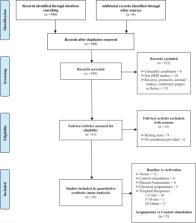
          Literature searches were conducted on online databases, including MEDLINE, Embase, Cochrane Library, Web of Science, China National Knowledge Infrastructure, Wanfang database, WeiPu database, and China Biology Medicine, for task-based fMRI studies of acupuncture at ST36 in healthy subjects. Brain regions activated by ST36 acupuncture were systematically evaluated and subjected to seed-based d mapping meta-analysis. Subgroup analysis was conducted on control procedures, manual acupuncture, electrical acupuncture (EA), and acupuncture-specific activations. Meta-regression analysis was performed to explore the effects of needle retention time on brain activities following ST36 acupuncture stimulation. The activated brain regions were further decoded and mapped on large-scale functional networks to further decipher the clinical relevance of acupuncturing at ST36.

          Results

          A total of sixteen studies, involving a total of 401 right-handed healthy participants, that satisfied the inclusion criteria were included in the present meta-analysis. Meta-analysis showed that acupuncturing on ST36 positively activates the opercular part of the right inferior frontal gyrus (IFG.R), left superior temporal gyrus (STG.L), and right median cingulate/paracingulate gyri (MCG.R) regions. Needle retention time in an acupuncture session positively correlates with the activation of the left olfactory cortex, as shown in meta-regression analysis. Subgroup analysis revealed that EA stimulation may be a source of heterogeneity in the pooled results. Functional network mappings showed that the activated areas were mapped to the auditory network and salience network. Further functional decoding analysis showed that acupuncture on ST36 was associated with pain, secondary somatosensory, sound and language processing, and mood regulation.

          Conclusion

          Acupuncture at ST36 in healthy individuals positively activates the opercular part of IFG.R, STG.L, and MCG.R. The left olfactory cortex may exhibit positive needle retention time-dependent activities. Our findings may have clinical implications for acupuncture in analgesia, language processing, and mood disorders.

          Systematic Review Registration

          https://inplasy.com/inplasy-2021-12-0035.

          Related collections

          Most cited references66

          • Record: found
          • Abstract: found
          • Article: not found

          Methodological index for non-randomized studies (minors): development and validation of a new instrument.

          Because of specific methodological difficulties in conducting randomized trials, surgical research remains dependent predominantly on observational or non-randomized studies. Few validated instruments are available to determine the methodological quality of such studies either from the reader's perspective or for the purpose of meta-analysis. The aim of the present study was to develop and validate such an instrument. After an initial conceptualization phase of a methodological index for non-randomized studies (MINORS), a list of 12 potential items was sent to 100 experts from different surgical specialties for evaluation and was also assessed by 10 clinical methodologists. Subsequent testing involved the assessment of inter-reviewer agreement, test-retest reliability at 2 months, internal consistency reliability and external validity. The final version of MINORS contained 12 items, the first eight being specifically for non-comparative studies. Reliability was established on the basis of good inter-reviewer agreement, high test-retest reliability by the kappa-coefficient and good internal consistency by a high Cronbach's alpha-coefficient. External validity was established in terms of the ability of MINORS to identify excellent trials. MINORS is a valid instrument designed to assess the methodological quality of non-randomized surgical studies, whether comparative or non-comparative. The next step will be to determine its external validity when used in a large number of studies and to compare it with other existing instruments.
            Bookmark
            • Record: found
            • Abstract: found
            • Article: not found

            Decoding subject-driven cognitive states with whole-brain connectivity patterns.

            Decoding specific cognitive states from brain activity constitutes a major goal of neuroscience. Previous studies of brain-state classification have focused largely on decoding brief, discrete events and have required the timing of these events to be known. To date, methods for decoding more continuous and purely subject-driven cognitive states have not been available. Here, we demonstrate that free-streaming subject-driven cognitive states can be decoded using a novel whole-brain functional connectivity analysis. Ninety functional regions of interest (ROIs) were defined across 14 large-scale resting-state brain networks to generate a 3960 cell matrix reflecting whole-brain connectivity. We trained a classifier to identify specific patterns of whole-brain connectivity as subjects rested quietly, remembered the events of their day, subtracted numbers, or (silently) sang lyrics. In a leave-one-out cross-validation, the classifier identified these 4 cognitive states with 84% accuracy. More critically, the classifier achieved 85% accuracy when identifying these states in a second, independent cohort of subjects. Classification accuracy remained high with imaging runs as short as 30-60 s. At all temporal intervals assessed, the 90 functionally defined ROIs outperformed a set of 112 commonly used structural ROIs in classifying cognitive states. This approach should enable decoding a myriad of subject-driven cognitive states from brief imaging data samples.
              Bookmark
              • Record: found
              • Abstract: found
              • Article: not found

              Ten simple rules for neuroimaging meta-analysis

              Neuroimaging has evolved into a widely used method to investigate the functional neuroanatomy, brain-behaviour relationships, and pathophysiology of brain disorders, yielding a literature of more than 30,000 papers. With such an explosion of data, it is increasingly difficult to sift through the literature and distinguish spurious from replicable findings. Furthermore, due to the large number of studies, it is challenging to keep track of the wealth of findings. A variety of meta-analytical methods (coordinate-based and image-based) have been developed to help summarise and integrate the vast amount of data arising from neuroimaging studies. However, the field lacks specific guidelines for the conduct of such meta-analyses. Based on our combined experience, we propose best-practice recommendations that researchers from multiple disciplines may find helpful. In addition, we provide specific guidelines and a checklist that will hopefully improve the transparency, traceability, replicability and reporting of meta-analytical results of neuroimaging data.
                Bookmark

                Author and article information

                Contributors
                Journal
                Front Neurol
                Front Neurol
                Front. Neurol.
                Frontiers in Neurology
                Frontiers Media S.A.
                1664-2295
                22 July 2022
                2022
                : 13
                : 930753
                Affiliations
                [1] 1The First Clinical Medical College, Guangzhou University of Chinese Medicine , Guangzhou, China
                [2] 2Department of Radiology, The First Affiliated Hospital of Guangzhou University of Chinese Medicine , Guangzhou, China
                [3] 3Department of Geriatrics, The First Affiliated Hospital, Guangzhou University of Chinese Medicine , Guangzhou, China
                [4] 4Department of Rehabilitation, The First Affiliated Hospital of Guangzhou University of Chinese Medicine , Guangzhou, China
                [5] 5Department of Emergency, The First Affiliated Hospital of Guangzhou University of Chinese Medicine , Guangzhou, China
                [6] 6The First Comprehensive Department, The Second Affiliated Hospital, Guangzhou University of Chinese Medicine (Guangdong Province Hospital of Chinese Medicine) , Guangzhou, China
                [7] 7Department of Chinese Medicine, The First Affiliated Hospital, School of Clinical Medicine of Guangdong Pharmaceutical University , Guangzhou, China
                Author notes

                Edited by: Yong Tang, Chengdu University of Traditional Chinese Medicine, China

                Reviewed by: Jeungchan Lee, Spaulding Rehabilitation Hospital, United States; Im Quah-Smith, Royal Hospital for Women, Australia

                *Correspondence: Shijun Qiu qiu-sj@ 123456163.com

                This article was submitted to Experimental Therapeutics, a section of the journal Frontiers in Neurology

                †These authors have contributed equally to this work

                Article
                10.3389/fneur.2022.930753
                9373901
                35968313
                8eb011c3-4631-45f4-a5f8-a3ec27cd51dc
                Copyright © 2022 Huang, Yue, Huang, Long, Kang, Rao, Zeng, Zuo, Wang, Li, Wang, Qiu and Zhao.

                This is an open-access article distributed under the terms of the Creative Commons Attribution License (CC BY). The use, distribution or reproduction in other forums is permitted, provided the original author(s) and the copyright owner(s) are credited and that the original publication in this journal is cited, in accordance with accepted academic practice. No use, distribution or reproduction is permitted which does not comply with these terms.

                History
                : 28 April 2022
                : 23 June 2022
                Page count
                Figures: 4, Tables: 4, Equations: 0, References: 66, Pages: 18, Words: 9791
                Funding
                Funded by: National Natural Science Foundation of China, doi 10.13039/501100001809;
                Funded by: Guangdong Medical Research Foundation, doi 10.13039/501100003785;
                Funded by: Guangdong Pharmaceutical University, doi 10.13039/501100008366;
                Funded by: Traditional Chinese Medicine Bureau of Guangdong Province, doi 10.13039/501100010883;
                Categories
                Neurology
                Systematic Review

                Neurology
                acupuncture,task-based fmri,st36 (zusanli),brain activation,systematic review
                Neurology
                acupuncture, task-based fmri, st36 (zusanli), brain activation, systematic review

                Comments

                Comment on this article