3
views
0
recommends
+1 Recommend
0 collections
    0
    shares
      • Record: found
      • Abstract: found
      • Article: found
      Is Open Access

      Glutaminase-1 (GLS1) inhibition limits metastatic progression in osteosarcoma

      research-article

      Read this article at

      Bookmark
          There is no author summary for this article yet. Authors can add summaries to their articles on ScienceOpen to make them more accessible to a non-specialist audience.

          Abstract

          Background

          Osteosarcoma (OS) is a malignant bone tumor that often develops during the period of rapid growth associated with adolescence. Despite successful primary tumor control accompanied by adjuvant chemotherapy, death from pulmonary metastases occurs in approximately 30% of patients within 5 years. As overall survival in patients remains unchanged over the last 30 years, urgent needs for novel therapeutic strategies exist. Cancer metastasis is characterized by complex molecular events which result from alterations in gene and protein expression/function. Recent studies suggest that metabolic adaptations, or “metabolic reprogramming,” may similarly contribute to cancer metastasis. The goal of this study was to specifically interrogate the metabolic vulnerabilities of highly metastatic OS cell lines in a series of in vitro and in vivo experiments, in order to identify a tractable metabolically targeted therapeutic strategy for patients.

          Methods

          Nutrient deprivation and drug treatment experiments were performed in MG63.3, 143B, and K7M2 OS cell lines to identify the impact of glutaminase-1 (GLS1) inhibition and metformin treatment on cell proliferation. We functionally validated the impact of drug treatment with extracellular flux analysis, nuclear magnetic resonance (NMR) spectroscopy, and mass spectrometry. 13C-glucose and 13C-glutamine tracing was employed to identify specific contributions of these nutrients to the global metabolic profiles generated with GLS1 inhibition and metformin treatment in vivo.

          Results

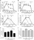
          Highly metastatic OS cell lines require glutamine for proliferation, and exposure to CB-839, in combination with metformin, induces both primary tumor growth inhibition and a distinct reduction in metastatic outgrowth in vivo. Further, combination-treated OS cells showed a reduction in cellular mitochondrial respiration, while NMR confirmed the pharmacodynamic effects of glutaminase inhibition in tumor tissues. We observed global decreases in glycolysis and tricarboxylic acid (TCA) cycle functionality, alongside an increase in fatty acid oxidation and pyrimidine catabolism.

          Conclusions

          This data suggests combination-treated cells cannot compensate for metformin-induced electron transport chain inhibition by upregulating glutaminolysis to generate TCA cycle intermediates required for cell proliferation, translating into significant reductions in tumor growth and metastatic progression. This therapeutic approach could be considered for future clinical development for OS patients presenting with or at high risk of developing metastasis.

          Related collections

          Most cited references33

          • Record: found
          • Abstract: found
          • Article: not found

          Metastasis: a question of life or death.

          The metastatic process is highly inefficient--very few of the many cells that migrate from the primary tumour successfully colonize distant sites. One proposed mechanism to explain this inefficiency is provided by the cancer stem cell model, which hypothesizes that micrometastases can only be established by tumour stem cells, which are few in number. However, recent in vitro and in vivo observations indicate that apoptosis is an important process regulating metastasis. Here we stress that the inhibition of cell death, apart from its extensively described function in primary tumour development, is a crucial characteristic of metastatic cancer cells.
            Bookmark
            • Record: found
            • Abstract: found
            • Article: found
            Is Open Access

            Metformin inhibits mitochondrial complex I of cancer cells to reduce tumorigenesis

            Recent epidemiological and laboratory-based studies suggest that the anti-diabetic drug metformin prevents cancer progression. How metformin diminishes tumor growth is not fully understood. In this study, we report that in human cancer cells, metformin inhibits mitochondrial complex I (NADH dehydrogenase) activity and cellular respiration. Metformin inhibited cellular proliferation in the presence of glucose, but induced cell death upon glucose deprivation, indicating that cancer cells rely exclusively on glycolysis for survival in the presence of metformin. Metformin also reduced hypoxic activation of hypoxia-inducible factor 1 (HIF-1). All of these effects of metformin were reversed when the metformin-resistant Saccharomyces cerevisiae NADH dehydrogenase NDI1 was overexpressed. In vivo, the administration of metformin to mice inhibited the growth of control human cancer cells but not those expressing NDI1. Thus, we have demonstrated that metformin's inhibitory effects on cancer progression are cancer cell autonomous and depend on its ability to inhibit mitochondrial complex I. DOI: http://dx.doi.org/10.7554/eLife.02242.001
              Bookmark
              • Record: found
              • Abstract: found
              • Article: found

              Antitumor activity of the glutaminase inhibitor CB-839 in triple-negative breast cancer.

              Glutamine serves as an important source of energy and building blocks for many tumor cells. The first step in glutamine utilization is its conversion to glutamate by the mitochondrial enzyme glutaminase. CB-839 is a potent, selective, and orally bioavailable inhibitor of both splice variants of glutaminase (KGA and GAC). CB-839 had antiproliferative activity in a triple-negative breast cancer (TNBC) cell line, HCC-1806, that was associated with a marked decrease in glutamine consumption, glutamate production, oxygen consumption, and the steady-state levels of glutathione and several tricarboxylic acid cycle intermediates. In contrast, no antiproliferative activity was observed in an estrogen receptor-positive cell line, T47D, and only modest effects on glutamine consumption and downstream metabolites were observed. Across a panel of breast cancer cell lines, GAC protein expression and glutaminase activity were elevated in the majority of TNBC cell lines relative to receptor positive cells. Furthermore, the TNBC subtype displayed the greatest sensitivity to CB-839 treatment and this sensitivity was correlated with (i) dependence on extracellular glutamine for growth, (ii) intracellular glutamate and glutamine levels, and (iii) GAC (but not KGA) expression, a potential biomarker for sensitivity. CB-839 displayed significant antitumor activity in two xenograft models: as a single agent in a patient-derived TNBC model and in a basal like HER2(+) cell line model, JIMT-1, both as a single agent and in combination with paclitaxel. Together, these data provide a strong rationale for the clinical investigation of CB-839 as a targeted therapeutic in patients with TNBC and other glutamine-dependent tumors.
                Bookmark

                Author and article information

                Contributors
                amy.leblanc@nih.gov
                Journal
                Cancer Metab
                Cancer Metab
                Cancer & Metabolism
                BioMed Central (London )
                2049-3002
                5 March 2020
                5 March 2020
                2020
                : 8
                : 4
                Affiliations
                [1 ]ISNI 0000 0004 1936 8075, GRID grid.48336.3a, Comparative Oncology Program, Center for Cancer Research, , National Cancer Institute, National Institutes of Health, ; Bethesda, MD 20892 USA
                [2 ]ISNI 0000 0004 1936 8075, GRID grid.48336.3a, Metabolomics Section, NeuroOncology Branch, Center for Cancer Research, , National Cancer Institute, National Institutes of Health, ; Bethesda, MD 20892 USA
                [3 ]ISNI 0000 0004 1936 8075, GRID grid.48336.3a, Pediatric Oncology Branch, Center for Cancer Research, , National Cancer Institute, National Institutes of Health, ; Bethesda, MD 20892 USA
                [4 ]ISNI 0000 0004 1936 8075, GRID grid.48336.3a, Laboratory of Human Carcinogenesis, Center for Cancer Research, , National Cancer Institute, National Institutes of Health, ; Bethesda, MD 20892 USA
                Article
                209
                10.1186/s40170-020-0209-8
                7057558
                32158544
                8de7cfb0-de9c-4ae1-8bcd-0c11d297573f
                © The Author(s). 2020

                Open AccessThis article is distributed under the terms of the Creative Commons Attribution 4.0 International License ( http://creativecommons.org/licenses/by/4.0/), which permits unrestricted use, distribution, and reproduction in any medium, provided you give appropriate credit to the original author(s) and the source, provide a link to the Creative Commons license, and indicate if changes were made. The Creative Commons Public Domain Dedication waiver ( http://creativecommons.org/publicdomain/zero/1.0/) applies to the data made available in this article, unless otherwise stated.

                History
                : 6 May 2019
                : 8 January 2020
                Funding
                Funded by: Center for Cancer Research, National Cancer Institute, National Institutes of Health
                Award ID: Z01-BC006161
                Categories
                Research
                Custom metadata
                © The Author(s) 2020

                osteosarcoma,metastasis,metabolism,glutaminase,metformin
                osteosarcoma, metastasis, metabolism, glutaminase, metformin

                Comments

                Comment on this article