8
views
0
recommends
+1 Recommend
0 collections
    0
    shares
      • Record: found
      • Abstract: found
      • Article: found
      Is Open Access

      Konjac ceramide (kCer) regulates keratinocyte migration by Sema3A-like repulsion mechanism

      research-article
      a , * , b , b , c , d , e , f , f , g , a
      Biochemistry and Biophysics Reports
      Elsevier
      NGF, nerve growth factor, Cer, ceramide, GlcCer, glucosylceramide, kCer, konjac ceramide, C16Cer, N-hexadecanoyl-D-erythro-sphingosine, C18Cer, N-octadecanoyl-D-erythro-sphingosine, C24Cer, N-tetracosanoyl-D-erythro-sphingosine, EGCase I, endoglycoceramidase I, p-Cofilin, phospho-Cofilin, CRMP2, collapsin response mediator protein 2, p-CRMP2, phospho-collapsin response mediator protein 2, Sema3A, semaphorin 3A, Nrp1, neuropilin1, hGPCR, histamine G-coupled receptor, His, histamine, H1R, histamine G-coupled receptor 1, H4R, histamine G-coupled receptor 4, AP-Sema3A, alkaline phosphatase-fused Sema3A, BSA, bovine serum albumin, PBS, phosphate-buffered saline, DMEM, Dulbecco's modified Eagle's medium, GAPDH, Glyceraldehyde 3-phosphate dehydrogenase, HRs, histamine G-coupled receptors

      Read this article at

      Bookmark
          There is no author summary for this article yet. Authors can add summaries to their articles on ScienceOpen to make them more accessible to a non-specialist audience.

          Abstract

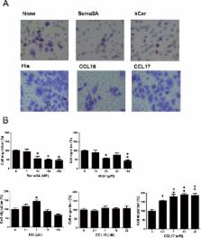
          Previously, we proposed the following mechanism for konjac ceramide (kCer)-mediated neurite outgrowth inhibition: kCer binds to Nrp as a Sema3A agonist, resulting in Nrp1/PlexA complex formation and activation of the Sema3A signaling pathway to induce phosphorylation of CRMP2 and microtubule depolymerization. The Sema3A/Nrp1 signaling pathway is known to be also expressed in normal human keratinocytes. To determine whether kCer can function in human keratinocytes as it does in neurites, that is, if it can bind to Nrp1 in place of Sema3A, we studied the effect of kCer on HaCaT cell migration activity. Using a trans-well chamber assay, we compared the effects of Sema3A and kCer on serum-derived cell migration activity. kCer showed Sema3A-like suppression of cell migration activity and induction of cellular Cofilin phosphorylation. In addition, kCer and Sema3A inhibited histamine (His)-enhanced migration of immature HaCaT cells. We have demonstrated that kCer does not interact with histaime receptors H1R or H4R directly, but we speculate that kCer may transduce a signal downstream of the His signaling pathway.

          Highlights

          • Konjac ceramide (kCer) inhibits HaCaT cell migration by Sema3A-like action.

          • kCer displaces the binding of Sema3A to cell surface receptor.

          • kCer inhibits histamine-induced stimulation of HaCaT cell migration.

          • kCer does not interact with histamine receptors, H1R and H4R.

          Related collections

          Most cited references15

          • Record: found
          • Abstract: found
          • Article: not found

          Itch and nerve fibers with special reference to atopic dermatitis: therapeutic implications.

          Nerve density in the epidermis is partly involved in itch sensitization in pruritic skin diseases, such as atopic dermatitis (AD). Epidermal innervation is thought to be regulated by the balance between nerve elongation factors (e.g. nerve growth factor) and nerve repulsion factors (e.g. semaphorin 3A). Semaphorin 3A (Sema3A) has been shown to inhibit nerve growth factor (NGF)-induced sprouting of sensory nerves, and epidermal Sema3A levels are lower in AD patients, concomitant with an increase in epidermal nerve density. In addition, treatment with anti-NGF, Sema3A replacement, and several existing treatments, such as ultraviolet-based therapies, normalized the hyperinnervation in AD, resulting in suppression of itching. This review expands knowledge regarding potential therapeutic strategies for ameliorating intractable pruritus in AD.
            Bookmark
            • Record: found
            • Abstract: found
            • Article: not found

            Plexin/neuropilin complexes mediate repulsion by the axonal guidance signal semaphorin 3A.

            In the developing nervous system axons navigate with great precision over large distances to reach their target areas. Chemorepulsive signals such as the semaphorins play an essential role in this process. The effects of one of these repulsive cues, semaphorin 3A (Sema3A), are mediated by the membrane protein neuropilin-1 (Npn-1). Recent work has shown that neuropilin-1 is essential but not sufficient to form functional Sema3A receptors and indicates that additional components are required to transduce signals from the cell surface to the cytoskeleton. Here we show that members of the plexin family interact with the neuropilins and act as co-receptors for Sema3A. Neuropilin/plexin interaction restricts the binding specificity of neuropilin-1 and allows the receptor complex to discriminate between two different semaphorins. Deletion of the highly conserved cytoplasmic domain of Plexin-A1 or -A2 creates a dominant negative Sema3A receptor that renders sensory axons resistant to the repulsive effects of Sema3A when expressed in sensory ganglia. These data suggest that functional semaphorin receptors contain plexins as signal-transducing and neuropilins as ligand-binding subunits.
              Bookmark
              • Record: found
              • Abstract: found
              • Article: not found

              Structure of the semaphorin-3A receptor binding module.

              The semaphorins are a large group of extracellular proteins involved in a variety of processes during development, including neuronal migration and axon guidance. Their distinctive feature is a conserved 500 amino acid semaphorin domain, a ligand-receptor interaction module also present in plexins and scatter-factor receptors. We report the crystal structure of a secreted 65 kDa form of Semaphorin-3A (Sema3A), containing the full semaphorin domain. Unexpectedly, the semaphorin fold is a variation of the beta propeller topology. Analysis of the Sema3A structure and structure-based mutagenesis data identify the neuropilin binding site and suggest a potential plexin interaction site. Based on the structure, we present a model for the initiation of semaphorin signaling and discuss potential similarities with the signaling mechanisms of other beta propeller cell surface receptors, such as integrins and the LDL receptor.
                Bookmark

                Author and article information

                Contributors
                Journal
                Biochem Biophys Rep
                Biochem Biophys Rep
                Biochemistry and Biophysics Reports
                Elsevier
                2405-5808
                03 January 2019
                March 2019
                03 January 2019
                : 17
                : 132-138
                Affiliations
                [a ]Lipid Biofunction Section, Frontier Research Center for Advanced Material and Life Science, Faculty of Advanced Life Science, Hokkaido University, Sapporo, Hokkaido, Japan
                [b ]Bioproduction Research Institute, National Institute of Advanced Industrial Science and Technology (AIST), Sapporo, Hokkaido, Japan
                [c ]Department of Biochemistry and Molecular Genetics, Ehime University Graduate School of Medicine, Shitsukawa, Ehime, Japan
                [d ]Department of Neuropathology, Institute of Brain Science, Hirosaki University Graduate School of Medicine, Hirosaki, Aomori, Japan
                [e ]Department of Applied Biochemistry and Food Science, Faculty of Agriculture, Saga University, Saga, Japan
                [f ]Graduate School of Pharmaceutical Sciences, Tohoku University, Sendai, Miyagi, Japan
                [g ]R&D Headquarters, Daicel Corporation, Tokyo, Japan
                Author notes
                [* ]Correspondence to: Lipid Biofunction Section, Frontier Research Center for Advanced Material and Life Science, Faculty of Advanced Life Science, Hokkaido University, Kita21, Nishi11, Kita Ward, Sapporo, Hokkaido 001-0021, Japan. susuki@ 123456sci.hokudai.edu
                Article
                S2405-5808(18)30146-8
                10.1016/j.bbrep.2018.11.004
                6319021
                8dd4a458-b0c4-40df-bd1f-37cc19efa13e
                © 2018 Published by Elsevier B.V.

                This is an open access article under the CC BY-NC-ND license (http://creativecommons.org/licenses/by-nc-nd/4.0/).

                History
                : 19 June 2018
                : 2 October 2018
                : 12 November 2018
                Categories
                Research Article

                ngf, nerve growth factor,cer, ceramide,glccer, glucosylceramide,kcer, konjac ceramide,c16cer, n-hexadecanoyl-d-erythro-sphingosine,c18cer, n-octadecanoyl-d-erythro-sphingosine,c24cer, n-tetracosanoyl-d-erythro-sphingosine,egcase i, endoglycoceramidase i,p-cofilin, phospho-cofilin,crmp2, collapsin response mediator protein 2,p-crmp2, phospho-collapsin response mediator protein 2,sema3a, semaphorin 3a,nrp1, neuropilin1,hgpcr, histamine g-coupled receptor,his, histamine,h1r, histamine g-coupled receptor 1,h4r, histamine g-coupled receptor 4,ap-sema3a, alkaline phosphatase-fused sema3a,bsa, bovine serum albumin,pbs, phosphate-buffered saline,dmem, dulbecco's modified eagle's medium,gapdh, glyceraldehyde 3-phosphate dehydrogenase,hrs, histamine g-coupled receptors

                Comments

                Comment on this article