6
views
0
recommends
+1 Recommend
0 collections
    0
    shares
      • Record: found
      • Abstract: found
      • Article: found
      Is Open Access

      Differentiation of human male germ cells from Wharton's jelly-derived mesenchymal stem cells

      research-article

      Read this article at

      Bookmark
          There is no author summary for this article yet. Authors can add summaries to their articles on ScienceOpen to make them more accessible to a non-specialist audience.

          Abstract

          Objective

          Recapitulation of the spermatogenesis process in vitro is a tool for studying the biology of germ cells, and may lead to promising therapeutic strategies in the future. In this study, we attempted to transdifferentiate Wharton's jelly-derived mesenchymal stem cells (WJ-MSCs) into male germ cells using all- trans retinoic acid and Sertoli cell-conditioned medium.

          Methods

          Human WJ-MSCs were propagated by the explant culture method, and cells at the second passage were induced with differentiation medium containing all- trans retinoic acid for 2 weeks. Putative germ cells were cultured with Sertoli cell-conditioned medium at 36℃ for 3 more weeks.

          Results

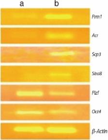
          The gene expression profile was consistent with the stage-specific development of germ cells. The expression of Oct4 and Plzf (early germ cell markers) was diminished, while Stra8 (a premeiotic marker), Scp3 (a meiotic marker), and Acr and Prm1 (postmeiotic markers) were upregulated during the induction period. In morphological studies, approximately 5% of the cells were secondary spermatocytes that had completed two stages of acrosome formation (the Golgi phase and the cap phase). A few spermatid-like cells that had undergone the initial stage of tail formation were also noted.

          Conclusion

          Human WJ-MSCs can be transdifferentiated into more advanced stages of germ cells by a simple two-step induction protocol using retinoic acid and Sertoli cell-conditioned medium.

          Related collections

          Most cited references36

          • Record: found
          • Abstract: found
          • Article: not found

          Spermatogenic cells of the prepuberal mouse: isolation and morphological characterization

          A procedure is described which permits the isolation from the prepuberal mouse testis of highly purified populations of primitive type A spermatogonia, type A spermatogonia, type B spermatogonia, preleptotene primary spermatocytes, leptotene and zygotene primary spermatocytes, pachytene primary spermatocytes and Sertoli cells. The successful isolation of these prepuberal cell types was accomplished by: (a) defining distinctive morphological characteristics of the cells, (b) determining the temporal appearance of spermatogenic cells during prepuberal development, (c) isolating purified seminiferous cords, after dissociation of the testis with collagenase, (d) separating the trypsin-dispersed seminiferous cells by sedimentation velocity at unit gravity, and (e) assessing the identity and purity of the isolated cell types by microscopy. The seminiferous epithelium from day 6 animals contains only primitive type A spermatogonia and Sertoli cells. Type A and type B spermatogonia are present by day 8. At day 10, meiotic prophase is initiated, with the germ cells reaching the early and late pachytene stages by 14 and 18, respectively. Secondary spermatocytes and haploid spermatids appear throughout this developmental period. The purity and optimum day for the recovery of specific cell types are as follows: day 6, Sertoli cells (purity>99 percent) and primitive type A spermatogonia (90 percent); day 8, type A spermatogonia (91 percent) and type B spermatogonia (76 percent); day 18, preleptotene spermatocytes (93 percent), leptotene/zygotene spermatocytes (52 percent), and pachytene spermatocytes (89 percent), leptotene/zygotene spermatocytes (52 percent), and pachytene spermatocytes (89 percent).
            Bookmark
            • Record: found
            • Abstract: found
            • Article: not found

            The central role of Sertoli cells in spermatogenesis.

            Sertoli cells are the somatic cells of the testis that are essential for testis formation and spermatogenesis. Sertoli cells facilitate the progression of germ cells to spermatozoa via direct contact and by controlling the environment milieu within the seminiferous tubules. The regulation of spermatogenesis by FSH and testosterone occurs by the action of these hormones on the Sertoli cells. While the action of testosterone is necessary for spermatogenesis, the action of FSH minimally serves to promote spermatogenic output by increasing the number of Sertoli cells. Copyright 1998 Academic Press.
              Bookmark
              • Record: found
              • Abstract: found
              • Article: not found

              Embryonic stem cells can form germ cells in vitro.

              Knock-in embryonic stem (ES) cells, in which GFP or lacZ was expressed from the endogenous mouse vasa homolog (Mvh), which is specifically expressed in differentiating germ cells, were used to visualize germ cell production during in vitro differentiation. The appearance of MVH-positive germ cells depended on embryoid body formation and was greatly enhanced by the inductive effects of bone morphogenic protein 4-producing cells. The ES-derived MVH-positive cells could participate in spermatogenesis when transplanted into reconstituted testicular tubules, demonstrating that ES cells can produce functional germ cells in vitro. In vitro germ cell differentiation provides a paradigm for studying the molecular basis of germ line establishment, as well as for developing new approaches to reproductive engineering.
                Bookmark

                Author and article information

                Journal
                Clin Exp Reprod Med
                Clin Exp Reprod Med
                CERM
                Clinical and Experimental Reproductive Medicine
                The Korean Society for Reproductive Medicine
                2233-8233
                2233-8241
                June 2018
                29 June 2018
                : 45
                : 2
                : 75-81
                Affiliations
                [1 ]Department of Obstetrics and Gynaecology, Faculty of Medicine, University of Kelaniya, Ragama, Sri Lanka.
                [2 ]StemCure Pvt. Ltd., Ahmedabad, India.
                Author notes
                Corresponding author: DMAB Dissanayake. Department of Obstetrics and Gynaecology, Faculty of Medicine, University of Kelaniya, No. 6, Talagolla Rd, Ragama, Sri Lanka. Tel: +94-714119634, Fax: +94-112958337, anuradissa@ 123456kln.ac.lk
                Article
                10.5653/cerm.2018.45.2.75
                6030615
                29984207
                8d672c07-d2fd-4967-9b40-8c36c5f417e0
                Copyright © 2018. The Korean Society for Reproductive Medicine

                This is an Open Access article distributed under the terms of the Creative Commons Attribution Non-Commercial License ( http://creativecommons.org/licenses/by-nc/4.0/) which permits unrestricted non-commercial use, distribution, and reproduction in any medium, provided the original work is properly cited.

                History
                : 01 October 2017
                : 13 March 2018
                : 03 May 2018
                Funding
                Funded by: Higher Education for the Twenty-First Century (HETC) project;
                Award ID: KLN/O-Med-R/1
                Categories
                Original Article

                Obstetrics & Gynecology
                all-trans retinoic acid,germ cells,mesenchymal stromal cells
                Obstetrics & Gynecology
                all-trans retinoic acid, germ cells, mesenchymal stromal cells

                Comments

                Comment on this article