16
views
0
recommends
+1 Recommend
0 collections
    0
    shares
      • Record: found
      • Abstract: found
      • Article: not found

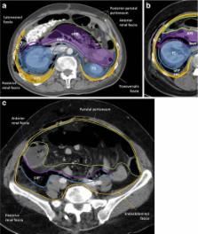
      The anatomical compartments and their connections as demonstrated by ectopic air

      review-article

      Read this article at

      ScienceOpenPublisherPMC
      Bookmark
          There is no author summary for this article yet. Authors can add summaries to their articles on ScienceOpen to make them more accessible to a non-specialist audience.

          Abstract

          Air/gas outside the aero-digestive tract is abnormal; depending on its location, it is usually called emphysema, referring to trapped air/gas in tissues, or ectopic air/gas. It can be associated to a wide range of disorders, and although it usually is an innocuous condition, it should prompt a search for the underlying aetiology, since some of its causes impose an urgent treatment. In rare instances, it may itself represent a life-threatening condition, depending on the site involved and how quickly it evolves. Abnormal air/gas beyond viscera and serosal spaces, reaches its location following some anatomic boundaries, such as fascia, which may help search the source; however if the air pressure exceeds the strength of the tissues, or the time between the aggression and the imaging is too long, the air/gas is almost everywhere, which may hinder its cause. Good knowledge of the anatomic spaces and how they connect between them facilitates the quick detection of the cause.

          Teaching points

          Ectopic air can be depicted on conventional radiographs; but CT is more sensitive and accurate

          Visceral and retropharyngeal spaces directly communicate with mediastinum

          Renal fascia is a single multilaminated structure, which contains potential space

          Related collections

          Most cited references32

          • Record: found
          • Abstract: found
          • Article: not found

          Subcutaneous and mediastinal emphysema. Pathophysiology, diagnosis, and management.

          Subcutaneous emphysema and pneumomediastinum occur frequently in critically ill patients in association with blunt or penetrating trauma, soft-tissue infections, or any condition that creates a gradient between intra-alveolar and perivascular interstitial pressures. A continuum of fascial planes connects cervical soft tissues with the medlastinum and retroperitoneum, permitting aberrant air arising in any one of these areas to spread elsewhere. Diagnosis is made in the appropriate clinical setting by careful physical examination and inspection of the chest roentgenogram. While the presence of air in subcutaneous or mediastinal tissue is not dangerous in itself, prompt recognition of the underlying cause is essential. Certain trauma-related causes may require surgical intervention, but the routine use of chest tubes tracheostomy, or mediastinal drains is not recommended.
            Bookmark
            • Record: found
            • Abstract: found
            • Article: not found

            Emphysematous infections of the abdomen and pelvis: a pictorial review.

            Emphysematous (gas-forming) infections of the abdomen and pelvis represent potentially life-threatening conditions that require aggressive medical and often surgical management. The initial clinical manifestation of these entities may be insidious, but rapid progression to sepsis will occur in the absence of early therapeutic intervention. Conventional radiography and ultrasonography are often the initial imaging modalities used to evaluate patients with abdominopelvic complaints. However, when a differential diagnosis remains, or if further localization or confirmation of tentative findings is needed, computed tomography (CT) should be considered the imaging modality of choice. CT is both highly sensitive and specific in the detection of abnormal gas and well suited to reliable depiction of the anatomic location and extent of the gas. Of equal importance may be the capability of CT to help reliably identify benign sources of gas, because treatment (if any) varies dramatically depending on the source. Knowledge of the pathophysiologic characteristics, common predisposing conditions, and typical imaging features associated with gas-forming infections of the gallbladder, stomach, pancreas, and genitourinary system will help make early diagnosis and successful treatment possible. In addition, such knowledge will aid in further diagnostic work-up, surveillance of potential complications, and evaluation of therapeutic response. Copyright RSNA, 2002
              Bookmark
              • Record: found
              • Abstract: found
              • Article: not found

              Pneumomediastinum revisited.

              Pneumomediastinum may result from a variety of causes that may be either intrathoracic (eg, narrowed or plugged airway, straining against a closed glottis, blunt chest trauma, alveolar rupture) or extrathoracic (eg, sinus fracture, iatrogenic manipulation in dental extraction, perforation of a hollow viscus [corrected]. The radiographic signs of pneumomediastinum depend on the depiction of normal anatomic structures that are outlined by the air as it leaves the mediastinum. These signs include the thymic sail sign, "ring around the artery" sign, tubular artery sign, double bronchial wall sign, continuous diaphragm sign, and extrapleural sign. In distal esophageal rupture, air may migrate from the mediastinum into the pulmonary ligament. Pneumomediastinum may be difficult to differentiate from medial pneumothorax and pneumopericardium. Occasionally, normal anatomic structures (eg, major fissure, anterior junction line) may simulate air within the mediastinum. Iatrogenic entities that may simulate pneumomediastinum include helium in the balloon of an intraaortic assist device. In addition, pneumomediastinum may be simulated by the Mach band effect, which manifests as a region of lucency adjacent to structures with convex borders. The absence of an opaque line, which is typically seen in pneumomediastinum, can aid in differentiation. Computed tomographic (CT) digital radiography and conventional CT can also be helpful in establishing or confirming the diagnosis.
                Bookmark

                Author and article information

                Contributors
                +351-96-6611711 , +351-256-379721 , ana_rita_couto@hotmail.com
                Journal
                Insights Imaging
                Insights Imaging
                Insights into Imaging
                Springer Berlin Heidelberg (Berlin/Heidelberg )
                1869-4101
                25 September 2013
                25 September 2013
                December 2013
                : 4
                : 6
                : 759-772
                Affiliations
                [ ]Department of Imagiology, Hospital-Escola da Universidade Fernando Pessoa, Gondomar, Porto Portugal
                [ ]Department of Radiology, Centro Hospitalar de Entre Douro e Vouga, Rua Dr Cândido Pinho, 4520-211 Santa Maria da Feira, Portugal
                Article
                278
                10.1007/s13244-013-0278-0
                3846937
                24065628
                8d5e612a-4331-4f44-b788-b35b2e3cf26f
                © The Author(s) 2013

                Open Access This article is distributed under the terms of the Creative Commons Attribution License which permits any use, distribution, and reproduction in any medium, provided the original author(s) and the source are credited.

                History
                : 4 April 2013
                : 10 July 2013
                : 17 July 2013
                Categories
                Pictorial Review
                Custom metadata
                © The Author(s) 2013

                Radiology & Imaging
                subcutaneous emphysema,pneumomediastinum,pneumoretroperitoneum,fascia,anatomy
                Radiology & Imaging
                subcutaneous emphysema, pneumomediastinum, pneumoretroperitoneum, fascia, anatomy

                Comments

                Comment on this article