19
views
0
recommends
+1 Recommend
0 collections
    0
    shares
      • Record: found
      • Abstract: found
      • Article: found
      Is Open Access

      Fundus Autofluorescence in Lamellar Macular Holes and Pseudoholes: A Review

      review-article
      Journal of Ophthalmology
      Hindawi

      Read this article at

      Bookmark
          There is no author summary for this article yet. Authors can add summaries to their articles on ScienceOpen to make them more accessible to a non-specialist audience.

          Abstract

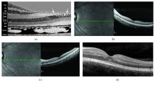
          Macular pseudoholes (MPHs) and lamellar macular holes (LMHs) have been recently defined according to spectral domain optical coherence tomography (SD-OCT) criteria. A major feature for differentiating an MPH from an LMH remains the loss of foveal tissue. The anatomy of the foveola is peculiar with the macular pigment (MP) embedded in a very thin layer of tissue underlying the internal limiting membrane and mainly constituted of a specialized group of Müller cells and Henle's fibers. Despite the near microscopic resolution (≈5–7  μm) and the capability to visualize the outer retina in detail, SD-OCT may fail to ascertain whether a very small loss of this foveolar tissue has occurred. Blue-fundus autofluorescence (B-FAF) imaging is useful in this respect because even very small loss of MP can be identified, suggesting a corresponding localized loss of the innermost layers of the foveola. A definition of MP loss would help differentiating an LMH from an MPH where B-FAF imaging will be negative.

          Related collections

          Most cited references30

          • Record: found
          • Abstract: found
          • Article: not found

          The International Vitreomacular Traction Study Group classification of vitreomacular adhesion, traction, and macular hole.

          The International Vitreomacular Traction Study (IVTS) Group was convened to develop an optical coherence tomography (OCT)-based anatomic classification system for diseases of the vitreomacular interface (VMI).
            Bookmark
            • Record: found
            • Abstract: found
            • Article: not found

            Proposed lexicon for anatomic landmarks in normal posterior segment spectral-domain optical coherence tomography: the IN•OCT consensus.

            To develop a consensus nomenclature for the classification of retinal and choroidal layers and bands visible on spectral-domain optical coherence tomography (SD-OCT) images of a normal eye.
              Bookmark
              • Record: found
              • Abstract: found
              • Article: not found

              In vivo fluorescence of the ocular fundus exhibits retinal pigment epithelium lipofuscin characteristics.

              To characterize the intrinsic fluorescence (autofluorescence) of the human ocular fundus with regard to its excitation and emission spectra, age relationship, retinal location, and topography, and to identify the dominant fluorophore among the fundus layers. Using a novel fundus spectrophotometer, fluorescence measurements were made at 7 degrees temporal to the fovea and at the fovea in 30 normal subjects and in 3 selected patients. Topographic measurements were made in 3 subjects. Ex vivo measurements of fluorescence of human retinal pigment epithelium (RPE) were obtained and compared to in vivo data. Fundus fluorescence reveals a broad band of emission between 500 and 750 nm, a maximum of approximately 630 nm, and optimal excitation of approximately 510 nm. Exhibiting a significant increase with age, this fluorescence is highest at 7 degrees to 15 degrees from the fovea, shows a well-defined foveal minimum, and decreases toward the periphery. In vivo fluorescence spectra are consistent with those obtained ex vivo on human RPE. Measurements with short wavelength excitation are strongly influenced by ocular media absorption and reveal an additional minor fluorophore in the fovea. Spectral characteristics, correlation with age, topographic distribution, and retinal location between the choriocapillaris and the photoreceptors suggest that the dominant fundus fluorophore is RPE lipofuscin. The minor fluorophore is probably in the neurosensory retina but has not been identified.
                Bookmark

                Author and article information

                Contributors
                Journal
                J Ophthalmol
                J Ophthalmol
                JOPH
                Journal of Ophthalmology
                Hindawi
                2090-004X
                2090-0058
                2019
                10 February 2019
                : 2019
                : 4948212
                Affiliations
                Eye Clinic, Department of Clinical Science “Luigi Sacco”, Sacco Hospital, University of Milan, Milan, Italy
                Author notes

                Guest Editor: Akihiro Ohira

                Author information
                http://orcid.org/0000-0002-3737-0238
                Article
                10.1155/2019/4948212
                6387722
                8cc7fda8-5712-4534-b9d9-34e981b00817
                Copyright © 2019 Ferdinando Bottoni.

                This is an open access article distributed under the Creative Commons Attribution License, which permits unrestricted use, distribution, and reproduction in any medium, provided the original work is properly cited.

                History
                : 20 November 2018
                : 26 December 2018
                : 3 January 2019
                Categories
                Review Article

                Ophthalmology & Optometry
                Ophthalmology & Optometry

                Comments

                Comment on this article