8
views
0
recommends
+1 Recommend
0 collections
    0
    shares
      • Record: found
      • Abstract: found
      • Article: found
      Is Open Access

      Identification of the ESKAPE pathogens by mass spectrometric analysis of microbial membrane glycolipids

      research-article

      Read this article at

      Bookmark
          There is no author summary for this article yet. Authors can add summaries to their articles on ScienceOpen to make them more accessible to a non-specialist audience.

          Abstract

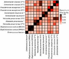
          Rapid diagnostics that enable identification of infectious agents improve patient outcomes, antimicrobial stewardship, and length of hospital stay. Current methods for pathogen detection in the clinical laboratory include biological culture, nucleic acid amplification, ribosomal protein characterization, and genome sequencing. Pathogen identification from single colonies by matrix-assisted laser desorption/ionization time-of-flight mass spectrometry (MALDI-TOF-MS) analysis of high abundance proteins is gaining popularity in clinical laboratories. Here, we present a novel and complementary approach that utilizes essential microbial glycolipids as chemical fingerprints for identification of individual bacterial species. Gram-positive and negative bacterial glycolipids were extracted using a single optimized protocol. Extracts of the clinically significant ESKAPE pathogens: E nterococcus faecium, S taphylococcus aureus, K lebsiella pneumoniae, A cinetobacter baumannii, P seudomonas aeruginosa, and E nterobacter spp. were analyzed by MALDI-TOF-MS in negative ion mode to obtain glycolipid mass spectra. A library of glycolipid mass spectra from 50 microbial entries was developed that allowed bacterial speciation of the ESKAPE pathogens, as well as identification of pathogens directly from blood bottles without culture on solid medium and determination of antimicrobial peptide resistance. These results demonstrate that bacterial glycolipid mass spectra represent chemical barcodes that identify pathogens, potentially providing a useful alternative to existing diagnostics.

          Related collections

          Most cited references22

          • Record: found
          • Abstract: found
          • Article: not found

          Ongoing revolution in bacteriology: routine identification of bacteria by matrix-assisted laser desorption ionization time-of-flight mass spectrometry.

          Matrix-assisted laser desorption ionization time-of-flight (MALDI-TOF) mass spectrometry accurately identifies both selected bacteria and bacteria in select clinical situations. It has not been evaluated for routine use in the clinic. We prospectively analyzed routine MALDI-TOF mass spectrometry identification in parallel with conventional phenotypic identification of bacteria regardless of phylum or source of isolation. Discrepancies were resolved by 16S ribosomal RNA and rpoB gene sequence-based molecular identification. Colonies (4 spots per isolate directly deposited on the MALDI-TOF plate) were analyzed using an Autoflex II Bruker Daltonik mass spectrometer. Peptidic spectra were compared with the Bruker BioTyper database, version 2.0, and the identification score was noted. Delays and costs of identification were measured. Of 1660 bacterial isolates analyzed, 95.4% were correctly identified by MALDI-TOF mass spectrometry; 84.1% were identified at the species level, and 11.3% were identified at the genus level. In most cases, absence of identification (2.8% of isolates) and erroneous identification (1.7% of isolates) were due to improper database entries. Accurate MALDI-TOF mass spectrometry identification was significantly correlated with having 10 reference spectra in the database (P=.01). The mean time required for MALDI-TOF mass spectrometry identification of 1 isolate was 6 minutes for an estimated 22%-32% cost of current methods of identification. MALDI-TOF mass spectrometry is a cost-effective, accurate method for routine identification of bacterial isolates in or =10 reference spectra per bacterial species and a 1.9 identification score (Brucker system). It may replace Gram staining and biochemical identification in the near future.
            Bookmark
            • Record: found
            • Abstract: found
            • Article: not found

            Matrix-assisted laser desorption ionization-time of flight mass spectrometry: a fundamental shift in the routine practice of clinical microbiology.

            Within the past decade, clinical microbiology laboratories experienced revolutionary changes in the way in which microorganisms are identified, moving away from slow, traditional microbial identification algorithms toward rapid molecular methods and mass spectrometry (MS). Historically, MS was clinically utilized as a high-complexity method adapted for protein-centered analysis of samples in chemistry and hematology laboratories. Today, matrix-assisted laser desorption ionization-time of flight (MALDI-TOF) MS is adapted for use in microbiology laboratories, where it serves as a paradigm-shifting, rapid, and robust method for accurate microbial identification. Multiple instrument platforms, marketed by well-established manufacturers, are beginning to displace automated phenotypic identification instruments and in some cases genetic sequence-based identification practices. This review summarizes the current position of MALDI-TOF MS in clinical research and in diagnostic clinical microbiology laboratories and serves as a primer to examine the "nuts and bolts" of MALDI-TOF MS, highlighting research associated with sample preparation, spectral analysis, and accuracy. Currently available MALDI-TOF MS hardware and software platforms that support the use of MALDI-TOF with direct and precultured specimens and integration of the technology into the laboratory workflow are also discussed. Finally, this review closes with a prospective view of the future of MALDI-TOF MS in the clinical microbiology laboratory to accelerate diagnosis and microbial identification to improve patient care.
              Bookmark
              • Record: found
              • Abstract: found
              • Article: found
              Is Open Access

              Survey of bacterial diversity in chronic wounds using Pyrosequencing, DGGE, and full ribosome shotgun sequencing

              Background Chronic wound pathogenic biofilms are host-pathogen environments that colonize and exist as a cohabitation of many bacterial species. These bacterial populations cooperate to promote their own survival and the chronic nature of the infection. Few studies have performed extensive surveys of the bacterial populations that occur within different types of chronic wound biofilms. The use of 3 separate16S-based molecular amplifications followed by pyrosequencing, shotgun Sanger sequencing, and denaturing gradient gel electrophoresis were utilized to survey the major populations of bacteria that occur in the pathogenic biofilms of three types of chronic wound types: diabetic foot ulcers (D), venous leg ulcers (V), and pressure ulcers (P). Results There are specific major populations of bacteria that were evident in the biofilms of all chronic wound types, including Staphylococcus, Pseudomonas, Peptoniphilus, Enterobacter, Stenotrophomonas, Finegoldia, and Serratia spp. Each of the wound types reveals marked differences in bacterial populations, such as pressure ulcers in which 62% of the populations were identified as obligate anaerobes. There were also populations of bacteria that were identified but not recognized as wound pathogens, such as Abiotrophia para-adiacens and Rhodopseudomonas spp. Results of molecular analyses were also compared to those obtained using traditional culture-based diagnostics. Only in one wound type did culture methods correctly identify the primary bacterial population indicating the need for improved diagnostic methods. Conclusion If clinicians can gain a better understanding of the wound's microbiota, it will give them a greater understanding of the wound's ecology and will allow them to better manage healing of the wound improving the prognosis of patients. This research highlights the necessity to begin evaluating, studying, and treating chronic wound pathogenic biofilms as multi-species entities in order to improve the outcomes of patients. This survey will also foster the pioneering and development of new molecular diagnostic tools, which can be used to identify the community compositions of chronic wound pathogenic biofilms and other medical biofilm infections.
                Bookmark

                Author and article information

                Contributors
                rkernst@umaryland.edu
                dgoodlett@rx.umaryland.edu
                Journal
                Sci Rep
                Sci Rep
                Scientific Reports
                Nature Publishing Group UK (London )
                2045-2322
                25 July 2017
                25 July 2017
                2017
                : 7
                : 6403
                Affiliations
                [1 ]Department of Microbial Pathogenesis, University of Maryland, Baltimore, MD 21201 USA
                [2 ]Center for Vascular and Inflammatory Diseases, University of Maryland, Baltimore, MD 21201 USA
                [3 ]ISNI 0000 0004 1936 9000, GRID grid.21925.3d, Division of Infectious Diseases, , University of Pittsburgh, ; Pittsburgh, PA 15261 USA
                [4 ]Department of Pathology, University of Maryland, Baltimore, MD 21201 USA
                [5 ]Department of Pharmaceutical Sciences, University of Maryland, Baltimore, MD 21201 USA
                Article
                4793
                10.1038/s41598-017-04793-4
                5526941
                28743946
                8bff5591-b0c5-4abe-9486-76e1e2a1de95
                © The Author(s) 2017

                Open Access This article is licensed under a Creative Commons Attribution 4.0 International License, which permits use, sharing, adaptation, distribution and reproduction in any medium or format, as long as you give appropriate credit to the original author(s) and the source, provide a link to the Creative Commons license, and indicate if changes were made. The images or other third party material in this article are included in the article’s Creative Commons license, unless indicated otherwise in a credit line to the material. If material is not included in the article’s Creative Commons license and your intended use is not permitted by statutory regulation or exceeds the permitted use, you will need to obtain permission directly from the copyright holder. To view a copy of this license, visit http://creativecommons.org/licenses/by/4.0/.

                History
                : 17 February 2017
                : 19 May 2017
                Categories
                Article
                Custom metadata
                © The Author(s) 2017

                Uncategorized
                Uncategorized

                Comments

                Comment on this article