3
views
0
recommends
+1 Recommend
0 collections
    0
    shares
      • Record: found
      • Abstract: found
      • Article: found
      Is Open Access

      Chemically programmable bacterial probes for the recognition of cell surface proteins

      research-article

      Read this article at

      Bookmark
          There is no author summary for this article yet. Authors can add summaries to their articles on ScienceOpen to make them more accessible to a non-specialist audience.

          Abstract

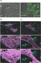
          Common methods to label cell surface proteins (CSPs) involve the use of fluorescently modified antibodies (Abs) or small-molecule-based ligands. However, optimizing the labeling efficiency of such systems, for example, by modifying them with additional fluorophores or recognition elements, is challenging. Herein we show that effective labeling of CSPs overexpressed in cancer cells and tissues can be obtained with fluorescent probes based on chemically modified bacteria. The bacterial probes (B-probes) are generated by non-covalently linking a bacterial membrane protein to DNA duplexes appended with fluorophores and small-molecule binders of CSPs overexpressed in cancer cells. We show that B-probes are exceptionally simple to prepare and modify because they are generated from self-assembled and easily synthesized components, such as self-replicating bacterial scaffolds and DNA constructs that can be readily appended, at well-defined positions, with various types of dyes and CSP binders. This structural programmability enabled us to create B-probes that can label different types of cancer cells with distinct colors, as well as generate very bright B-probes in which the multiple dyes are spatially separated along the DNA scaffold to avoid self-quenching. This enhancement in the emission signal enabled us to label the cancer cells with greater sensitivity and follow the internalization of the B-probes into these cells. The potential to apply the design principles underlying B-probes in therapy or inhibitor screening is also discussed here.

          Graphical abstract

          Decorating bacteria with modified DNA duplexes is presented as a general strategy to obtain fluorescent bacterial probes (B-probes) that can selectively identify cancer cells. The high structural programmability of such systems enabled the creation of B-probes that can selectively label cancer cells and tissues with different emission colors and with enhanced brightness.

          Related collections

          Most cited references76

          • Record: found
          • Abstract: found
          • Article: not found

          The fluorescent toolbox for assessing protein location and function.

          Advances in molecular biology, organic chemistry, and materials science have recently created several new classes of fluorescent probes for imaging in cell biology. Here we review the characteristic benefits and limitations of fluorescent probes to study proteins. The focus is on protein detection in live versus fixed cells: determination of protein expression, localization, activity state, and the possibility for combination of fluorescent light microscopy with electron microscopy. Small organic fluorescent dyes, nanocrystals ("quantum dots"), autofluorescent proteins, small genetic encoded tags that can be complexed with fluorochromes, and combinations of these probes are highlighted.
            Bookmark
            • Record: found
            • Abstract: found
            • Article: found
            Is Open Access

            Overview of multiplex immunohistochemistry/immunofluorescence techniques in the era of cancer immunotherapy

            Abstract Conventional immunohistochemistry (IHC) is a widely used diagnostic technique in tissue pathology. However, this technique is associated with a number of limitations, including high inter‐observer variability and the capacity to label only one marker per tissue section. This review details various highly multiplexed techniques that have emerged to circumvent these constraints, allowing simultaneous detection of multiple markers on a single tissue section and the comprehensive study of cell composition, cellular functional and cell‐cell interactions. Among these techniques, multiplex Immunohistochemistry/Immunofluorescence (mIHC/IF) has emerged to be particularly promising. mIHC/IF provides high‐throughput multiplex staining and standardized quantitative analysis for highly reproducible, efficient and cost‐effective tissue studies. This technique has immediate potential for translational research and clinical practice, particularly in the era of cancer immunotherapy.
              Bookmark
              • Record: found
              • Abstract: found
              • Article: found
              Is Open Access

              Bacteria-cancer interactions: bacteria-based cancer therapy

              Recent advances in cancer therapeutics, such as targeted therapy and immunotherapy, have raised the hope for cures for many cancer types. However, there are still ongoing challenges to the pursuit of novel therapeutic approaches, including high toxicity to normal tissue and cells, difficulties in treating deep tumor tissue, and the possibility of drug resistance in tumor cells. The use of live tumor-targeting bacteria provides a unique therapeutic option that meets these challenges. Compared with most other therapeutics, tumor-targeting bacteria have versatile capabilities for suppressing cancer. Bacteria preferentially accumulate and proliferate within tumors, where they can initiate antitumor immune responses. Bacteria can be further programmed via simple genetic manipulation or sophisticated synthetic bioengineering to produce and deliver anticancer agents based on clinical needs. Therapeutic approaches using live tumor-targeting bacteria can be applied either as a monotherapy or in combination with other anticancer therapies to achieve better clinical outcomes. In this review, we introduce and summarize the potential benefits and challenges of this anticancer approach. We further discuss how live bacteria interact with tumor microenvironments to induce tumor regression. We also provide examples of different methods for engineering bacteria to improve efficacy and safety. Finally, we introduce past and ongoing clinical trials involving tumor-targeting bacteria.
                Bookmark

                Author and article information

                Contributors
                Journal
                Mater Today Bio
                Mater Today Bio
                Materials Today Bio
                Elsevier
                2590-0064
                23 May 2023
                June 2023
                23 May 2023
                : 20
                : 100669
                Affiliations
                [a ]Department of Chemical and Structural Biology, Weizmann Institute of Science Rehovot, 7610001, Israel
                [b ]Life Sciences Core Facilities, Weizmann Institute of Science, Rehovot, 7610001, Israel
                [c ]Department of Veterinary Resources, Weizmann Institute of Science, Rehovot, 7610001, Israel
                Author notes
                []Corresponding author. . david.margulies@ 123456weizmann.ac.il
                [∗∗ ]Corresponding author. . leila.motiei@ 123456weizmann.ac.il
                Article
                S2590-0064(23)00129-1 100669
                10.1016/j.mtbio.2023.100669
                10275978
                8b7c39a7-f764-4c71-9c5c-f0b801279de0
                © 2023 The Authors

                This is an open access article under the CC BY license (http://creativecommons.org/licenses/by/4.0/).

                History
                : 25 January 2023
                : 1 May 2023
                : 17 May 2023
                Categories
                Full Length Article

                self-assembly,dna nanotechnology,cell surface engineering,supramolecular chemistry,fluorescent probes

                Comments

                Comment on this article