21
views
0
recommends
+1 Recommend
0 collections
    0
    shares
      • Record: found
      • Abstract: found
      • Article: found
      Is Open Access

      Extracellular Vesicles in Prostate Cancer Carcinogenesis, Diagnosis, and Management

      review-article

      Read this article at

      Bookmark
          There is no author summary for this article yet. Authors can add summaries to their articles on ScienceOpen to make them more accessible to a non-specialist audience.

          Abstract

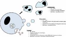
          Extracellular vesicles (EVs), especially exosomes, are now well recognized as major ways by which cancer cells interact with each other and stromal cells. The meaningful messages transmitted by the EVs are carried by all components of the EVs, i.e., the membrane lipids and the cargo (DNAs, RNAs, microRNAs, long non-coding RNAs, proteins). They are clearly part of the armed arsenal by which cancer cells obtain and share more and more advantages to grow and conquer new spaces. Identification of these messages offers a significant opportunity to better understand how a cancer occurs and then develops both locally and distantly. But it also provides a powerful means by which cancer progression can be detected and monitored. In the last few years, significant research efforts have been made to precisely identify how the EV trafficking is modified in cancer cells as compared to normal cells and how this trafficking is altered during cancer progression. Prostate cancer has not escaped this trend. The aim of this review is to describe the results obtained when assessing the meaningful content of prostate cancer- and stromal-derived EVs in terms of a better comprehension of the cellular and molecular mechanisms underlying prostate cancer occurrence and development. This review also deals with the use of EVs as powerful tools to diagnose non-indolent prostate cancer as early as possible and to accurately define, in a personalized approach, its present and potential aggressiveness, its response to treatment (androgen deprivation, chemotherapy, radiation, surgery), and the overall patients’ prognosis.

          Related collections

          Most cited references139

          • Record: found
          • Abstract: found
          • Article: not found

          Biogenesis and secretion of exosomes.

          Although observed for several decades, the release of membrane-enclosed vesicles by cells into their surrounding environment has been the subject of increasing interest in the past few years, which led to the creation, in 2012, of a scientific society dedicated to the subject: the International Society for Extracellular Vesicles. Convincing evidence that vesicles allow exchange of complex information fuelled this rise in interest. But it has also become clear that different types of secreted vesicles co-exist, with different intracellular origins and modes of formation, and thus probably different compositions and functions. Exosomes are one sub-type of secreted vesicles. They form inside eukaryotic cells in multivesicular compartments, and are secreted when these compartments fuse with the plasma membrane. Interestingly, different families of molecules have been shown to allow intracellular formation of exosomes and their subsequent secretion, which suggests that even among exosomes different sub-types exist. Copyright © 2014 Elsevier Ltd. All rights reserved.
            Bookmark
            • Record: found
            • Abstract: found
            • Article: not found

            Electron microscopic evidence for externalization of the transferrin receptor in vesicular form in sheep reticulocytes

            Using ferritin-labeled protein A and colloidal gold-labeled anti-rabbit IgG, the fate of the sheep transferrin receptor has been followed microscopically during reticulocyte maturation in vitro. After a few minutes of incubation at 37 degrees C, the receptor is found on the cell surface or in simple vesicles of 100-200 nm, in which the receptor appears to line the limiting membrane of the vesicles. With time (60 min or longer), large multivesicular elements (MVEs) appear whose diameter may reach 1-1.5 micron. Inside these large MVEs are round bodies of approximately 50-nm diam that bear the receptor at their external surfaces. The limiting membrane of the large MVEs is relatively free from receptor. When the large MVEs fuse with the plasma membrane, their contents, the 50-nm bodies, are released into the medium. The 50-nm bodies appear to arise by budding from the limiting membrane of the intracellular vesicles. Removal of surface receptor with pronase does not prevent exocytosis of internalized receptor. It is proposed that the exocytosis of the approximately 50-nm bodies represents the mechanism by which the transferrin receptor is shed during reticulocyte maturation.
              Bookmark
              • Record: found
              • Abstract: found
              • Article: not found

              High Levels of Exosomes Expressing CD63 and Caveolin-1 in Plasma of Melanoma Patients

              Background Metastatic melanoma is an untreatable cancer lacking reliable and non-invasive markers of disease progression. Exosomes are small vesicles secreted by normal as well as tumor cells. Human tumor-derived exosomes are involved in malignant progression and we evaluated the presence of exosomes in plasma of melanoma patients as a potential tool for cancer screening and follow-up. Methodology/Principal Findings We designed an in-house sandwich ELISA (Exotest) to capture and quantify exosomes in plasma based on expression of housekeeping proteins (CD63 and Rab-5b) and a tumor-associated marker (caveolin-1). Western blot and flow cytometry analysis of exosomes were used to confirm the Exotest-based findings. The Exotest allowed sensitive detection and quantification of exosomes purified from human tumor cell culture supernatants and plasma from SCID mice engrafted with human melanoma. Plasma levels of exosomes in melanoma-engrafted SCID mice correlated to tumor size. We evaluated the levels of plasma exosomes expressing CD63 and caveolin-1 in melanoma patients (n = 90) and healthy donors (n = 58). Consistently, plasma exosomes expressing CD63 (504±315) or caveolin-1 (619±310) were significantly increased in melanoma patients as compared to healthy donors (223±125 and 228±102, respectively). While the Exotest for CD63+ plasma exosomes had limited sensitivity (43%) the Exotest for detection of caveolin-1+ plasma exosomes showed a higher sensitivity (68%). Moreover, caveolin-1+ plasma exosomes were significantly increased with respect to CD63+ exosomes in the patients group. Conclusions/Significance We describe a new non-invasive assay allowing detection and quantification of human exosomes in plasma of melanoma patients. Our results suggest that the Exotest for detection of plasma exosomes carrying tumor-associated antigens may represent a novel tool for clinical management of cancer patients.
                Bookmark

                Author and article information

                Contributors
                URI : https://frontiersin.org/people/u/129094
                Journal
                Front Oncol
                Front Oncol
                Front. Oncol.
                Frontiers in Oncology
                Frontiers Media S.A.
                2234-943X
                13 June 2018
                2018
                : 8
                : 222
                Affiliations
                [1] 1Medical Unit of Molecular Oncology and Transfer, Department of Biochemistry and Molecular Biology, Centre Hospitalier Lyon-Sud, Hospices Civils of Lyon , Pierre-Bénite, France
                [2] 2Cancer Research Centre of Lyon, U1052 INSERM, CNRS 5286, Claude Bernard University Lyon 1, Léon Bérard Centre , Lyon, France
                Author notes

                Edited by: Stéphane Terry, Institut Gustave Roussy, France

                Reviewed by: Stefano Fais, Istituto Superiore di Sanità, Italy; Xavier Filella, Hospital Clínic de Barcelona, Spain

                *Correspondence: Virginie Vlaeminck-Guillem, virginie.vlaeminck-guillem@ 123456univ-lyon1.fr

                Specialty section: This article was submitted to Genitourinary Oncology, a section of the journal Frontiers in Oncology

                Article
                10.3389/fonc.2018.00222
                6008571
                29951375
                8a5e4b18-3582-4231-a5a4-407ecb55f333
                Copyright © 2018 Vlaeminck-Guillem.

                This is an open-access article distributed under the terms of the Creative Commons Attribution License (CC BY). The use, distribution or reproduction in other forums is permitted, provided the original author(s) and the copyright owner are credited and that the original publication in this journal is cited, in accordance with accepted academic practice. No use, distribution or reproduction is permitted which does not comply with these terms.

                History
                : 09 December 2017
                : 29 May 2018
                Page count
                Figures: 1, Tables: 3, Equations: 0, References: 229, Pages: 22, Words: 19096
                Categories
                Oncology
                Review

                Oncology & Radiotherapy
                extracellular vesicles,prostate cancer,microenvironment,diagnosis,theranostic,exosomes

                Comments

                Comment on this article