82
views
0
recommends
+1 Recommend
0 collections
    0
    shares
      • Record: found
      • Abstract: found
      • Article: found
      Is Open Access

      Experimental Differentiation of Intraocular Masses Using Ultrahigh-Field Magnetic Resonance Imaging – A Case Series

      research-article

      Read this article at

      Bookmark
          There is no author summary for this article yet. Authors can add summaries to their articles on ScienceOpen to make them more accessible to a non-specialist audience.

          Abstract

          Purpose

          The case reports presented here were compiled to demonstrate the potential for improved diagnosis and monitoring of disease progress of intraocular lesions using ultrahigh-field magnetic resonance microscopy (MRM) at 7.1 Tesla.

          Methods

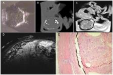
          High-resolution ex vivo ocular magnetic resonance (MR) images were acquired on an ultrahigh-field MR system (7.1 Tesla, ClinScan, Bruker BioScan, Germany) using a 2-channel coil with 4 coil elements and T2-weighted turbo spin echo (TSE) sequences of human eyes enucleated because of different intraocular lesions. Imaging parameters were: 40×40 mm field of view, 512×512 matrix, and 700 µm slice thickness. The results were correlated with in vivo ultrasound and histology of the enucleated eyes.

          Results

          Imaging was performed in enucleated eyes with choroidal melanoma, malignant melanoma of iris and ciliary body with scleral perforation, ciliary body melanoma, intraocular metastasis of esophageal cancer, subretinal bleeding in the presence of perforated corneal ulcer, hemorrhagic choroidal detachment, and premature retinopathy with phthisis and ossification of bulbar structures. MR imaging allowed differentiation between solid and cystic tumor components. In case of hemorrhage, fluid-fluid levels were identified. Melanin and calcifications caused significant hypointensity. Microstructural features of eye lesions identified by MRM were confirmed by histology.

          Conclusion

          This study demonstrates the potential of MRM for the visualization and differential diagnosis of intraocular lesions. At present, the narrow bore of the magnet still limits the use of this technology in humans in vivo. Further advances in ultrahigh-field MR imaging will permit visualization of tumor extent and evaluation of nonclassified intraocular structures in the near future.

          Related collections

          Most cited references26

          • Record: found
          • Abstract: found
          • Article: not found

          Intraocular pressure reduction after phacoemulsification with intraocular lens implantation in glaucomatous and nonglaucomatous eyes: evaluation of a causal relationship between the natural lens and open-angle glaucoma.

          To study the long-term effects of phacoemulsification with intraocular lens (IOL) implantation in nonglaucomatous and glaucomatous eyes. Phillips Eye Institute, Minneapolis, Minnesota, and private practice, Savannah, Georgia, USA. Intraocular pressure (IOP) after phacoemulsification with IOL implantation was retrospectively reviewed. Eyes were divided into 5 groups by preoperative IOP. Data were recorded preoperatively, 1 year postoperatively, and at the final check. Analysis included preoperative IOP versus IOP at 1 year and final IOP, percentage of eyes with elevated or reduced IOP postoperatively, patient age at surgery, and years of postoperative follow-up. The study comprised 124 eyes. The final mean IOP reduction was 8.5 mm Hg (34%) in the 29 to 23 mm Hg group, 4.6 mm Hg (22%) in the 22 to 20 mm Hg group, 3.4 mm Hg (18%) in the 19 to 18 mm Hg group, and 1.1 mm Hg (10%) in the 17 to 15 mm Hg group. In the 14 to 5 mm Hg group, IOP increased by 1.7 mm Hg (15%). Intraocular pressure reduction was proportional to preoperative IOP; the highest preoperative IOPs decreased the most and the lowest increased slightly. One-year IOP reductions were sustained for 10 years and were similar in patients of all ages. The IOP reductions were similar to previously reported reductions in nonglaucomatous eyes, indicating that the aging crystalline lens may be a major cause of ocular hypertension and glaucoma and that phacoemulsification with IOL implantation may help prevent and treat adult glaucoma.
            Bookmark
            • Record: found
            • Abstract: found
            • Article: not found

            Choroidal thickness in Behcet's uveitis: an enhanced depth imaging-optical coherence tomography and its association with angiographic changes.

            To evaluate the change in subfoveal choroidal thickness between active and quiescent phases of Behcet's posterior uveitis and compare this with the healthy population using enhanced depth imaging optical coherence tomography (EDI-OCT). Thirty eyes from 30 patients with Behcet's posterior uveitis (mean age, 47.03 ± 11.01 years) were retrospectively enrolled in the study. Their subfoveal choroidal thickness was measured using EDI-OCT in the active and quiescent phases of Behcet's uveitis, and compared with the age, sex, and spherical equivalent-matched healthy population. Changes in retinal vascular leakage on fluorescein angiography (FA) were correlated with the changes in subfoveal choroidal thickness. Mean subfoveal choroidal thickness in the acute phase of Behcet's uveitis was significantly greater than that in the quiescent phase (398.77 ± 155.59 μm versus 356.72 ± 141.09 μm; P = 0.004). Subfoveal choroidal thickness in the quiescent phase was also significantly greater than that of the healthy population (259.96 ± 65.16 μm; P < 0.0001). There was a statistically significant association between the change in subfoveal choroidal thickness and the change in vascular leakage revealed by FA (ρ = 0.381, P = 0.046). Subfoveal choroidal thickness in the uninvolved fellow eyes of patients with unilateral Behcet's uveitis was also evaluated and it was significantly greater than that of the healthy population (n = 13 eyes; P = 0.001) CONCLUSIONS: This study found choroidal thickening during the active phase of Behcet's posterior uveitis. Subfoveal choroidal thickness during the quiescent phase was also significantly greater than in normal eyes. The degree of reduction in choroidal thickening was significantly correlated with improvement in retinal vascular leakage as revealed by FA.
              Bookmark
              • Record: found
              • Abstract: found
              • Article: not found

              Review of optical coherence tomography for intraocular tumors.

              Optical coherence tomography has assumed an important role in the management of numerous ocular conditions. With regard to ocular oncology, optical coherence tomography can illustrate retinal changes overlying choroidal tumors. Some of these features include photoreceptor loss, intraretinal edema, and retinal thinning overlying choroidal nevus; fresh subretinal fluid with preservation of photoreceptors overlying choroidal melanoma; and intraretinal edema, retinoschisis, and retinal thinning overlying irradiated choroidal melanoma. The optical coherence tomography features of tumors of the retinal pigment epithelium include typical findings of peaked vitreoretinal traction and retinal disorganization with combined hamartoma of the retina and retinal pigment epithelium, full-thickness retinal shadowing with congenital simple hamartoma, and photoreceptor loss and retinal thinning overlying congenital hypertrophy of the retinal pigment epithelium. Optical coherence tomography of retinal tumors, such as retinoblastoma and astrocytic hamartoma, reveals full-thickness replacement of the retinal anatomic layers with the tumor and shadowing corresponding to the intralesional calcification. For all intraocular tumors, optical coherence tomography provides valuable information regarding the status of the retina and the retinal pigment epithelium and can be useful in ascertaining reasons for visual loss.
                Bookmark

                Author and article information

                Contributors
                Role: Editor
                Journal
                PLoS One
                PLoS ONE
                plos
                plosone
                PLoS ONE
                Public Library of Science (San Francisco, USA )
                1932-6203
                2013
                9 December 2013
                : 8
                : 12
                : e81284
                Affiliations
                [1 ]Department of Ophthalmology, University of Rostock, Rostock, Germany
                [2 ]Institute of Diagnostic Radiology and Neuroradiology, Greifswald University Hospital, Greifswald, Germany
                [3 ]Institute of Pathology, University of Rostock, Rostock, Germany
                Medical University Graz, Austria
                Author notes

                Competing Interests: The authors have declared that no competing interests exist.

                Conceived and designed the experiments: PCK NH RFG SL OS. Performed the experiments: KF PCK NH SL OS. Analyzed the data: KF PCK NH AZ SL OS. Contributed reagents/materials/analysis tools: KF PCK NH RFG SL. Wrote the paper: KF PCK.

                Article
                PONE-D-13-25273
                10.1371/journal.pone.0081284
                3857191
                8a0eac2e-b220-45a8-8c77-a8b02b4e9d4a
                Copyright @ 2013

                This is an open-access article distributed under the terms of the Creative Commons Attribution License, which permits unrestricted use, distribution, and reproduction in any medium, provided the original author and source are credited.

                History
                : 18 June 2013
                : 20 October 2013
                Page count
                Pages: 6
                Funding
                The authors have no support or funding to report.
                Categories
                Research Article

                Uncategorized
                Uncategorized

                Comments

                Comment on this article