6
views
0
recommends
+1 Recommend
0 collections
    0
    shares
      • Record: found
      • Abstract: found
      • Article: found
      Is Open Access

      Heterogeneity and plasticity of epithelial–mesenchymal transition (EMT) in cancer metastasis: Focusing on partial EMT and regulatory mechanisms

      review-article

      Read this article at

      Bookmark
          There is no author summary for this article yet. Authors can add summaries to their articles on ScienceOpen to make them more accessible to a non-specialist audience.

          Abstract

          Epithelial–mesenchymal transition (EMT) or mesenchymal–epithelial transition (MET) plays critical roles in cancer metastasis. Recent studies, especially those based on single‐cell sequencing, have revealed that EMT is not a binary process, but a heterogeneous and dynamic disposition with intermediary or partial EMT states. Multiple double‐negative feedback loops involved by EMT‐related transcription factors (EMT‐TFs) have been identified. These feedback loops between EMT drivers and MET drivers finely regulate the EMT transition state of the cell. In this review, the general characteristics, biomarkers and molecular mechanisms of different EMT transition states were summarized. We additionally discussed the direct and indirect roles of EMT transition state in tumour metastasis. More importantly, this article provides direct evidence that the heterogeneity of EMT is closely related to the poor prognosis in gastric cancer. Notably, a seesaw model was proposed to explain how tumour cells regulate themselves to remain in specific EMT transition states, including epithelial state, hybrid/intermediate state and mesenchymal state. Additionally, this article also provides a review of the current status, limitations and future perspectives of EMT signalling in clinical applications.

          Abstract

          • The general biomarkers in EMT signalling pathway were updated.

          • Cellular EMT transition state is continuous, including epithelial state, diverse hybrid states (p‐EMT states) and mesenchymal state. Tumour cells in p‐EMT state may play a role in hematogenous metastasis due to their extreme plasticity.

          • We propose a seesaw model between EMT drivers and MET drivers to explain how cells regulate their own EMT transformation state. The expression of EMT drivers and MET drivers is finely regulated by the double negative feedback loop in EMT signalling.

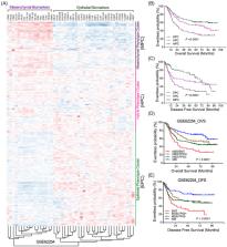
          • Tumour heterogeneity should be partly attributed to the EMT plasticity of tumour cells. The inter‐tumour heterogeneity is clinical associated with poor prognosis in gastric cancer.

          • EMT signalling plays crucial roles in cancer metastasis, stemness, chemoresistance and immune suppression. Targeting EMT signalling is a promising anti‐tumour strategy that kills numerous birds with one stone.

          Related collections

          Most cited references161

          • Record: found
          • Abstract: found
          • Article: not found

          Molecular mechanisms of epithelial-mesenchymal transition.

          The transdifferentiation of epithelial cells into motile mesenchymal cells, a process known as epithelial-mesenchymal transition (EMT), is integral in development, wound healing and stem cell behaviour, and contributes pathologically to fibrosis and cancer progression. This switch in cell differentiation and behaviour is mediated by key transcription factors, including SNAIL, zinc-finger E-box-binding (ZEB) and basic helix-loop-helix transcription factors, the functions of which are finely regulated at the transcriptional, translational and post-translational levels. The reprogramming of gene expression during EMT, as well as non-transcriptional changes, are initiated and controlled by signalling pathways that respond to extracellular cues. Among these, transforming growth factor-β (TGFβ) family signalling has a predominant role; however, the convergence of signalling pathways is essential for EMT.
            Bookmark
            • Record: found
            • Abstract: found
            • Article: not found

            EMT Transition States during Tumor Progression and Metastasis

            Epithelial-mesenchymal transition (EMT) is a process in which epithelial cells acquire mesenchymal features. In cancer, EMT is associated with tumor initiation, invasion, metastasis, and resistance to therapy. Recently, it has been demonstrated that EMT is not a binary process, but occurs through distinct cellular states. Here, we review the recent studies that demonstrate the existence of these different EMT states in cancer and the mechanisms regulating their functions. We discuss the different functional characteristics, such as proliferation, propagation, plasticity, invasion, and metastasis associated with the distinct EMT states. We summarize the role of the transcriptional and epigenetic landscapes, gene regulatory network and their surrounding niche in controlling the transition through the different EMT states.
              Bookmark
              • Record: found
              • Abstract: found
              • Article: found

              A perspective on cancer cell metastasis.

              Metastasis causes most cancer deaths, yet this process remains one of the most enigmatic aspects of the disease. Building on new mechanistic insights emerging from recent research, we offer our perspective on the metastatic process and reflect on possible paths of future exploration. We suggest that metastasis can be portrayed as a two-phase process: The first phase involves the physical translocation of a cancer cell to a distant organ, whereas the second encompasses the ability of the cancer cell to develop into a metastatic lesion at that distant site. Although much remains to be learned about the second phase, we feel that an understanding of the first phase is now within sight, due in part to a better understanding of how cancer cell behavior can be modified by a cell-biological program called the epithelial-to-mesenchymal transition.
                Bookmark

                Author and article information

                Contributors
                lwd35@163.com
                qinss77@163.com , qinss77@hbmu.edu.cn
                Journal
                Cell Prolif
                Cell Prolif
                10.1111/(ISSN)1365-2184
                CPR
                Cell Proliferation
                John Wiley and Sons Inc. (Hoboken )
                0960-7722
                1365-2184
                19 February 2023
                June 2023
                : 56
                : 6 ( doiID: 10.1111/cpr.v56.6 )
                : e13423
                Affiliations
                [ 1 ] Department of Stomatology, Taihe Hospital and Hubei Key Laboratory of Embryonic Stem Cell Research, School of Basic Medical Sciences Hubei University of Medicine Shiyan China
                [ 2 ] Laboratory of Tumor Biology, Academy of Bio‐medicine Research Hubei University of Medicine Shiyan China
                Author notes
                [*] [* ] Correspondence

                Shanshan Qin and Weidong Leng, Department of Stomatology, Taihe Hospital and Hubei Key Laboratory of Embryonic Stem Cell Research, School of Basic Medical Sciences, Hubei University of Medicine, Shiyan 442000, Hubei, China. Email: qinss77@ 123456163.com ; qinss77@ 123456hbmu.edu.cn and lwd35@ 123456163.com

                Author information
                https://orcid.org/0000-0002-8527-5278
                Article
                CPR13423
                10.1111/cpr.13423
                10280150
                36808651
                8946ea0c-1057-4e04-9076-2de4519b8d18
                © 2023 The Authors. Cell Proliferation published by Beijing Institute for Stem Cell and Regenerative Medicine and John Wiley & Sons Ltd.

                This is an open access article under the terms of the http://creativecommons.org/licenses/by/4.0/ License, which permits use, distribution and reproduction in any medium, provided the original work is properly cited.

                History
                : 05 January 2023
                : 30 August 2022
                : 27 January 2023
                Page count
                Figures: 8, Tables: 0, Pages: 14, Words: 11213
                Funding
                Funded by: National Natural Science Foundation of China , doi 10.13039/501100001809;
                Award ID: 81802375
                Award ID: 82203829
                Award ID: 82273451
                Funded by: Natural Science Foundation of Hubei Province , doi 10.13039/501100003819;
                Award ID: 2022CFB448
                Award ID: 2022CFB911
                Categories
                Review
                Review
                Custom metadata
                2.0
                June 2023
                Converter:WILEY_ML3GV2_TO_JATSPMC version:6.2.9 mode:remove_FC converted:20.06.2023

                Cell biology
                Cell biology

                Comments

                Comment on this article