11
views
0
recommends
+1 Recommend
0 collections
    0
    shares
      • Record: found
      • Abstract: found
      • Article: found
      Is Open Access

      Cancer-derived exosomal miR-25-3p promotes pre-metastatic niche formation by inducing vascular permeability and angiogenesis

      research-article

      Read this article at

      Bookmark
          There is no author summary for this article yet. Authors can add summaries to their articles on ScienceOpen to make them more accessible to a non-specialist audience.

          Abstract

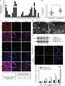
          Cancer-derived exosomes are considered a major driver of cancer-induced pre-metastatic niche formation at foreign sites, but the mechanisms remain unclear. Here, we show that miR-25-3p, a metastasis-promoting miRNA of colorectal cancer (CRC), can be transferred from CRC cells to endothelial cells via exosomes. Exosomal miR-25-3p regulates the expression of VEGFR2, ZO-1, occludin and Claudin5 in endothelial cells by targeting KLF2 and KLF4, consequently promotes vascular permeability and angiogenesis. In addition, exosomal miR-25-3p from CRC cells dramatically induces vascular leakiness and enhances CRC metastasis in liver and lung of mice. Moreover, the expression level of miR-25-3p from circulating exosomes is significantly higher in CRC patients with metastasis than those without metastasis. Our work suggests that exosomal miR-25-3p is involved in pre-metastatic niche formation and may be used as a blood-based biomarker for CRC metastasis.

          Abstract

          The mechanisms underlying pre-metastatic niche formation by cancer derived exosomes is unclear. Here they show that colorectal cancer (CRC) derived exosomal miR-25-3p promotes vascular leakiness and angiogenesis, CRC metastasis, and is upregulated in CRC pateints with metastasis, and suggest miR-25-3p as a biomarker for CRC metastasis.

          Related collections

          Most cited references21

          • Record: found
          • Abstract: found
          • Article: not found

          The pre-metastatic niche: finding common ground.

          It is rapidly becoming evident that the formation of tumor-promoting pre-metastatic niches in secondary organs adds a previously unrecognized degree of complexity to the challenge of curing metastatic disease. Primary tumor cells orchestrate pre-metastatic niche formation through secretion of a variety of cytokines and growth factors that promote mobilization and recruitment of bone marrow-derived cells to future metastatic sites. Hypoxia within the primary tumor, and secretion of specific microvesicles termed exosomes, are emerging as important processes and vehicles for tumor-derived factors to modulate pre-metastatic sites. It has also come to light that reduced immune surveillance is a novel mechanism through which primary tumors create favorable niches in secondary organs. This review provides an overview of our current understanding of underlying mechanisms of pre-metastatic niche formation and highlights the common links as well as discrepancies between independent studies. Furthermore, the possible clinical implications, links to metastatic persistence and dormancy, and novel approaches for treatment of metastatic disease through reversal of pre-metastatic niche formation are identified and explored.
            Bookmark
            • Record: found
            • Abstract: found
            • Article: not found

            KLF4-dependent perivascular cell plasticity mediates pre-metastatic niche formation and metastasis

            A deeper understanding of the metastatic process is required for the development of new therapies that improve patient survival. Metastatic tumor cell growth and survival in distant organs is facilitated by the formation of a pre-metastatic niche composed of hematopoietic cells, stromal cells, and extracellular matrix (ECM). Perivascular cells, including vascular smooth muscle cells (vSMCs) and pericytes, are involved in new vessel formation and in promoting stem cell maintenance and proliferation. Given the well-described plasticity of perivascular cells, we hypothesize that perivascular cells similarly regulate tumor cell fate at metastatic sites. Using perivascular cell-specific and pericyte-specific lineage-tracing models, we trace the fate of perivascular cells in the pre-metastatic and metastatic microenvironments. We show that perivascular cells lose the expression of traditional vSMC/pericyte markers in response to tumor-secreted factors and exhibit increased proliferation, migration, and ECM synthesis. Increased expression of the pluripotency gene Klf4 in these phenotypically-switched perivascular cells promotes a less differentiated state characterized by enhanced ECM production that establishes a pro-metastatic fibronectin-rich environment. Genetic inactivation of Klf4 in perivascular cells decreases pre-metastatic niche formation and metastasis. Our data reveal a previously unidentified role for perivascular cells in pre-metastatic niche formation and uncover novel strategies for limiting metastasis.
              Bookmark
              • Record: found
              • Abstract: found
              • Article: not found

              The exosomes in tumor immunity.

              Exosomes are a kind of nanometric membrane vesicles and can be released by almost all kinds of cells, including cancer cells. As the important mediators in intercellular communications, exosomes mediate exchange of protein and genetic material derived from parental cells. Emerging evidences show that exosomes secreted by either host cells or cancer cells are involved in tumor initiation, growth, invasion and metastasis. Moreover, communications between immune cells and cancer cells via exosomes play dual roles in modulating tumor immunity. In this review, we focus on exosome-mediated immunosuppression via inhibition of antitumor responses elicited by immune cells (DCs, NK cells, CD4(+) and CD8(+) T cells, etc.) and induction of immunosuppressive or regulatory cell populations (MDSCs, Tregs and Bregs). Transfer of cytokines, microRNAs (miRNAs) and functional mRNAs by tumor-derived exosomes (TEXs) is crucial in the immune escape. Furthermore, exosomes secreted from several kinds of immune cells (DCs, CD4(+) and CD8(+) Tregs) also participate in immunosuppression. On the other hand, we summarize the current application of DC-derived and modified tumor-derived exosomes as tumor vaccines. The potential challenges about exosome-based vaccines for clinical application are also discussed.
                Bookmark

                Author and article information

                Contributors
                redsnow007@hotmail.com
                Journal
                Nat Commun
                Nat Commun
                Nature Communications
                Nature Publishing Group UK (London )
                2041-1723
                19 December 2018
                19 December 2018
                2018
                : 9
                : 5395
                Affiliations
                [1 ]GRID grid.416466.7, Department of Pathology, , Nanfang Hospital, Southern Medical University, ; Guangzhou, 510515 Guangdong Province People’s Republic of China
                [2 ]ISNI 0000 0000 8877 7471, GRID grid.284723.8, Department of Pathology, , Southern Medical University, ; Guangzhou, 510515 Guangdong Province People’s Republic of China
                [3 ]Guangdong Province Key Laboratory of Molecular Tumor Pathology, Guangzhou, 510515 Guangdong province People’s Republic of China
                [4 ]GRID grid.440180.9, Department of Pathology, , Dongguan People’s hospital, ; Dongguan, Guangdong Province People’s Republic of China
                [5 ]GRID grid.413107.0, Department of General Surgery, , The Third Affiliated Hospital of Southern Medical University, ; Guangdong Province, People’s Republic of China
                [6 ]ISNI 0000 0000 8877 7471, GRID grid.284723.8, Department of General Surgery, Nanfang Hospital, , Southern Medical University, ; Guangzhou, 510515 Guangdong Province People’s Republic of China
                Article
                7810
                10.1038/s41467-018-07810-w
                6300604
                30568162
                884ac459-98ab-4498-b469-e596871da71f
                © The Author(s) 2018

                Open Access This article is licensed under a Creative Commons Attribution 4.0 International License, which permits use, sharing, adaptation, distribution and reproduction in any medium or format, as long as you give appropriate credit to the original author(s) and the source, provide a link to the Creative Commons license, and indicate if changes were made. The images or other third party material in this article are included in the article’s Creative Commons license, unless indicated otherwise in a credit line to the material. If material is not included in the article’s Creative Commons license and your intended use is not permitted by statutory regulation or exceeds the permitted use, you will need to obtain permission directly from the copyright holder. To view a copy of this license, visit http://creativecommons.org/licenses/by/4.0/.

                History
                : 12 October 2017
                : 22 November 2018
                Categories
                Article
                Custom metadata
                © The Author(s) 2018

                Uncategorized
                Uncategorized

                Comments

                Comment on this article