20
views
0
recommends
+1 Recommend
0 collections
    0
    shares
      • Record: found
      • Abstract: found
      • Article: found
      Is Open Access

      Midbrain circuit regulation of individual alcohol drinking behaviors in mice

      research-article

      Read this article at

      Bookmark
          There is no author summary for this article yet. Authors can add summaries to their articles on ScienceOpen to make them more accessible to a non-specialist audience.

          Abstract

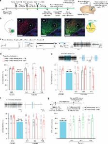
          Alcohol-use disorder (AUD) is the most prevalent substance-use disorder worldwide. There is substantial individual variability in alcohol drinking behaviors in the population, the neural circuit mechanisms of which remain elusive. Utilizing in vivo electrophysiological techniques, we find that low alcohol drinking (LAD) mice have dramatically higher ventral tegmental area (VTA) dopamine neuron firing and burst activity. Unexpectedly, VTA dopamine neuron activity in high alcohol drinking (HAD) mice does not differ from alcohol naive mice. Optogenetically enhancing VTA dopamine neuron burst activity in HAD mice decreases alcohol drinking behaviors. Circuit-specific recordings reveal that spontaneous activity of nucleus accumbens-projecting VTA (VTA-NAc) neurons is selectively higher in LAD mice. Specifically activating this projection is sufficient to reduce alcohol consumption in HAD mice. Furthermore, we uncover ionic and cellular mechanisms that suggest unique neuroadaptations between the alcohol drinking groups. Together, these data identify a neural circuit responsible for individual alcohol drinking behaviors.

          Abstract

          Mice exposed to a two-bottle alcohol choice paradigm can be divided into high and low drinking groups. Here, the authors show that stimulating VTA neurons to induce higher phasic activity patterns that are observed in low alcohol drinking mice, suppresses alcohol drinking in mice that are high alcohol drinking.

          Related collections

          Most cited references41

          • Record: found
          • Abstract: not found
          • Article: not found

          Neurobiologic Advances from the Brain Disease Model of Addiction.

            Bookmark
            • Record: found
            • Abstract: found
            • Article: not found

            Rapid regulation of depression-related behaviors by control of midbrain dopamine neurons

            Ventral tegmental area (VTA) dopamine (DA) neurons in the brain’s reward circuit play a crucial role in mediating stress responses 1–4 including determining susceptibility vs. resilience to social stress-induced behavioural abnormalities 5 . VTA DA neurons exhibit two in vivo patterns of firing: low frequency tonic firing and high frequency phasic firing 6–8 . Phasic firing of the neurons, which is well known to encode reward signals 6,7,9 , is upregulated by repeated social defeat stress, a highly validated mouse model of depression 5,8,10–13 . Surprisingly, this pathophysiological effect is seen in susceptible mice only, with no change in firing rate apparent in resilient individuals 5,8 . However, direct evidence linking—in real-time—DA neuron phasic firing in promoting the susceptible (depression-like) phenotype is lacking. Here, we took advantage of the temporal precision and cell type- and projection pathway-specificity of optogenetics to demonstrate that enhanced phasic firing of these neurons mediates susceptibility to social defeat stress in freely behaving mice. We show that optogenetic induction of phasic, but not tonic, firing, in VTA DA neurons of mice undergoing a subthreshold social defeat paradigm rapidly induced a susceptible phenotype as measured by social avoidance and decreased sucrose preference. Optogenetic phasic stimulation of these neurons also quickly induced a susceptible phenotype in previously resilient mice that had been subjected to repeated social defeat stress. Furthermore, we show differences in projection pathway-specificity in promoting stress susceptibility: phasic activation of VTA neurons projecting to the nucleus accumbens (NAc), but not to the medial prefrontal cortex (mPFC), induced susceptibility to social defeat stress. Conversely, optogenetic inhibition of the VTA-NAc projection induced resilience, while inhibition of the VTA-mPFC projection promoted susceptibility. Overall, these studies reveal novel firing pattern- and neural circuit-specific mechanisms of depression.
              Bookmark
              • Record: found
              • Abstract: found
              • Article: not found

              Unique properties of mesoprefrontal neurons within a dual mesocorticolimbic dopamine system.

              The mesocorticolimbic dopamine system is essential for cognitive and emotive brain functions and is thus an important target in major brain diseases like schizophrenia, drug addiction, and attention deficit hyperactivity disorder. However, the cellular basis for the diversity in behavioral functions and associated dopamine-release pattern within the mesocorticolimbic system has remained unclear. Here, we report the identification of a type of dopaminergic neuron within the mesocorticolimbic dopamine system with unconventional fast-firing properties and small DAT/TH mRNA expression ratios that selectively projects to prefrontal cortex and nucleus accumbens core and medial shell as well as to basolateral amygdala. In contrast, well-described conventional slow-firing dopamine midbrain neurons only project to the lateral shell of the nucleus accumbens and the dorsolateral striatum. Among this dual dopamine midbrain system defined in this study by converging anatomical, electrophysiological, and molecular properties, mesoprefrontal dopaminergic neurons are unique, as only they do not possess functional somatodendritic Girk2-coupled dopamine D2 autoreceptors.
                Bookmark

                Author and article information

                Contributors
                ming-hu.han@mssm.edu
                Journal
                Nat Commun
                Nat Commun
                Nature Communications
                Nature Publishing Group UK (London )
                2041-1723
                20 December 2017
                20 December 2017
                2017
                : 8
                : 2220
                Affiliations
                [1 ]ISNI 0000 0001 0670 2351, GRID grid.59734.3c, Department of Pharmacological Sciences and Institute for Systems Biomedicine, , Icahn School of Medicine at Mount Sinai, ; New York, NY 10029 USA
                [2 ]ISNI 0000 0001 0670 2351, GRID grid.59734.3c, Fishberg Department of Neuroscience and Friedman Brain Institute, , Icahn School of Medicine at Mount Sinai, ; New York, NY 10029 USA
                [3 ]ISNI 0000 0000 9927 0537, GRID grid.417303.2, Jiangsu Province Key Laboratory of Anesthesiology, , Xuzhou Medical University, ; Xuzhou, 221002 Jiangsu China
                [4 ]ISNI 0000 0004 0533 7147, GRID grid.420090.f, Intramural Research Program, , National Institute on Drug Abuse, National Institutes of Health, ; Baltimore, MD 21224 USA
                [5 ]ISNI 0000 0000 8954 1233, GRID grid.279863.1, Department of Physiology, Alcohol and Drug Abuse Center of Excellence, , Neuroscience Center of Excellence, Louisiana State University Health Sciences Center, ; New Orleans, LA 70112 USA
                [6 ]ISNI 0000000122199231, GRID grid.214007.0, Department of Molecular and Cellular Neuroscience, , The Scripps Research Institute, ; San Diego, CA 92037 USA
                [7 ]ISNI 0000 0001 0670 2351, GRID grid.59734.3c, Department of Medicine, , Icahn School of Medicine at Mount Sinai, ; New York, CA 10029 USA
                [8 ]ISNI 0000 0001 2180 1673, GRID grid.255381.8, Department of Health Sciences, , College of Public Health, East Tennessee State University, ; Johnson City, TN 37614 USA
                [9 ]ISNI 0000 0001 2264 7217, GRID grid.152326.1, Department of Pharmacology, , Vanderbilt University, ; Nashville, TN 37232 USA
                [10 ]GRID grid.440573.1, Division of Science, , New York University Abu Dhabi (NYUAD), Saadiyat Island Campus, ; Abu Dhabi, PO Box 129188 United Arab Emirates
                [11 ]ISNI 0000 0001 2183 6649, GRID grid.257167.0, Department of Biological Sciences, , Hunter College, City University of New York, ; New York, NY 10065 USA
                Author information
                http://orcid.org/0000-0002-8190-3011
                Article
                2365
                10.1038/s41467-017-02365-8
                5738419
                28232747
                88300897-6cca-477d-a973-58be423e4865
                © The Author(s) 2017

                Open Access This article is licensed under a Creative Commons Attribution 4.0 International License, which permits use, sharing, adaptation, distribution and reproduction in any medium or format, as long as you give appropriate credit to the original author(s) and the source, provide a link to the Creative Commons license, and indicate if changes were made. The images or other third party material in this article are included in the article’s Creative Commons license, unless indicated otherwise in a credit line to the material. If material is not included in the article’s Creative Commons license and your intended use is not permitted by statutory regulation or exceeds the permitted use, you will need to obtain permission directly from the copyright holder. To view a copy of this license, visit http://creativecommons.org/licenses/by/4.0/.

                History
                : 19 September 2016
                : 23 November 2017
                Categories
                Article
                Custom metadata
                © The Author(s) 2017

                Uncategorized
                Uncategorized

                Comments

                Comment on this article