20
views
0
recommends
+1 Recommend
0 collections
    0
    shares
      • Record: found
      • Abstract: found
      • Article: found
      Is Open Access

      Toxoplasma gondii in small exotic felids from zoos in Europe and the Middle East: serological prevalence and risk factors

      research-article

      Read this article at

      Bookmark
          There is no author summary for this article yet. Authors can add summaries to their articles on ScienceOpen to make them more accessible to a non-specialist audience.

          Abstract

          Background

          Toxoplasma gondii infections and cases of clinical toxoplasmosis have been recorded in zoo animals. Wild felids in human care can serve as definitive hosts that shed oocysts, but also as intermediate hosts for the parasite. Some felid species, such as the Pallas’s cat ( Otocolobus manul) or sand cat ( Felis margarita), may suffer from clinically apparent toxoplasmosis. In the present study, our main aim was to assess risk factors for T. gondii infections in small exotic felids.

          Methods

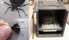
          A seroepidemiological study was conducted using the reduviid bug Dipetalogaster maxima for blood sample collection, a method previously evaluated on domestic cats. A total of 336 samples from 17 felid species were collected in 51 institutions, 48 of which were within Europe and the remaining three in the Middle East (United Arabic Emirates and Qatar). These samples were analyzed for T. gondii antibodies by immunoblotting and an immunofluorescent antibody test. Potential risk factors in zoos for seropositivity regarding T. gondii among members of the European Association of Zoos and Aquaria (EAZA) were evaluated using a questionnaire and individual data from the Zoological Information Management System (ZIMS).

          Results

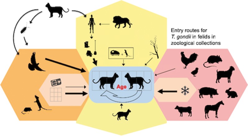
          The sampled felids showed an overall seroprevalence for T. gondii of 63%. The risk factor study including data of 311 small exotic cats of 10 species resulted in a final generalized linear mixed model comprised of five variables: the likelihood of seropositivity increased statistically significantly with “Age”, while feeding “Cattle: frozen” relative to “Cattle: fresh”, “Outdoor housing fenced in on all sides”, “Mesh size 2–5 cm” relative to “Mesh size > 5 cm” and “Wearing gloves: yes” had statistically significant protective effects.

          Conclusions

          Wild felids, including endangered species, kept in human care in European and Middle Eastern institutions, are widely exposed to T. gondii. Risk factor analysis revealed that feeding previously frozen tissues, keeping animals in enclosures that are fenced on all sides using fences with small mesh sizes, and wearing gloves when working inside enclosures seem to be the most relevant protective measures to prevent T. gondii infections in these animals

          .

          Related collections

          Most cited references87

          • Record: found
          • Abstract: found
          • Article: found
          Is Open Access

          Toxoplasma gondii infection and toxoplasmosis in farm animals: Risk factors and economic impact

          The protozoan parasite Toxoplasma gondii is a zoonotic parasite that can be transmitted from animals to humans. Felids, including domestic cats, are definitive hosts that can shed oocysts with their feces. In addition to infections that occur by accidental oral uptake of food or water contaminated with oocysts, it is assumed that a large proportion of affected humans may have become infected by consuming meat or other animal products that contained infective parasitic stages of T. gondii. Since farm animals represent a direct source of infection for humans, but also a possible reservoir for the parasite, it is important to control T. gondii infections in livestock. Moreover, T. gondii may also be pathogenic to livestock where it could be responsible for considerable economic losses in some regions and particular farming systems, e.g. in areas where the small ruminant industry is relevant. This review aims to summarize actual knowledge on the prevalence and effects of infections with T. gondii in the most important livestock species and on the effects of toxoplasmosis on livestock. It also provides an overview on potential risk factors favoring infections of livestock with T. gondii. Knowledge on potential risk factors is prerequisite to implement effective biosecurity measures on farms to prevent T. gondii infections. Risk factors identified by many studies are cat-related, but also those associated with a potential contamination of fodder or water, and with access to a potentially contaminated environment. Published information on the costs T. gondii infections cause in livestock production, is scarce. The most recent peer reviewed reports from Great Britain and Uruguay suggest annual cost of about 5–15 million US $ per country. Since these estimates are outdated, future studies are needed to estimate the present costs due to toxoplasmosis in livestock. Further, the fact that T. gondii infections in livestock may affect human health needs to be considered and the respective costs should also be estimated, but this is beyond the scope of this article.
            Bookmark
            • Record: found
            • Abstract: found
            • Article: not found

            Association of CD4+ T cell-dependent, interferon-gamma-mediated necrosis of the small intestine with genetic susceptibility of mice to peroral infection with Toxoplasma gondii

            Since there is a remarkable difference in susceptibility to peroral infection with Toxoplasma gondii among inbred strains of mice, we performed studies to examine the mechanism(s) of this difference in susceptibility. After peroral infection with the ME49 strain of T. gondii, C57BL/6 (B6) mice all died whereas BALB/c mice all survived. At day 7 of infection (when B6 mice began dying), massive necrosis of the villi and mucosal cells in the ilea were observed in B6 but not in BALB/c mice. To analyze the role of T cells in resistance against death and development of necrosis in the ilea after infection, studies were performed using athymic nude and euthymic control B6 and BALB/c mice. Athymic B6 mice all died after infection, but surprisingly, they survived significantly longer than control B6 mice, indicating that T cells predispose to early death in these mice. Necrosis in the ilea was observed in control B6 but not in athymic B6 mice; however, significantly less numbers of tachyzoites were observed in the ilea of the former than the latter mice. These results indicate that necrosis in the ilea of the B6 mice was not due to destruction of tissue by tachyzoites but was mediated by T cells. This deleterious effect of T cells appears to contribute to early death in these mice. In contrast, T cells conferred resistance against death in BALB/c mice but did not cause necrosis in their ilea. To analyze the T cell subset(s) that induces necrosis of the ilea in B6 mice, we examined histological changes of the small intestines after infection of mutant mice deficient in different T cell subsets (with the same H-2b haplotype as B6 mice). Mice deficient in alpha/beta or CD4+ T cells did not develop necrosis in the ilea, whereas wild-type control mice and mice deficient in gamma/delta or CD8+ T cells did, suggesting that the cells that induce necrosis in the ilea after infection are CD4+ alpha/beta T cells. Since interferon (IFN)-gamma has been shown to be critical for survival of BALB/c mice after infection with T. gondii, we examined the role of this cytokine in resistance/susceptibility of infected B6 mice. Treatment of B6 mice with anti-IFN-gamma monoclonal antibody shortly before they developed illness prolonged time to death and prevented necrosis in the ilea in these mice. These results indicate that IFN- gamma mediates necrosis in the ilea of B6 mice after infection. This CD4+ T cell-dependent, IFN-gamma-mediated necrosis of the small intestines appears to be a mechanism that underlies the genetic susceptibility of B6 mice to peroral infection with T. gondii, whereas the same cytokine plays a critical role in the resistance of genetically resistant BALB/c mice.
              Bookmark
              • Record: found
              • Abstract: found
              • Article: not found

              Duration of immunity to shedding of Toxoplasma gondii oocysts by cats.

              Cats that have shed Toxoplasma gondii oocysts are considered to be immune to reshedding of oocysts. To investigate if this immunity persists in cats for 6 yr, 12 4-6-mo-old cats without T. gondii antibodies were inoculated orally with tissue cysts of the ME-49 strain (6 cats) and the TS-2 strain (6 cats) of T. gondii. All of them shed > or = 20 million oocysts between 4 and 13 days after feeding tissue cysts. Two cats became ill between 11 and 13 days after primary infection; 1 died on the 13th day, and the other had to be killed on the 11th day because of generalized acute toxoplasmosis. Toxoplasma gondii oocysts were not found on the hair of 10 cats examined 7 days after cats had shed millions of oocysts. On day 39 after primary infection, 5 cats (2 infected with the ME-49 strain and 3 infected with the TS-2 strain) were challenged orally with tissue cysts of the ME-49 strain. None of the challenged cats shed oocysts. One cat died due to causes unrelated to toxoplasmosis. Seventy-seven months after primary infection, the remaining 9 cats were challenged orally with tissue cysts of the P89 strain of T. gondii. Four of these 9 cats re-shed T. gondii oocysts; 3 of them had been challenged also at 39 days after primary infection. Two control cats housed together with chronically infected cats for 6 yr remained seronegative for T. gondii; both of these shed oocysts after challenge with the P89 strain.
                Bookmark

                Author and article information

                Contributors
                maike.luecht@hellabrunn.de
                julia.stagegaard@reepark.dk
                franz.conraths@fli.de
                gereon.schares@fli.de
                Journal
                Parasit Vectors
                Parasit Vectors
                Parasites & Vectors
                BioMed Central (London )
                1756-3305
                11 September 2019
                11 September 2019
                2019
                : 12
                : 449
                Affiliations
                [1 ]GRID grid.417834.d, Friedrich-Loeffler-Institut, Institute of Epidemiology, Federal Research Institute for Animal Health, ; Südufer 10, 17493 Greifswald-Insel Riems, Germany
                [2 ]Münchener Tierpark; Hellabrunn AG, Tierparkstr. 30, 81543 Munich, Germany
                [3 ]Ree Park-Safari, Stubbe Søvej 15, 8400 Ebeltoft, Denmark
                Author information
                http://orcid.org/0000-0002-3217-289X
                Article
                3706
                10.1186/s13071-019-3706-2
                6737647
                31511050
                87ad08de-0bb7-4a18-b6f5-b08bd83f13a5
                © The Author(s) 2019

                Open AccessThis article is distributed under the terms of the Creative Commons Attribution 4.0 International License ( http://creativecommons.org/licenses/by/4.0/), which permits unrestricted use, distribution, and reproduction in any medium, provided you give appropriate credit to the original author(s) and the source, provide a link to the Creative Commons license, and indicate if changes were made. The Creative Commons Public Domain Dedication waiver ( http://creativecommons.org/publicdomain/zero/1.0/) applies to the data made available in this article, unless otherwise stated.

                History
                : 13 May 2019
                : 5 September 2019
                Funding
                Funded by: Ree Park – Safari (Ebeltoft), Randers Regnskov, BHJ-Fonden
                Categories
                Research
                Custom metadata
                © The Author(s) 2019

                Parasitology
                toxoplasma gondii,serology,felinae,captive felids,zoological gardens,epidemiology,risk analysis,risk factors,triatominae,dipetalogaster maxima

                Comments

                Comment on this article