21
views
0
recommends
+1 Recommend
0 collections
    0
    shares
      • Record: found
      • Abstract: found
      • Article: found
      Is Open Access

      Glutaminase-containing microvesicles from HIV-1-infected macrophages and immune-activated microglia induce neurotoxicity

      research-article

      Read this article at

      Bookmark
          There is no author summary for this article yet. Authors can add summaries to their articles on ScienceOpen to make them more accessible to a non-specialist audience.

          Abstract

          Background

          HIV-1-infected and/or immune-activated microglia and macrophages are pivotal in the pathogenesis of HIV-1-associated neurocognitive disorders (HAND). Glutaminase, a metabolic enzyme that facilitates glutamate generation, is upregulated and may play a pathogenic role in HAND. Our previous studies have demonstrated that glutaminase is released to the extracellular fluid during HIV-1 infection and neuroinflammation. However, key molecular mechanisms that regulate glutaminase release remain unknown. Recent advances in understanding intercellular trafficking have identified microvesicles (MVs) as a novel means of shedding cellular contents. We posit that during HIV-1 infection and immune activation, microvesicles may mediate glutaminase release, generating excessive and neurotoxic levels of glutamate.

          Results

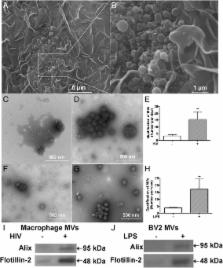
          MVs isolated through differential centrifugation from cell-free supernatants of monocyte-derived macrophages (MDM) and BV2 microglia cell lines were first confirmed in electron microscopy and immunoblotting. As expected, we found elevated number of MVs, glutaminase immunoreactivities, as well as glutaminase enzyme activity in the supernatants of HIV-1 infected MDM and lipopolysaccharide (LPS)-activated microglia when compared with controls. The elevated glutaminase was blocked by GW4869, a neutral sphingomyelinase inhibitor known to inhibit MVs release, suggesting a critical role of MVs in mediating glutaminase release. More importantly, MVs from HIV-1-infected MDM and LPS-activated microglia induced significant neuronal injury in rat cortical neuron cultures. The MV neurotoxicity was blocked by a glutaminase inhibitor or GW4869, suggesting that the neurotoxic potential of HIV-1-infected MDM and LPS-activated microglia is dependent on the glutaminase-containing MVs.

          Conclusions

          These findings support MVs as a potential pathway/mechanism of excessive glutamate generation and neurotoxicity in HAND and therefore MVs may serve as a novel therapeutic target.

          Electronic supplementary material

          The online version of this article (doi:10.1186/s13024-015-0058-z) contains supplementary material, which is available to authorized users.

          Related collections

          Most cited references40

          • Record: found
          • Abstract: found
          • Article: found
          Is Open Access

          Extracellular vesicles as mediators of neuron-glia communication

          In the nervous system, glia cells maintain homeostasis, synthesize myelin, provide metabolic support, and participate in immune defense. The communication between glia and neurons is essential to synchronize these diverse functions with brain activity. Evidence is accumulating that secreted extracellular vesicles (EVs), such as exosomes and shedding microvesicles, are key players in intercellular signaling. The cells of the nervous system secrete EVs, which potentially carry protein and RNA cargo from one cell to another. After delivery, the cargo has the ability to modify the target cell phenotype. Here, we review the recent advances in understanding the role of EV secretion by astrocytes, microglia, and oligodendrocytes in the central nervous system. Current work has demonstrated that oligodendrocytes transfer exosomes to neurons as a result of neurotransmitter signaling suggesting that these vesicles may mediate glial support of neurons.
            Bookmark
            • Record: found
            • Abstract: found
            • Article: not found

            Regulation of glutaminase activity and glutamine metabolism.

            Glutamine is synthesized primarily in skeletal muscle, lungs, and adipose tissue. Plasma glutamine plays an important role as a carrier of nitrogen, carbon, and energy between organs and is used for hepatic urea synthesis, for renal ammoniagenesis, for gluconeogenesis in both liver and kidney, and as a major respiratory fuel for many cells. The catabolism of glutamine is initiated by either of two isoforms of the mitochondrial glutaminase. Liver-type glutaminase is expressed only in periportal hepatocytes of the postnatal liver, where it effectively couples ammonia production with urea synthesis. Kidney-type glutaminase is abundant in kidney, brain, intestine, fetal liver, lymphocytes, and transformed cells, where the resulting ammonia is released without further metabolism. The two isoenzymes have different structural and kinetic properties that contribute to their function and short-term regulation. Although there is a high degree of identity in amino acid sequences, the two glutaminases are the products of different but related genes. The two isoenzymes are also subject to long-term regulation. Hepatic glutaminase is increased during starvation, diabetes, and feeding a high-protein diet, whereas kidney-type glutaminase is increased only in kidney in response to metabolic acidosis. The adaptations in hepatic glutaminase are mediated by changes in the rate of transcription, whereas kidney-type glutaminase is regulated at a posttranscriptional level.
              Bookmark
              • Record: found
              • Abstract: found
              • Article: not found

              IL-1β and TNF-α induce neurotoxicity through glutamate production: a potential role for neuronal glutaminase.

              Glutaminase 1 is the main enzyme responsible for glutamate production in mammalian cells. The roles of macrophage and microglia glutaminases in brain injury, infection, and inflammation are well documented. However, little is known about the regulation of neuronal glutaminase, despite neurons being a predominant cell type of glutaminase expression. Using primary rat and human neuronal cultures, we confirmed that interleukin-1β (IL-1β) and tumor necrosis factor-α (TNF-α), two pro-inflammatory cytokines that are typically elevated in neurodegenerative disease states, induced neuronal death and apoptosis in vitro. Furthermore, both intracellular and extracellular glutamate levels were significantly elevated following IL-1β and/or TNF-α treatment. Pre-treatment with N-Methyl-D-aspartate (NMDA) receptor antagonist MK-801 blocked cytokine-induced glutamate production and alleviated the neurotoxicity, indicating that IL-1β and/or TNF-α induce neurotoxicity through glutamate. To determine the potential source of excess glutamate production in the culture during inflammation, we investigated the neuronal glutaminase and found that treatment with IL-1β or TNF-α significantly upregulated the kidney-type glutaminase (KGA), a glutaminase 1 isoform, in primary human neurons. The up-regulation of neuronal glutaminase was also demonstrated in situ in a murine model of HIV-1 encephalitis. In addition, IL-1β or TNF-α treatment increased the levels of KGA in cytosol and TNF-α specifically increased KGA levels in the extracellular fluid, away from its main residence in mitochondria. Together, these findings support neuronal glutaminase as a potential component of neurotoxicity during inflammation and that modulation of glutaminase may provide therapeutic avenues for neurodegenerative diseases. © 2013 International Society for Neurochemistry.
                Bookmark

                Author and article information

                Contributors
                beiqing.wu2@unmc.edu
                402-559-5656 , yhuan1@unmc.edu
                alexander.braun@unmc.edu
                zenghan.tong@unmc.edu
                runze.zhao@unmc.edu
                402-559-5656 , jzheng@unmc.edu
                Journal
                Mol Neurodegener
                Mol Neurodegener
                Molecular Neurodegeneration
                BioMed Central (London )
                1750-1326
                6 November 2015
                6 November 2015
                2015
                : 10
                : 61
                Affiliations
                [ ]Laboratory of Neuroimmunology and Regenerative Therapy, Departments of Pharmacology and Experimental Neuroscience, University of Nebraska Medical Center, Omaha, NE 68198-5930 USA
                [ ]Departments of Pathology and Microbiology, University of Nebraska Medical Center, Omaha, NE 68198-5930 USA
                [ ]Center for Translational Neurodegeneration and Regenerative Therapy, Shanghai Tenth People’s Hospital, Tongji University School of Medicine, Shanghai, 200025 China
                Article
                58
                10.1186/s13024-015-0058-z
                4635976
                26546362
                877d7993-9750-4e19-b799-f036e59da8ee
                © Wu et al. 2015

                Open AccessThis article is distributed under the terms of the Creative Commons Attribution 4.0 International License ( http://creativecommons.org/licenses/by/4.0/), which permits unrestricted use, distribution, and reproduction in any medium, provided you give appropriate credit to the original author(s) and the source, provide a link to the Creative Commons license, and indicate if changes were made. The Creative Commons Public Domain Dedication waiver ( http://creativecommons.org/publicdomain/zero/1.0/) applies to the data made available in this article, unless otherwise stated.

                History
                : 21 April 2015
                : 2 November 2015
                Categories
                Research Article
                Custom metadata
                © The Author(s) 2015

                Neurosciences
                hand,hiv,glutamate,glutaminase,microvesicles,neurotoxicity
                Neurosciences
                hand, hiv, glutamate, glutaminase, microvesicles, neurotoxicity

                Comments

                Comment on this article