10
views
0
recommends
+1 Recommend
1 collections
    0
    shares
      • Record: found
      • Abstract: found
      • Article: not found

      Protein Expression Profile of ACE2 in the Normal and COVID-19-Affected Human Brain

      research-article

      Read this article at

      ScienceOpenPublisherPMC
      Bookmark
          There is no author summary for this article yet. Authors can add summaries to their articles on ScienceOpen to make them more accessible to a non-specialist audience.

          Abstract

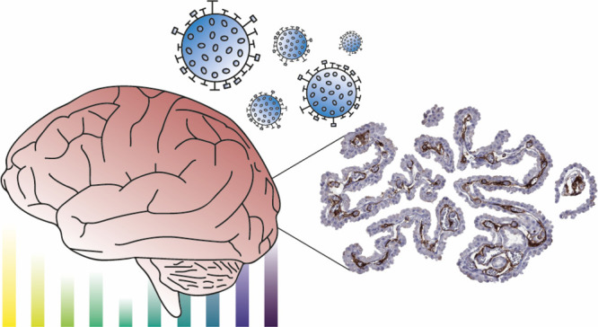
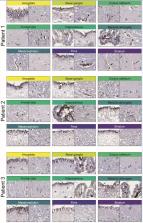
          SARS-coronavirus 2 (SARS-CoV-2) that caused the coronavirus disease 2019 (COVID-19) pandemic has posed to be a global challenge. An increasing number of neurological symptoms have been linked to the COVID-19 disease, but the underlying mechanisms of such symptoms and which patients could be at risk are not yet established. The suggested key receptor for host cell entry is angiotensin I converting enzyme 2 (ACE2). Previous studies on limited tissue material have shown no or low protein expression of ACE2 in the normal brain. Here, we used stringently validated antibodies and immunohistochemistry to examine the protein expression of ACE2 in all major regions of the normal brain. The expression pattern was compared with the COVID-19-affected brain of patients with a varying degree of neurological symptoms. In the normal brain, the expression was restricted to the choroid plexus and ependymal cells with no expression in any other brain cell types. Interestingly, in the COVID-19-affected brain, an upregulation of ACE2 was observed in endothelial cells of certain patients, most prominently in the white matter and with the highest expression observed in the patient with the most severe neurological symptoms. The data shows differential expression of ACE2 in the diseased brain and highlights the need to further study the role of endothelial cells in COVID-19 disease in relation to neurological symptoms.

          Related collections

          Most cited references37

          • Record: found
          • Abstract: found
          • Article: found

          COVID-19: consider cytokine storm syndromes and immunosuppression

          As of March 12, 2020, coronavirus disease 2019 (COVID-19) has been confirmed in 125 048 people worldwide, carrying a mortality of approximately 3·7%, 1 compared with a mortality rate of less than 1% from influenza. There is an urgent need for effective treatment. Current focus has been on the development of novel therapeutics, including antivirals and vaccines. Accumulating evidence suggests that a subgroup of patients with severe COVID-19 might have a cytokine storm syndrome. We recommend identification and treatment of hyperinflammation using existing, approved therapies with proven safety profiles to address the immediate need to reduce the rising mortality. Current management of COVID-19 is supportive, and respiratory failure from acute respiratory distress syndrome (ARDS) is the leading cause of mortality. 2 Secondary haemophagocytic lymphohistiocytosis (sHLH) is an under-recognised, hyperinflammatory syndrome characterised by a fulminant and fatal hypercytokinaemia with multiorgan failure. In adults, sHLH is most commonly triggered by viral infections 3 and occurs in 3·7–4·3% of sepsis cases. 4 Cardinal features of sHLH include unremitting fever, cytopenias, and hyperferritinaemia; pulmonary involvement (including ARDS) occurs in approximately 50% of patients. 5 A cytokine profile resembling sHLH is associated with COVID-19 disease severity, characterised by increased interleukin (IL)-2, IL-7, granulocyte-colony stimulating factor, interferon-γ inducible protein 10, monocyte chemoattractant protein 1, macrophage inflammatory protein 1-α, and tumour necrosis factor-α. 6 Predictors of fatality from a recent retrospective, multicentre study of 150 confirmed COVID-19 cases in Wuhan, China, included elevated ferritin (mean 1297·6 ng/ml in non-survivors vs 614·0 ng/ml in survivors; p 39·4°C 49 Organomegaly None 0 Hepatomegaly or splenomegaly 23 Hepatomegaly and splenomegaly 38 Number of cytopenias * One lineage 0 Two lineages 24 Three lineages 34 Triglycerides (mmol/L) 4·0 mmol/L 64 Fibrinogen (g/L) >2·5 g/L 0 ≤2·5 g/L 30 Ferritin ng/ml 6000 ng/ml 50 Serum aspartate aminotransferase <30 IU/L 0 ≥30 IU/L 19 Haemophagocytosis on bone marrow aspirate No 0 Yes 35 Known immunosuppression † No 0 Yes 18 The Hscore 11 generates a probability for the presence of secondary HLH. HScores greater than 169 are 93% sensitive and 86% specific for HLH. Note that bone marrow haemophagocytosis is not mandatory for a diagnosis of HLH. HScores can be calculated using an online HScore calculator. 11 HLH=haemophagocytic lymphohistiocytosis. * Defined as either haemoglobin concentration of 9·2 g/dL or less (≤5·71 mmol/L), a white blood cell count of 5000 white blood cells per mm3 or less, or platelet count of 110 000 platelets per mm3 or less, or all of these criteria combined. † HIV positive or receiving longterm immunosuppressive therapy (ie, glucocorticoids, cyclosporine, azathioprine).
            Bookmark
            • Record: found
            • Abstract: found
            • Article: not found

            Proteomics. Tissue-based map of the human proteome.

            Resolving the molecular details of proteome variation in the different tissues and organs of the human body will greatly increase our knowledge of human biology and disease. Here, we present a map of the human tissue proteome based on an integrated omics approach that involves quantitative transcriptomics at the tissue and organ level, combined with tissue microarray-based immunohistochemistry, to achieve spatial localization of proteins down to the single-cell level. Our tissue-based analysis detected more than 90% of the putative protein-coding genes. We used this approach to explore the human secretome, the membrane proteome, the druggable proteome, the cancer proteome, and the metabolic functions in 32 different tissues and organs. All the data are integrated in an interactive Web-based database that allows exploration of individual proteins, as well as navigation of global expression patterns, in all major tissues and organs in the human body. Copyright © 2015, American Association for the Advancement of Science.
              Bookmark
              • Record: found
              • Abstract: found
              • Article: found
              Is Open Access

              Integrated analysis of multimodal single-cell data

              Summary The simultaneous measurement of multiple modalities represents an exciting frontier for single-cell genomics and necessitates computational methods that can define cellular states based on multimodal data. Here, we introduce “weighted-nearest neighbor” analysis, an unsupervised framework to learn the relative utility of each data type in each cell, enabling an integrative analysis of multiple modalities. We apply our procedure to a CITE-seq dataset of 211,000 human peripheral blood mononuclear cells (PBMCs) with panels extending to 228 antibodies to construct a multimodal reference atlas of the circulating immune system. Multimodal analysis substantially improves our ability to resolve cell states, allowing us to identify and validate previously unreported lymphoid subpopulations. Moreover, we demonstrate how to leverage this reference to rapidly map new datasets and to interpret immune responses to vaccination and coronavirus disease 2019 (COVID-19). Our approach represents a broadly applicable strategy to analyze single-cell multimodal datasets and to look beyond the transcriptome toward a unified and multimodal definition of cellular identity.
                Bookmark

                Author and article information

                Journal
                J Proteome Res
                J Proteome Res
                pr
                jprobs
                Journal of Proteome Research
                American Chemical Society
                1535-3893
                1535-3907
                28 July 2022
                : acs.jproteome.2c00184
                Affiliations
                []Department of Immunology, Genetics and Pathology, Rudbeck Laboratory, Uppsala University , 751 85 Uppsala, Sweden
                []Department of Neuroscience, Neurology, Uppsala University , 751 85 Uppsala, Sweden
                [§ ]Department of Surgical Sciences, Radiology, Uppsala University , 751 85 Uppsala, Sweden
                []Department of Neuroscience, Neurosurgery, Uppsala University , 751 85 Uppsala, Sweden
                []Department of Clinical Pathology and Cytology, Uppsala University Hospital , 751 85 Uppsala, Sweden
                Author notes
                Author information
                https://orcid.org/0000-0001-5611-1015
                https://orcid.org/0000-0001-9333-0110
                Article
                10.1021/acs.jproteome.2c00184
                9364976
                35901083
                8773943f-2dd3-41f2-84b4-3f20dd071674
                © 2022 The Authors. Published by American Chemical Society

                This article is made available via the PMC Open Access Subset for unrestricted RESEARCH re-use and analyses in any form or by any means with acknowledgement of the original source. These permissions are granted for the duration of the World Health Organization (WHO) declaration of COVID-19 as a global pandemic.

                History
                : 11 April 2022
                Funding
                Funded by: Knut och Alice Wallenbergs Stiftelse, doi 10.13039/501100004063;
                Award ID: NA
                Funded by: Swedish government and county councils, doi NA;
                Award ID: NA
                Categories
                Article
                Custom metadata
                pr2c00184
                pr2c00184

                Molecular biology
                sars-cov-2,covid-19,neurology,ace2,immunohistochemistry,proteomics,transcriptomics,antibodies,brain

                Comments

                Comment on this article