4
views
0
recommends
+1 Recommend
0 collections
    0
    shares
      • Record: found
      • Abstract: found
      • Article: found
      Is Open Access

      S1 Brain Connectivity in Carpal Tunnel Syndrome Underlies Median Nerve and Functional Improvement Following Electro-Acupuncture

      research-article

      Read this article at

      Bookmark
          There is no author summary for this article yet. Authors can add summaries to their articles on ScienceOpen to make them more accessible to a non-specialist audience.

          Abstract

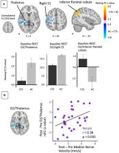
          Carpal Tunnel Syndrome (CTS) is a median nerve entrapment neuropathy that alters primary somatosensory cortex (S1) organization. While electro-acupuncture (EA), a form of peripheral neuromodulation, has been shown to improve clinical and neurophysiological CTS outcomes, the role of EA-evoked brain response during therapy (within and beyond S1) for improved outcomes is unknown. We investigated S1-associated whole brain fMRI connectivity during both a resting and sustained EA stimulation state in age-matched healthy controls ( N = 28) and CTS patients ( N = 64), at baseline and after 8 weeks of acupuncture therapy (local, distal, or sham EA). Compared to healthy controls, CTS patients at baseline showed decreased resting state functional connectivity between S1 and thalamic pulvinar nucleus. Increases in S1/pulvinar connectivity strength following verum EA therapy (combined local and distal) were correlated with improvements in median nerve velocity ( r = 0.38, p = 0.035). During sustained local EA, compared to healthy controls, CTS patients demonstrated increased functional connectivity between S1 and anterior hippocampus (aHipp). Following 8 weeks of local EA therapy, S1/aHipp connectivity significantly decreased and greater decrease was associated with improvement in patients' functional status ( r = 0.64, p = 0.01) and increased median nerve velocity ( r = −0.62, p = 0.013). Thus, connectivity between S1 and other brain areas is also disrupted in CTS patients and may be improved following EA therapy. Furthermore, stimulus-evoked fMRI connectivity adds therapy-specific, mechanistic insight to more common resting state connectivity approaches. Specifically, local EA modulates S1 connectivity to sensory and affective processing regions, linked to patient function and median nerve health.

          Related collections

          Most cited references47

          • Record: found
          • Abstract: not found
          • Article: not found

          Improved Optimization for the Robust and Accurate Linear Registration and Motion Correction of Brain Images

            Bookmark
            • Record: found
            • Abstract: found
            • Article: not found

            A component based noise correction method (CompCor) for BOLD and perfusion based fMRI.

            A component based method (CompCor) for the reduction of noise in both blood oxygenation level-dependent (BOLD) and perfusion-based functional magnetic resonance imaging (fMRI) data is presented. In the proposed method, significant principal components are derived from noise regions-of-interest (ROI) in which the time series data are unlikely to be modulated by neural activity. These components are then included as nuisance parameters within general linear models for BOLD and perfusion-based fMRI time series data. Two approaches for the determination of the noise ROI are considered. The first method uses high-resolution anatomical data to define a region of interest composed primarily of white matter and cerebrospinal fluid, while the second method defines a region based upon the temporal standard deviation of the time series data. With the application of CompCor, the temporal standard deviation of resting-state perfusion and BOLD data in gray matter regions was significantly reduced as compared to either no correction or the application of a previously described retrospective image based correction scheme (RETROICOR). For both functional perfusion and BOLD data, the application of CompCor significantly increased the number of activated voxels as compared to no correction. In addition, for functional BOLD data, there were significantly more activated voxels detected with CompCor as compared to RETROICOR. In comparison to RETROICOR, CompCor has the advantage of not requiring external monitoring of physiological fluctuations.
              Bookmark
              • Record: found
              • Abstract: found
              • Article: not found

              Symmetric diffeomorphic image registration with cross-correlation: evaluating automated labeling of elderly and neurodegenerative brain.

              One of the most challenging problems in modern neuroimaging is detailed characterization of neurodegeneration. Quantifying spatial and longitudinal atrophy patterns is an important component of this process. These spatiotemporal signals will aid in discriminating between related diseases, such as frontotemporal dementia (FTD) and Alzheimer's disease (AD), which manifest themselves in the same at-risk population. Here, we develop a novel symmetric image normalization method (SyN) for maximizing the cross-correlation within the space of diffeomorphic maps and provide the Euler-Lagrange equations necessary for this optimization. We then turn to a careful evaluation of our method. Our evaluation uses gold standard, human cortical segmentation to contrast SyN's performance with a related elastic method and with the standard ITK implementation of Thirion's Demons algorithm. The new method compares favorably with both approaches, in particular when the distance between the template brain and the target brain is large. We then report the correlation of volumes gained by algorithmic cortical labelings of FTD and control subjects with those gained by the manual rater. This comparison shows that, of the three methods tested, SyN's volume measurements are the most strongly correlated with volume measurements gained by expert labeling. This study indicates that SyN, with cross-correlation, is a reliable method for normalizing and making anatomical measurements in volumetric MRI of patients and at-risk elderly individuals.
                Bookmark

                Author and article information

                Contributors
                Journal
                Front Neurol
                Front Neurol
                Front. Neurol.
                Frontiers in Neurology
                Frontiers Media S.A.
                1664-2295
                27 October 2021
                2021
                : 12
                : 754670
                Affiliations
                [1] 1Department of Radiology, Athinoula A. Martinos Center for Biomedical Imaging, Harvard Medical School, Massachusetts General Hospital , Boston, MA, United States
                [2] 2Department of Radiology, Logan University , Chesterfield, MO, United States
                [3] 3Department of Physical Medicine and Rehabilitation, Spaulding Rehabilitation Hospital , Boston, MA, United States
                [4] 4Division of Clinical Medicine, Korea Institute of Oriental Medicine , Daejeon, South Korea
                [5] 5Department of Pain Medicine, Harvard Vanguard Medical Associates, Atrium Health , Boston, MA, United States
                Author notes

                Edited by: Younbyoung Chae, Kyung Hee University, South Korea

                Reviewed by: Junsuk Kim, Dong-Eui University, South Korea; Till Nierhaus, Freie Universität Berlin, Germany

                *Correspondence: Harrison Fisher hpfisher3@ 123456gmail.com

                This article was submitted to Applied Neuroimaging, a section of the journal Frontiers in Neurology

                Article
                10.3389/fneur.2021.754670
                8578723
                34777225
                874485f4-63d6-48fb-9107-afb8f6a4f116
                Copyright © 2021 Fisher, Sclocco, Maeda, Kim, Malatesta, Gerber, Audette, Kettner and Napadow.

                This is an open-access article distributed under the terms of the Creative Commons Attribution License (CC BY). The use, distribution or reproduction in other forums is permitted, provided the original author(s) and the copyright owner(s) are credited and that the original publication in this journal is cited, in accordance with accepted academic practice. No use, distribution or reproduction is permitted which does not comply with these terms.

                History
                : 06 August 2021
                : 28 September 2021
                Page count
                Figures: 3, Tables: 2, Equations: 0, References: 47, Pages: 12, Words: 8303
                Funding
                Funded by: National Center for Complementary and Integrative Health, doi 10.13039/100008460;
                Award ID: P01-AT009965
                Award ID: R01-AT004714
                Award ID: R01-AT007550
                Award ID: R61/R33-AT009306
                Funded by: National Center for Research Resources, doi 10.13039/100000097;
                Award ID: P41-RR14075
                Award ID: S10-RR021110
                Award ID: S10-RR023043
                Categories
                Neurology
                Original Research

                Neurology
                carpal tunnel syndrome,electro-acupuncture,functional magnetic resonance imaging,functional connectivity,peripheral nerve function

                Comments

                Comment on this article