372
views
0
recommends
+1 Recommend
0 collections
    0
    shares
      • Record: found
      • Abstract: found
      • Article: not found

      An Interferon-Inducible Neutrophil-Driven Blood Transcriptional Signature in Human Tuberculosis

      research-article

      Read this article at

      ScienceOpenPublisherPMC
      Bookmark
          There is no author summary for this article yet. Authors can add summaries to their articles on ScienceOpen to make them more accessible to a non-specialist audience.

          Abstract

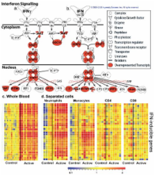
          Tuberculosis (TB), caused by infection with Mycobacterium tuberculosis ( M. tuberculosis), is a major cause of morbidity and mortality worldwide and efforts to control TB are hampered by difficulties with diagnosis, prevention and treatment 1, 2. Most people infected with M. tuberculosis remain asymptomatic, termed latent TB, with a 10% lifetime risk of developing active TB disease, but current tests cannot identify which individuals will develop disease 3. The immune response to M. tuberculosis is complex and incompletely characterized, hindering development of new diagnostics, therapies and vaccines 4, 5. We identified a whole blood 393 transcript signature for active TB in intermediate and high burden settings, correlating with radiological extent of disease and reverting to that of healthy controls following treatment. A subset of latent TB patients had signatures similar to those in active TB patients. We also identified a specific 86-transcript signature that discriminated active TB from other inflammatory and infectious diseases. Modular and pathway analysis revealed that the TB signature was dominated by a neutrophil-driven interferon (IFN)-inducible gene profile, consisting of both IFN-γ and Type I IFNαβ signalling. Comparison with transcriptional signatures in purified cells and flow cytometric analysis, suggest that this TB signature reflects both changes in cellular composition and altered gene expression. Although an IFN signature was also observed in whole blood of patients with Systemic Lupus Erythematosus (SLE), their complete modular signature differed from TB with increased abundance of plasma cell transcripts. Our studies demonstrate a hitherto under-appreciated role of Type I IFNαβ signalling in TB pathogenesis, which has implications for vaccine and therapeutic development. Our study also provides a broad range of transcriptional biomarkers with potential as diagnostic and prognostic tools to combat the TB epidemic.

          Related collections

          Most cited references35

          • Record: found
          • Abstract: found
          • Article: not found

          Interferon-inducible gene expression signature in peripheral blood cells of patients with severe lupus.

          Systemic lupus erythematosus (SLE) is a complex, inflammatory autoimmune disease that affects multiple organ systems. We used global gene expression profiling of peripheral blood mononuclear cells to identify distinct patterns of gene expression that distinguish most SLE patients from healthy controls. Strikingly, about half of the patients studied showed dysregulated expression of genes in the IFN pathway. Furthermore, this IFN gene expression "signature" served as a marker for more severe disease involving the kidneys, hematopoetic cells, and/or the central nervous system. These results provide insights into the genetic pathways underlying SLE, and identify a subgroup of patients who may benefit from therapies targeting the IFN pathway.
            Bookmark
            • Record: found
            • Abstract: found
            • Article: not found

            Genetic dissection of immunity to mycobacteria: the human model.

            Humans are exposed to a variety of environmental mycobacteria (EM), and most children are inoculated with live Bacille Calmette-Guérin (BCG) vaccine. In addition, most of the world's population is occasionally exposed to human-borne mycobacterial species, which are less abundant but more virulent. Although rarely pathogenic, mildly virulent mycobacteria, including BCG and most EM, may cause a variety of clinical diseases. Mycobacterium tuberculosis, M. leprae, and EM M. ulcerans are more virulent, causing tuberculosis, leprosy, and Buruli ulcer, respectively. Remarkably, only a minority of individuals develop clinical disease, even if infected with virulent mycobacteria. The interindividual variability of clinical outcome is thought to result in part from variability in the human genes that control host defense. In this well-defined microbiological and clinical context, the principles of mouse immunology and the methods of human genetics can be combined to facilitate the genetic dissection of immunity to mycobacteria. The natural infections are unique to the human model, not being found in any of the animal models of experimental infection. We review current genetic knowledge concerning the simple and complex inheritance of predisposition to mycobacterial diseases in humans. Rare patients with Mendelian disorders have been found to be vulnerable to BCG, a few EM, and M. tuberculosis. Most cases of presumed Mendelian susceptibility to these and other mycobacterial species remain unexplained. In the general population leprosy and tuberculosis have been shown to be associated with certain human genetic polymorphisms and linked to certain chromosomal regions. The causal vulnerability genes themselves have yet to be identified and their pathogenic alleles immunologically validated. The studies carried out to date have been fruitful, initiating the genetic dissection of protective immunity against a variety of mycobacterial species in natural conditions of infection. The human model has potential uses beyond the study of mycobacterial infections and may well become a model of choice for the investigation of immunity to infectious agents.
              Bookmark
              • Record: found
              • Abstract: found
              • Article: not found

              Interferon and Granulopoiesis Signatures in Systemic Lupus Erythematosus Blood

              Systemic lupus erythematosus (SLE) is a prototype systemic autoimmune disease characterized by flares of high morbidity. Using oligonucleotide microarrays, we now show that active SLE can be distinguished by a remarkably homogeneous gene expression pattern with overexpression of granulopoiesis-related and interferon (IFN)-induced genes. Using the most stringent statistical analysis (Bonferroni correction), 15 genes were found highly up-regulated in SLE patients, 14 of which are targets of IFN and one, defensin DEFA-3, a major product of immature granulocytes. A more liberal correction (Benjamini and Hochberg correction) yielded 18 additional genes, 12 of which are IFN-regulated and 4 granulocyte-specific. Indeed immature neutrophils were identified in a large fraction of SLE patients white blood cells. High dose glucocorticoids, a standard treatment of disease flares, shuts down the interferon signature, further supporting the role of this cytokine in SLE. The expression of 10 genes correlated with disease activity according to the SLEDAI. The most striking correlation (P < 0.001, r = 0.55) was found with the formyl peptide receptor-like 1 protein that mediates chemotactic activities of defensins. Therefore, while the IFN signature confirms the central role of this cytokine in SLE, microarray analysis of blood cells reveals that immature granulocytes may be involved in SLE pathogenesis.
                Bookmark

                Author and article information

                Journal
                0410462
                6011
                Nature
                Nature
                Nature
                0028-0836
                1476-4687
                15 June 2010
                19 August 2010
                08 November 2012
                : 466
                : 7309
                : 973-977
                Affiliations
                [1 ]Division of Immunoregulation, MRC National Institute for Medical Research, The Ridgeway, Mill Hill, London, NW7 1AA, UK.
                [2 ]Division of Mycobacterial Research, MRC National Institute for Medical Research, The Ridgeway, Mill Hill, London, NW7 1AA, UK.
                [3 ]Department of Respiratory Medicine, Imperial College Healthcare NHS Trust.
                [4 ]Institute of Infectious Diseases and Molecular Medicine Institute of Infectious Diseases and Molecular Medicine, University of Cape Town
                [5 ]Division of Medicine, Imperial College London.
                [6 ]Baylor Institute for Immunology Research-ANRS Center for Human Vaccines, INSERM U899, 3434 Live Oak St., Dallas, Texas 75204
                [7 ]Institute for Health Care Research and Improvement, Baylor Health Care System, Dallas, Texas 75204.
                [8 ]Department of Radiology, Imperial College Healthcare NHS Trust.
                [9 ]UT Southwestern Medical Center, Dallas, Texas.
                [10 ]Center for Vaccines and Immunity, The Research Institute at Nationwide Children's Hospital, 700 Children's Drive, Columbus, OH 43205, USA.
                Author notes
                [# ]Please address correspondence to AOG.
                [*]

                CG & FMcN contributed equally to this study.

                Author contributions

                M.B, D.C, O.M.K, and A.OG. designed the study on TB with input from JB and RW and for other diseases with input from V.P and O.R; M.B, S.B, T.O, K.W, J.C, A.M, R.B. and O.M.K recruited, sampled and collected patient data; M.B, R.B, A.M. and C.G processed whole blood for microarray experiments with help from J.S; C.G performed blood cell subset separations and processing for microarray experiments with help from J.S; M.B, C.G and Z.X performed microarray data analysis, with advice and input from JS, DC and VP; M.B. and Z.X. performed Ingenuity, Modular and Molecular Distance to Health Analyses; M.B. performed multiplex serum analyses; F.McN performed flow cytometry analysis; D.C., V.P and A.OG supervised data analysis; M.B and D.B performed statistical analysis; M.B, S.B, R.D and O.M.K performed analyses of radiology; A.OG and M.B wrote the manuscript, with early input from C.G, F.McN, J.B, D.C. and J.S, and subsequently all authors provided advice and approved the final manuscript.

                All microarray data are deposited in GEO under Accession Numbers GSE19491, GSE19444, GSE19443, GSE19442, GSE19439, GSE19435. Some of the work has been submitted as US Patent Application PCT 371: Blood Transcriptional Signature of Mycobacterium Tuberculosis Infection: Serial No: 12/602,488.

                Article
                EMS31044
                10.1038/nature09247
                3492754
                20725040
                87147a1f-c1d1-4cec-a381-d7b716a17c23

                Users may view, print, copy, download and text and data- mine the content in such documents, for the purposes of academic research, subject always to the full Conditions of use: http://www.nature.com/authors/editorial_policies/license.html#terms

                History
                Funding
                Funded by: Wellcome Trust :
                Award ID: 088316 || WT
                Funded by: Medical Research Council :
                Award ID: U117588499(88499) || MRC_
                Funded by: Medical Research Council :
                Award ID: U.1175.02.001.00006(65642) || MRC_
                Categories
                Article

                Uncategorized
                Uncategorized

                Comments

                Comment on this article