0
views
0
recommends
+1 Recommend
0 collections
    0
    shares
      • Record: found
      • Abstract: found
      • Article: found
      Is Open Access

      Not All Osteophytes Are Located on the Right Side of the Vertebrae in Diffuse Idiopathic Skeletal Hyperostosis: A Quantitative Analysis in Relation to the Position of Aorta

      other

      Read this article at

      Bookmark
          There is no author summary for this article yet. Authors can add summaries to their articles on ScienceOpen to make them more accessible to a non-specialist audience.

          Abstract

          Objective

          Diffuse idiopathic skeletal hyperostosis (DISH) is characterized by osteophytes in the anterior vertebrae, and the presence of aorta may have an impact on their formation. However, the anatomical positional relationship between the aorta and osteophytes in patients with DISH remains controversial. This study aimed to evaluate the position of osteophytes in relation to aorta in DISH, and the influence of aortic pulsation on the formation of osteophytes from the perspective of morphology.

          Methods

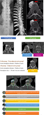
          We conducted a retrospective review of 101 patients diagnosed with DISH and symptomatic lumbar spinal stenosis between June 2018 and December 2021. A total of 637 segments with heterotopic ossification in DISH were used for quantitative measurements on CT scans. The Cartesian coordinate system was built up on the axial CT scans to reflect the relative position between aorta and osteophytes. Osteophytes were divided into adjacent aorta group (AD group) and non‐adjacent aorta group (N‐AD group). In terms of the morphology, osteophytes in the AD group were further divided into convex, flat, and concave types. The relative position between aorta and osteophytes, and the aorta‐osteophyte distance and morphology of osteophytes were compared. Univariate analysis of variance was performed for multiple groups, and two independent‐samples t‐tests were used for two groups.

          Results

          From T5 to L4, aorta gradually descended from left side to middle of vertebrae, and osteophytes gradually shifted from right side of vertebrae (T5‐T10) to bilateral sides (T11‐L4). Of 637 osteophytes in DISH, 60.1% (383/637) were in AD group, including convex type 0.6% (4/637), flat type 34.7% (221/637), and concave type 24.8% (158/637). The N‐AD group accounted for 39.9% (254/637). Flat osteophytes were concentrated in T5‐T12, while concave osteophytes in T11‐L4. Overall, the aorta‐osteophyte distance of concave type was significantly smaller than that of flat type.

          Conclusion

          Osteophytes are not always located on the right side of vertebrae, but move with the position of the descending aorta. Furthermore, the morphology of osteophytes varies by vertebral segment in DISH, which is related to aorta descending anteriorly in the spine.

          Abstract

          DISH is characterized by anterior heterotopic ossification of spine, avoiding the aorta. Segments with osteophytes were grouped according to the morphology in the study, in order to investigate the positional relationship between the aorta and osteophytes and the influence of the aorta on the formation of the osteophytes in DISH.

          Related collections

          Most cited references23

          • Record: found
          • Abstract: found
          • Article: not found

          Radiographic and pathologic features of spinal involvement in diffuse idiopathic skeletal hyperostosis (DISH).

          The vertebral involvement of DISH is described from an evaluation of 215 cadaveric spines and 100 patients with the disease. Radiographic features include linear new bone formation along the anterolateral aspect of the thoracic spine, a bumpy contour, subjacent radiolucency, and irregular and pointed bony excrescences at the superior and inferior vertebral margins in the cervical and lumbar regions. Pathologic features include focal and diffuse calcification and ossification in the anterior longitudinal ligament, paraspinal connective tissue, and annulus fibrosis, degeneration in the peripheral annulus fibrosis fibers, L-T-, and Y-shaped anterolateral extensions of fibrous tissue, hypervascularity, chronic inflammatory cellular infiltration, and periosteal new bone formation on the anterior surface of the vertebral bodies.
            Bookmark
            • Record: found
            • Abstract: found
            • Article: not found

            Diffuse idiopathic skeletal hyperostosis: clinical features and pathogenic mechanisms.

            Diffuse idiopathic skeletal hyperostosis (DISH) is a systemic condition characterized by the ossification and calcification of ligaments and entheses. DISH is observed on all continents and in all races, but most commonly in men over 50 years of age. Although DISH is asymptomatic in most individuals, the condition is often an indicator of underlying metabolic disease, and the presence of spinal or extraspinal ossifications can sometimes lead to symptoms including pain, stiffness, a reduced range of articular motion, and dysphagia, as well as increasing the risk of unstable spinal fractures. The aetiology of DISH is poorly understood, and the roles of the many factors that might be involved in the development of excess bone are not well delineated. The study of pathophysiological aspects of DISH is made difficult by the formal diagnosis requiring the presence of multiple contiguous fully formed bridging ossifications, which probably represent advanced stages of DISH. In this Review, the reader is provided with an up-to-date discussion of the epidemiological, aetiological and clinical aspects of DISH. Existing classification criteria (which, in the absence of diagnostic criteria, are used to establish a diagnosis of DISH) are also considered, together with the need for modified criteria that enable timely identification of early phases in the development of DISH.
              Bookmark
              • Record: found
              • Abstract: found
              • Article: not found
              Is Open Access

              The prevalence of diffuse idiopathic skeletal hyperostosis in Japan - the first report of measurement by CT and review of the literature.

              Diffuse idiopathic skeletal hyperostosis (DISH) is prone to be accompanied by a spinal column fracture which is resistant to conservative therapy. This major characteristic of DISH is not recognized adequately by physicians, because the disease's detailed pathological condition has not yet been investigated. Therefore, the purposes of this study were to investigate the prevalence of DISH using computed tomography (CT), and to validate the reliability of CT interpretation.
                Bookmark

                Author and article information

                Contributors
                drsunxu@163.com
                Journal
                Orthop Surg
                Orthop Surg
                10.1111/(ISSN)1757-7861
                OS
                Orthopaedic Surgery
                John Wiley & Sons Australia, Ltd (Melbourne )
                1757-7853
                1757-7861
                08 September 2023
                November 2023
                : 15
                : 11 ( doiID: 10.1111/os.v15.11 )
                : 2881-2888
                Affiliations
                [ 1 ] Division of Spine Surgery, Department of Orthopaedic Surgery Nanjing Drum Tower Hospital, Affiliated Hospital of Nanjing University Medical School Nanjing China
                [ 2 ] Division of Spine Surgery, Department of Orthopaedic Surgery Nanjing Drum Tower Hospital Clinical College of Jiangsu University Nanjing China
                Author notes
                [*] [* ] Address for correspondence Xu Sun, MD, Division of Spine Surgery, Department of Orthopaedic Surgery, Nanjing Drum Tower Hospital, Affiliated Hospital of Nanjing University Medical School, Zhongshan Road 321, Nanjing, China 210008; Tel.: +86‐2583105121; Fax: +86‐2583105121; Email: drsunxu@ 123456163.com

                Author information
                https://orcid.org/0009-0005-4225-5460
                https://orcid.org/0000-0003-4202-6092
                https://orcid.org/0000-0001-8569-3595
                https://orcid.org/0000-0002-2444-4744
                https://orcid.org/0000-0002-7181-877X
                Article
                OS13869
                10.1111/os.13869
                10622269
                37680188
                865e1278-bdf8-4e17-842c-0040cac28fba
                © 2023 The Authors. Orthopaedic Surgery published by Tianjin Hospital and John Wiley & Sons Australia, Ltd.

                This is an open access article under the terms of the http://creativecommons.org/licenses/by/4.0/ License, which permits use, distribution and reproduction in any medium, provided the original work is properly cited.

                History
                : 24 July 2023
                : 02 May 2023
                : 29 July 2023
                Page count
                Figures: 6, Tables: 3, Pages: 8, Words: 5131
                Funding
                Funded by: Jiangsu Provincial Medical Innovation Center of Orthopedic Surgery
                Award ID: CXZX202214
                Funded by: National Natural Science Foundation of China , doi 10.13039/501100001809;
                Award ID: 81772422
                Categories
                Clinical Article
                Clinical Articles
                Custom metadata
                2.0
                November 2023
                Converter:WILEY_ML3GV2_TO_JATSPMC version:6.3.4 mode:remove_FC converted:02.11.2023

                aorta,ct,diffuse idiopathic skeletal hyperostosis,morphology,osteophyte,position

                Comments

                Comment on this article