11
views
0
recommends
+1 Recommend
0 collections
    0
    shares
      • Record: found
      • Abstract: found
      • Article: found
      Is Open Access

      Simple X-ray versus ultrasonography examination in blunt chest trauma: effective tools of accurate diagnosis and considerations for rib fractures

      research-article

      Read this article at

      Bookmark
          There is no author summary for this article yet. Authors can add summaries to their articles on ScienceOpen to make them more accessible to a non-specialist audience.

          Abstract

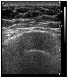
          Simple radiography is the best diagnostic tool for rib fractures caused by chest trauma, but it has some limitations. Thus, other tools are also being used. The aims of this study were to investigate the effectiveness of ultrasonography (US) for identifying rib fractures and to identify influencing factors of its effectiveness. Between October 2003 and August 2007, 201 patients with blunt chest trauma were available to undergo chest radiographic and US examinations for diagnosis of rib fractures. The two modalities were compared in terms of effectiveness based on simple radiographic readings and US examination results. We also investigated the factors that influenced the effectiveness of US examination. Rib fractures were detected on radiography in 69 patients (34.3%) but not in 132 patients. Rib fractures were diagnosed by using US examination in 160 patients (84.6%). Of the 132 patients who showed no rib fractures on radiography, 92 showed rib fractures on US. Among the 69 patients of rib fracture detected on radiography, 33 had additional rib fractures detected on US. Of the patients, 76 (37.8%) had identical radiographic and US results, and 125 (62.2%) had fractures detected on US that were previously undetected on radiography or additional fractures detected on US. Age, duration until US examination, and fracture location were not significant influencing factors. However, in the group without detected fractures on radiography, US showed a more significant effectiveness than in the group with detected fractures on radiography ( P=0.003). US examination could detect unnoticed rib fractures on simple radiography. US examination is especially more effective in the group without detected fractures on radiography. More attention should be paid to patients with chest trauma who have no detected fractures on radiography.

          Related collections

          Most cited references10

          • Record: found
          • Abstract: found
          • Article: not found

          Ultrasound in blunt abdominal and thoracic trauma.

          Between July 1989 and June 1991, 312 patients with blunt thoracic or abdominal injuries were examined prospectively. Sonographic examination was performed by surgeons in the emergency room using a mobile ultrasound unit. In 113 (36.2%) cases pathologic findings were demonstrated sonographically. These included 47 cases of hemothorax, 11 pericardial effusions, 52 cases of intra-abdominal fluid, 24 lesions of intra-abdominal organs, and 10 cases of retroperitoneal hematoma. Physical examination findings were positive in 96 (30.8%), negative in 63 (20.2%), and equivocal in 153 (49.0%). Two hundred thirty-nine patients had between one and eight injuries in addition to the blunt abdominal or thoracic trauma. These patients had an average Injury Severity Score (ISS) of 19.9 (range, 1 to 75). The 73 patients with isolated blunt trauma of the thorax or abdomen had an ISS of 4.9 (range, 0-25). None of the 66 patients (21.2%) with positive clinical findings and negative sonographic examination results had to be operated on later in the course of treatment, while 5 (36%) of 14 patients (4.5%) with negative physical examination findings and positive sonographic findings had to undergo surgery. The sensitivity for the demonstration of intra-abdominal fluid and organ lesions was 98.1% and 41.4%, respectively. The overall sensitivity and specificity of the ultrasonic examination were 90.0% and 99.5%, respectively.
            Bookmark
            • Record: found
            • Abstract: found
            • Article: not found

            Trauma ultrasound examination versus chest radiography in the detection of hemothorax.

            To compare the sensitivity, specificity, and accuracy of ultrasonography with those of the initial plain chest radiograph for detection of hemothorax in trauma patients. Data from a prior prospective study of trauma ultrasonography at a Level I trauma center were retrospectively analyzed. The medical records of a convenience sample of adult patients who presented with major blunt or penetrating torso trauma during a 17-month period were reviewed. Emergency physicians performed a trauma ultrasound examination, which included evaluation for pleural fluid. Ultrasound interpretations were recorded before other diagnostic tests were obtained and were not used in patient management decisions. Records of the study patients were reviewed for confirmation of the presence or absence of hemothorax by other diagnostic and therapeutic interventions. The chest radiograph and computed tomography (CT) scan interpretations were performed by attending radiologists who were not blinded to patient outcome. Five of the 245 patients enrolled in the study were excluded because tube thoracostomy was performed before the ultrasound examination was done. Altogether, 26 of the 240 study patients had hemothorax, as confirmed by tube thoracostomy or CT. Both ultrasound examination and the initial chest radiograph resulted in 0 false-positive, 1 false-negative, 25 true-positive, and 214 true-negative findings. Overall, both modailties were 96.2% sensitive, 100% specific, and 99.6% accurate. Ultrasonography is comparable to the initial chest radiograph for accuracy in detection of hemothorax and may expedite the diagnosis and treatment of this condition for patients with major trauma.
              Bookmark
              • Record: found
              • Abstract: found
              • Article: not found

              Sonography compared with radiography in revealing acute rib fracture.

              This study was undertaken to compare the sensitivities of sonography and radiography for revealing acute rib fracture. Chest radiography and rib sonography were performed on 50 patients with suspected rib fractures. Sonography was performed with a 9- or 12-MHz linear transducer. Fractures were identified by a disruption of the anterior margin of the rib, costochondral junction, or costal cartilage. The incidence, location, and degree of displacement of fractures revealed by radiography and sonography were compared. Sonography was performed again after 3 weeks in 37 subjects. At presentation, radiographs revealed eight rib fractures in six (12%) of 50 patients and sonography revealed 83 rib fractures in 39 (78%) of 50 patients. Seventy-four (89%) of the 83 sonographically detected fractures were located in the rib, four (5%) were located at the costochondral junction, and five (6%) in the costal cartilage. Repeated sonography after 3 weeks showed evidence of healing in all reexamined fractures. Combining sonography at presentation and after 3 weeks, 88% of subjects had sustained a fracture. Sonography reveals more fractures than does radiography and will reveal fractures in most patients presenting with suspected rib fracture. Further scientific studies are needed to clarify the appropriate role for sonography in rib fracture detection.
                Bookmark

                Author and article information

                Journal
                J Exerc Rehabil
                J Exerc Rehabil
                Journal of Exercise Rehabilitation
                Korean Society of Exercise Rehabilitation
                2288-176X
                2288-1778
                December 2016
                31 December 2016
                : 12
                : 6
                : 637-641
                Affiliations
                [1 ]Department of Thoracic and Cardiovascular Surgery, Gunpo G Sam General Hospital, Gunpo, Korea
                [2 ]Department of Computer Science and Statistics, Jeju National University, Jeju, Korea
                Author notes
                [* ]Corresponding author: Yunjung Lee, http://orcid.org/0000-0001-7394-1723, Department of Computer Science and Statistics, Jeju National University, Jeju National University,102 Jejudaehak-ro, Jeju 63243, Korea, Tel: +82-64-754-3597, Fax: +82-64-725-2579, E-mail: rheeyj@ 123456jejunu.ac.kr
                Article
                jer-12-6-637
                10.12965/jer.1632840.420
                5227329
                8638cb80-1d1b-4829-9252-92bc018c3cf4
                Copyright © 2016 Korean Society of Exercise Rehabilitation

                This is an Open Access article distributed under the terms of the Creative Commons Attribution Non-Commercial License ( http://creativecommons.org/licenses/by-nc/4.0/) which permits unrestricted non-commercial use, distribution, and reproduction in any medium, provided the original work is properly cited.

                History
                : 07 October 2016
                : 01 December 2016
                Categories
                Original Article

                blunt chest trauma,ultrasound examination,rib fracture

                Comments

                Comment on this article