22
views
0
recommends
+1 Recommend
0 collections
    0
    shares
      • Record: found
      • Abstract: found
      • Article: found
      Is Open Access

      Epigallocatechin-3-Gallate (EGCG): New Therapeutic Perspectives for Neuroprotection, Aging, and Neuroinflammation for the Modern Age

      , , , ,
      Biomolecules
      MDPI AG

      Read this article at

      Bookmark
          There is no author summary for this article yet. Authors can add summaries to their articles on ScienceOpen to make them more accessible to a non-specialist audience.

          Abstract

          Alzheimer’s and Parkinson’s diseases are the two most common forms of neurodegenerative diseases. The exact etiology of these disorders is not well known; however, environmental, molecular, and genetic influences play a major role in the pathogenesis of these diseases. Using Alzheimer’s disease (AD) as the archetype, the pathological findings include the aggregation of Amyloid Beta (Aβ) peptides, mitochondrial dysfunction, synaptic degradation caused by inflammation, elevated reactive oxygen species (ROS), and cerebrovascular dysregulation. This review highlights the neuroinflammatory and neuroprotective role of epigallocatechin-3-gallate (EGCG): the medicinal component of green tea, a known nutraceutical that has shown promise in modulating AD progression due to its antioxidant, anti-inflammatory, and anti-aging abilities. This report also re-examines the current literature and provides innovative approaches for EGCG to be used as a preventive measure to alleviate AD and other neurodegenerative disorders.

          Related collections

          Most cited references223

          • Record: found
          • Abstract: not found
          • Article: not found

          Alzheimer's disease.

            Bookmark
            • Record: found
            • Abstract: found
            • Article: not found

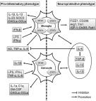
            The pro- and anti-inflammatory properties of the cytokine interleukin-6.

            Interleukin-6 is a cytokine not only involved in inflammation and infection responses but also in the regulation of metabolic, regenerative, and neural processes. In classic signaling, interleukin-6 stimulates target cells via a membrane bound interleukin-6 receptor, which upon ligand binding associates with the signaling receptor protein gp130. Gp130 dimerizes, leading to the activation of Janus kinases and subsequent phosphorylation of tyrosine residues within the cytoplasmic portion of gp130. This leads to the engagement of phosphatase Src homology domains containing tyrosin phosphatase-2 (SHP-2) and activation of the ras/raf/Mitogen-activated protein (MAP) kinase (MAPK) pathway. In addition, signal transducer and activator of transcription factors are recruited, which are phosphorylated, and consequently dimerize whereupon they translocate into the nucleus and activate target genes. Interestingly, only few cells express membrane bound interleukin-6 receptor whereas all cells display gp130 on the cell surface. While cells, which only express gp130, are not responsive to interleukin-6 alone, they can respond to a complex of interleukin-6 bound to a naturally occurring soluble form of the interleukin-6 receptor. Therefore, the generation of soluble form of the interleukin-6 receptor dramatically enlarges the spectrum of interleukin-6 target cells. This process has been named trans-signaling. Here, we review the involvement of both signaling modes in the biology of interleukin-6. It turns out that regenerative or anti-inflammatory activities of interleukin-6 are mediated by classic signaling whereas pro-inflammatory responses of interleukin-6 are rather mediated by trans-signaling. This is important since therapeutic blockade of interleukin-6 by the neutralizing anti-interleukin-6 receptor monoclonal antibody tocilizumab has recently been approved for the treatment of inflammatory diseases. This article is part of a Special Issue entitled: 11th European Symposium on Calcium. 2011 Elsevier B.V. All rights reserved.
              Bookmark
              • Record: found
              • Abstract: found
              • Article: found
              Is Open Access

              Neuroinflammation in neurodegenerative disorders: the roles of microglia and astrocytes

              Neuroinflammation is associated with neurodegenerative diseases, such as Alzheimer’s disease, Parkinson’s disease, and amyotrophic lateral sclerosis. Microglia and astrocytes are key regulators of inflammatory responses in the central nervous system. The activation of microglia and astrocytes is heterogeneous and traditionally categorized as neurotoxic (M1-phenotype microglia and A1-phenotype astrocytes) or neuroprotective (M2-phenotype microglia and A2-phenotype astrocytes). However, this dichotomized classification may not reflect the various phenotypes of microglia and astrocytes. The relationship between these activated glial cells is also very complicated, and the phenotypic distribution can change, based on the progression of neurodegenerative diseases. A better understanding of the roles of microglia and astrocytes in neurodegenerative diseases is essential for developing effective therapies. In this review, we discuss the roles of inflammatory response in neurodegenerative diseases, focusing on the contributions of microglia and astrocytes and their relationship. In addition, we discuss biomarkers to measure neuroinflammation and studies on therapeutic drugs that can modulate neuroinflammation.
                Bookmark

                Author and article information

                Contributors
                (View ORCID Profile)
                (View ORCID Profile)
                Journal
                BIOMHC
                Biomolecules
                Biomolecules
                MDPI AG
                2218-273X
                March 2022
                February 25 2022
                : 12
                : 3
                : 371
                Article
                10.3390/biom12030371
                35327563
                85a32ca6-1752-439d-ae55-0ced0bf2f181
                © 2022

                https://creativecommons.org/licenses/by/4.0/

                History

                Comments

                Comment on this article