19
views
0
recommends
+1 Recommend
0 collections
    0
    shares
      • Record: found
      • Abstract: found
      • Article: found
      Is Open Access

      Macrophages Are Key Regulators of Stem Cells during Skeletal Muscle Regeneration and Diseases

      review-article
      1 , 2 , 1 , 3 , 1 , 3 , 1 , 2 ,
      Stem Cells International
      Hindawi

      Read this article at

      Bookmark
          There is no author summary for this article yet. Authors can add summaries to their articles on ScienceOpen to make them more accessible to a non-specialist audience.

          Abstract

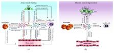
          Muscle regeneration is a closely regulated process that involves a variety of cell types such as satellite cells, myofibers, fibroadipogenic progenitors, endothelial cells, and inflammatory cells. Among these different cell types, macrophages emerged as a central actor coordinating the different cellular interactions and biological processes. Particularly, the transition of macrophages from their proinflammatory to their anti-inflammatory phenotype was shown to regulate inflammation, myogenesis, fibrosis, vascularization, and return to homeostasis. On the other hand, deregulation of macrophage accumulation or polarization in chronic degenerative muscle disorders was shown to impair muscle regeneration. Considering the key roles of macrophages in skeletal muscle, they represent an attractive target for new therapeutic approaches aiming at mitigating various muscle disorders. This review aims at summarizing the novel insights into macrophage heterogeneity, plasticity, and functions in skeletal muscle homeostasis, regeneration, and disease.

          Related collections

          Most cited references136

          • Record: found
          • Abstract: found
          • Article: not found

          Regulatory interactions between muscle and the immune system during muscle regeneration.

          Recent discoveries reveal complex interactions between skeletal muscle and the immune system that regulate muscle regeneration. In this review, we evaluate evidence that indicates that the response of myeloid cells to muscle injury promotes muscle regeneration and growth. Acute perturbations of muscle activate a sequence of interactions between muscle and inflammatory cells. The initial inflammatory response is a characteristic Th1 inflammatory response, first dominated by neutrophils and subsequently by CD68(+) M1 macrophages. M1 macrophages can propagate the Th1 response by releasing proinflammatory cytokines and cause further tissue damage through the release of nitric oxide. Myeloid cells in the early Th1 response stimulate the proliferative phase of myogenesis through mechanisms mediated by TNF-alpha and IL-6; experimental prolongation of their presence is associated with delayed transition to the early differentiation stage of myogenesis. Subsequent invasion by CD163(+)/CD206(+) M2 macrophages attenuates M1 populations through the release of anti-inflammatory cytokines, including IL-10. M2 macrophages play a major role in promoting growth and regeneration; their absence greatly slows muscle growth following injury or modified use and inhibits muscle differentiation and regeneration. Chronic muscle injury leads to profiles of macrophage invasion and function that differ from acute injuries. For example, mdx muscular dystrophy yields invasion of muscle by M1 macrophages, but their early invasion is accompanied by a subpopulation of M2a macrophages. M2a macrophages are IL-4 receptor(+)/CD206(+) cells that reduce cytotoxicity of M1 macrophages. Subsequent invasion of dystrophic muscle by M2c macrophages is associated with progression of the regenerative phase in pathophysiology. Together, these findings show that transitions in macrophage phenotype are an essential component of muscle regeneration in vivo following acute or chronic muscle damage.
            Bookmark
            • Record: found
            • Abstract: found
            • Article: not found

            Inflammatory processes in muscle injury and repair.

            Modified muscle use or injury can produce a stereotypic inflammatory response in which neutrophils rapidly invade, followed by macrophages. This inflammatory response coincides with muscle repair, regeneration, and growth, which involve activation and proliferation of satellite cells, followed by their terminal differentiation. Recent investigations have begun to explore the relationship between inflammatory cell functions and skeletal muscle injury and repair by using genetically modified animal models, antibody depletions of specific inflammatory cell populations, or expression profiling of inflamed muscle after injury. These studies have contributed to a complex picture in which inflammatory cells promote both injury and repair, through the combined actions of free radicals, growth factors, and chemokines. In this review, recent discoveries concerning the interactions between skeletal muscle and inflammatory cells are presented. New findings clearly show a role for neutrophils in promoting muscle damage soon after muscle injury or modified use. No direct evidence is yet available to show that neutrophils play a beneficial role in muscle repair or regeneration. Macrophages have also been shown capable of promoting muscle damage in vivo and in vitro through the release of free radicals, although other findings indicate that they may also play a role in muscle repair and regeneration through growth factors and cytokine-mediated signaling. However, this role for macrophages in muscle regeneration is still not definitive; other cells present in muscle can also produce the potentially regenerative factors, and it remains to be proven whether macrophage-derived factors are essential for muscle repair or regeneration in vivo. New evidence also shows that muscle cells can release positive and negative regulators of inflammatory cell invasion, and thereby play an active role in modulating the inflammatory process. In particular, muscle-derived nitric oxide can inhibit inflammatory cell invasion of healthy muscle and protect muscle from lysis by inflammatory cells in vivo and in vitro. On the other hand, muscle-derived cytokines can signal for inflammatory cell invasion, at least in vitro. The immediate challenge for advancing our current understanding of the relationships between muscle and inflammatory cells during muscle injury and repair is to place what has been learned in vitro into the complex and dynamic in vivo environment.
              Bookmark
              • Record: found
              • Abstract: found
              • Article: not found

              Two types of mouse T helper cell. IV. Th2 clones secrete a factor that inhibits cytokine production by Th1 clones

              A cytokine synthesis inhibitory factor (CSIF) is secreted by Th2 clones in response to Con A or antigen stimulation, but is absent in supernatants from Con A-induced Th1 clones. CSIF can inhibit the production of IL-2, IL-3, lymphotoxin (LT)/TNF, IFN-gamma, and granulocyte-macrophage CSF (GM-CSF) by Th1 cells responding to antigen and APC, but Th2 cytokine synthesis is not significantly affected. Transforming growth factor beta (TGF-beta) also inhibits IFN-gamma production, although less effectively than CSIF, whereas IL-2 and IL-4 partially antagonize the activity of CSIF. CSIF inhibition of cytokine synthesis is not complete, since early cytokine synthesis (before 8 h) is not significantly affected, whereas later synthesis is strongly inhibited. In the presence of CSIF, IFN-gamma mRNA levels are reduced slightly at 8, and strongly at 12 h after stimulation. Inhibition of cytokine expression by CSIF is not due to a general reduction in Th1 cell viability, since actin mRNA levels were not reduced, and proliferation of antigen-stimulated cells in response to IL-2, was unaffected. Biochemical characterization, mAbs, and recombinant or purified cytokines showed that CSIF is distinct from IL-1, IL-2, IL-3, IL-4, IL-5, IL-6, IL-7, IFN-gamma, GM-CSF, TGF-beta, TNF, LT, and P40. The potential role of CSIF in crossregulation of Th1 and Th2 responses is discussed.
                Bookmark

                Author and article information

                Contributors
                Journal
                Stem Cells Int
                Stem Cells Int
                SCI
                Stem Cells International
                Hindawi
                1687-966X
                1687-9678
                2019
                14 July 2019
                : 2019
                : 4761427
                Affiliations
                1CHU Sainte-Justine Research Center, Montreal, QC, Canada
                2School of Rehabilitation, Faculty of Medicine, Université de Montréal, Montreal, QC, Canada
                3Department of Pharmacology and Physiology, Faculty of Medicine, Université de Montréal, Montreal, QC, Canada
                Author notes

                Guest Editor: Fengjuan Lyu

                Author information
                https://orcid.org/0000-0002-2381-415X
                https://orcid.org/0000-0003-2184-9743
                https://orcid.org/0000-0002-7536-1666
                Article
                10.1155/2019/4761427
                6664695
                31396285
                8470b14e-b440-4fbd-a216-2a61b31f6a66
                Copyright © 2019 Junio Dort et al.

                This is an open access article distributed under the Creative Commons Attribution License, which permits unrestricted use, distribution, and reproduction in any medium, provided the original work is properly cited.

                History
                : 3 April 2019
                : 9 June 2019
                Funding
                Funded by: Stem Cell Network
                Funded by: ThéCell
                Funded by: Grand Défi Pierre Lavoie Foundation
                Funded by: Fonds de Recherche Québec-Santé
                Funded by: Natural Sciences and Engineering Research Council of Canada
                Funded by: Canadian Institutes of Health Research
                Funded by: CHU Sainte-Justine Foundation
                Categories
                Review Article

                Molecular medicine
                Molecular medicine

                Comments

                Comment on this article