14
views
0
recommends
+1 Recommend
0 collections
    0
    shares
      • Record: found
      • Abstract: found
      • Article: found
      Is Open Access

      PLCβ-Mediated Depletion of PIP 2 and ATP-Sensitive K + Channels Are Involved in Arginine Vasopressin-Induced Facilitation of Neuronal Excitability and LTP in the Dentate Gyrus

      research-article

      Read this article at

      Bookmark
          There is no author summary for this article yet. Authors can add summaries to their articles on ScienceOpen to make them more accessible to a non-specialist audience.

          Abstract

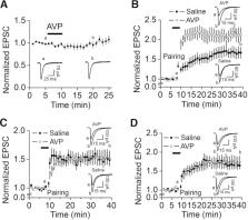
          Arginine vasopressin (AVP) serves as a neuromodulator in the brain. The hippocampus is one of the major targets for AVP, as it has been demonstrated that the hippocampus receives vasopressinergic innervation and expresses AVP receptors. The dentate gyrus (DG) granule cells (GCs) serve as a gate governing the inflow of information to the hippocampus. High densities of AVP receptors are expressed in the DG GCs. However, the roles and the underlying cellular and molecular mechanisms of AVP in the DG GCs have not been determined. We addressed this question by recording from the DG GCs in rat hippocampal slices. Our results showed that application of AVP concentration-dependently evoked an inward holding current recorded from the DG GCs. AVP depolarized the DG GCs and increased their action potential firing frequency. The excitatory effects of AVP were mediated by activation of V 1a receptors and required the function of phospholipase Cβ (PLCβ). Whereas intracellular Ca 2+ release and protein kinase C activity were unnecessary, PLCβ-induced depletion of phosphatidylinositol 4,5-bisphosphate was involved in AVP-evoked excitation of the DG GCs. AVP excited the DG GCs by depression of the ATP-sensitive K + channels, which were required for AVP-elicited facilitation of long-term potentiation at the perforant path–GC synapses. Our results may provide a cellular and molecular mechanism to explain the physiological functions of AVP, such as learning and memory, and pathologic disorders like anxiety.

          Related collections

          Most cited references98

          • Record: found
          • Abstract: found
          • Article: not found

          Are the dorsal and ventral hippocampus functionally distinct structures?

          One literature treats the hippocampus as a purely cognitive structure involved in memory; another treats it as a regulator of emotion whose dysfunction leads to psychopathology. We review behavioral, anatomical, and gene expression studies that together support a functional segmentation into three hippocampal compartments: dorsal, intermediate, and ventral. The dorsal hippocampus, which corresponds to the posterior hippocampus in primates, performs primarily cognitive functions. The ventral (anterior in primates) relates to stress, emotion, and affect. Strikingly, gene expression in the dorsal hippocampus correlates with cortical regions involved in information processing, while genes expressed in the ventral hippocampus correlate with regions involved in emotion and stress (amygdala and hypothalamus).
            Bookmark
            • Record: found
            • Abstract: found
            • Article: not found

            Functional organization of the hippocampal longitudinal axis.

            The precise functional role of the hippocampus remains a topic of much debate. The dominant view is that the dorsal (or posterior) hippocampus is implicated in memory and spatial navigation and the ventral (or anterior) hippocampus mediates anxiety-related behaviours. However, this 'dichotomy view' may need revision. Gene expression studies demonstrate multiple functional domains along the hippocampal long axis, which often exhibit sharply demarcated borders. By contrast, anatomical studies and electrophysiological recordings in rodents suggest that the long axis is organized along a gradient. Together, these observations suggest a model in which functional long-axis gradients are superimposed on discrete functional domains. This model provides a potential framework to explain and test the multiple functions ascribed to the hippocampus.
              Bookmark
              • Record: found
              • Abstract: found
              • Article: not found

              Reduced fear expression after lesions of the ventral hippocampus.

              The hippocampus has a critical role in several fundamental memory operations, including the conditioning of fear to contextual information. We show that the hippocampus is necessary also for unconditioned fear, and that the involved circuitry is at the ventral pole of the hippocampus. Rats with selective hippocampal lesions failed to avoid open arms in an elevated plus-maze and had decreased neuroendocrine stress responses during confinement to a brightly lit chamber. These effects were reproduced by lesions of the ventral half of the hippocampus, but not by damage to the dorsal three-quarters of the hippocampus or the amygdala. Ventral lesions failed to impair contextual fear conditioning or spatial navigation, suggesting that the ventral hippocampus may specifically influence some types of defensive fear-related behavior.
                Bookmark

                Author and article information

                Journal
                eNeuro
                eNeuro
                eneuro
                eNeuro
                eNeuro
                Society for Neuroscience
                2373-2822
                4 July 2022
                18 July 2022
                Jul-Aug 2022
                : 9
                : 4
                : ENEURO.0120-22.2022
                Affiliations
                [1]Department of Biomedical Sciences, School of Medicine and Health Sciences, University of North Dakota , Grand Forks, North Dakota 58203
                Author notes

                The authors declare no competing financial interests.

                Author contributions: S.L. designed research; S.L., C.A.B., and M.M. performed research; S.L. and C.A.B. analyzed data; S.L. wrote the paper.

                This work was supported by the National Institute of General Medical Sciences and National Institute of Mental Health Grant R01-MH-118258 to S.L.

                Correspondence should be addressed to Saobo Lei at saobo.lei@ 123456und.edu .
                Article
                eN-NWR-0120-22
                10.1523/ENEURO.0120-22.2022
                9298963
                35788107
                84285b7e-8d10-43f0-8277-b48dfc42370a
                Copyright © 2022 Lei et al.

                This is an open-access article distributed under the terms of the Creative Commons Attribution 4.0 International license, which permits unrestricted use, distribution and reproduction in any medium provided that the original work is properly attributed.

                History
                : 20 March 2022
                : 3 June 2022
                : 21 June 2022
                Page count
                Figures: 6, Tables: 0, Equations: 0, References: 98, Pages: 12, Words: 00
                Funding
                Funded by: HHS | NIH | National Institute of Mental Health (NIMH), doi 10.13039/100000025;
                Award ID: R01MH118258
                Categories
                6
                Research Article: New Research
                Neuronal Excitability
                Custom metadata
                July/August 2022

                action potential,depolarization,hippocampus,peptide,receptors,signal transduction

                Comments

                Comment on this article