39
views
0
recommends
+1 Recommend
0 collections
    0
    shares
      • Record: found
      • Abstract: found
      • Article: found
      Is Open Access

      Effects of dexamphetamine-induced dopamine release on resting-state network connectivity in recreational amphetamine users and healthy controls

      research-article

      Read this article at

      Bookmark
          There is no author summary for this article yet. Authors can add summaries to their articles on ScienceOpen to make them more accessible to a non-specialist audience.

          Abstract

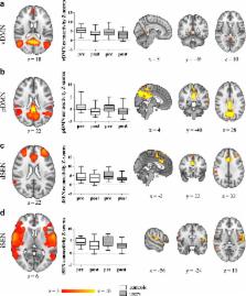
          Dexamphetamine (dAMPH) is not only used for the treatment of attention deficit hyperactivity disorder (ADHD), but also as a recreational drug. Acutely, dAMPH induces release of predominantly dopamine (DA) in the striatum, and in the cortex both DA and noradrenaline. Recent animal studies have shown that chronic dAMPH administration can induce changes in the DA system following long-term exposure, as evidenced by reductions in DA transporters, D 2/3 receptors and endogenous DA levels. However, only a limited number of studies have investigated the effects of dAMPH in the human brain. We used a combination of resting-state functional magnetic resonance imaging (rs-fMRI) and [ 123I]IBZM single-photon emission computed tomography (SPECT) (to assess baseline D 2/3 receptor binding and DA release) in 15 recreational AMPH users and 20 matched healthy controls to investigate the short-, and long-term effects of AMPH before and after an acute intravenous challenge with dAMPH. We found that acute dAMPH administration reduced functional connectivity in the cortico-striatal-thalamic network. dAMPH-induced DA release, but not DA D 2/3 receptor binding, was positively associated with connectivity changes in this network. In addition, acute dAMPH reduced connectivity in default mode networks and salience-executive-networks networks in both groups. In contrast to our hypothesis, no significant group differences were found in any of the rs-fMRI networks investigated, possibly due to lack of sensitivity or compensatory mechanisms. Our findings thus support the use of ICA-based resting-state functional connectivity as a tool to investigate acute, but not chronic, alterations induced by dAMPH on dopaminergic processing in the striatum.

          Related collections

          Most cited references37

          • Record: found
          • Abstract: found
          • Article: not found

          The neural basis of addiction: a pathology of motivation and choice.

          A primary behavioral pathology in drug addiction is the overpowering motivational strength and decreased ability to control the desire to obtain drugs. In this review the authors explore how advances in neurobiology are approaching an understanding of the cellular and circuitry underpinnings of addiction, and they describe the novel pharmacotherapeutic targets emerging from this understanding. Findings from neuroimaging of addicts are integrated with cellular studies in animal models of drug seeking. While dopamine is critical for acute reward and initiation of addiction, end-stage addiction results primarily from cellular adaptations in anterior cingulate and orbitofrontal glutamatergic projections to the nucleus accumbens. Pathophysiological plasticity in excitatory transmission reduces the capacity of the prefrontal cortex to initiate behaviors in response to biological rewards and to provide executive control over drug seeking. Simultaneously, the prefrontal cortex is hyperresponsive to stimuli predicting drug availability, resulting in supraphysiological glutamatergic drive in the nucleus accumbens, where excitatory synapses have a reduced capacity to regulate neurotransmission. Cellular adaptations in prefrontal glutamatergic innervation of the accumbens promote the compulsive character of drug seeking in addicts by decreasing the value of natural rewards, diminishing cognitive control (choice), and enhancing glutamatergic drive in response to drug-associated stimuli.
            Bookmark
            • Record: found
            • Abstract: found
            • Article: found
            Is Open Access

            Amphetamine, past and present – a pharmacological and clinical perspective

            Amphetamine was discovered over 100 years ago. Since then, it has transformed from a drug that was freely available without prescription as a panacea for a broad range of disorders into a highly restricted Controlled Drug with therapeutic applications restricted to attention deficit hyperactivity disorder (ADHD) and narcolepsy. This review describes the relationship between chemical structure and pharmacology of amphetamine and its congeners. Amphetamine’s diverse pharmacological actions translate not only into therapeutic efficacy, but also into the production of adverse events and liability for recreational abuse. Accordingly, the balance of benefit/risk is the key challenge for its clinical use. The review charts advances in pharmaceutical development from the introduction of once-daily formulations of amphetamine through to lisdexamfetamine, which is the first d-amphetamine prodrug approved for the management of ADHD in children, adolescents and adults. The unusual metabolic route for lisdexamfetamine to deliver d-amphetamine makes an important contribution to its pharmacology. How lisdexamfetamine’s distinctive pharmacokinetic/pharmacodynamic profile translates into sustained efficacy as a treatment for ADHD and its reduced potential for recreational abuse is also discussed.
              Bookmark
              • Record: found
              • Abstract: found
              • Article: not found

              Dopamine-dependent changes in the functional connectivity between basal ganglia and cerebral cortex in humans.

              We test the hypothesis that interaction between the human basal ganglia and cerebral cortex involves activity in multiple functional circuits characterized by their frequency of oscillation, phase characteristics, dopamine dependency and topography. To this end we took recordings from macroelectrodes (MEs) inserted into the subthalamic nucleus (STN) in eight awake patients following functional neurosurgery for Parkinson's disease. An EEG was also recorded, as were the signals from MEs in the globus pallidus interna (GPi) in two of the cases. Coherence between EEG and ME potentials was apparent in three major frequency bands, 2-10 Hz, 10-30 Hz and 70-85 Hz. These rhythmic activities differed in their cortical topography, although coherence was always strongest over the midline. Coherence between EEG and ME potentials in the 70-85 Hz band was only recorded in patients treated with levodopa. Cortical activity phase led that in the basal ganglia in those oscillatory activities with frequencies <30 Hz. In contrast, STN and GPi phase led cortex in the 70-85 Hz band. The temporal differences in the way in which cortical activity led or lagged behind that in STN/GPi were similar, around 20 ms, regardless of the overall direction of information flow and frequency band. We conclude that the basal ganglia may receive multiple cortical inputs at frequencies <30 Hz and, in the presence of dopaminergic activity, produce a high frequency drive back to the cerebral cortex, in particular the supplementary motor area (SMA).
                Bookmark

                Author and article information

                Contributors
                020-5668322 , a.g.schrantee@amc.uva.nl
                Journal
                Brain Imaging Behav
                Brain Imaging Behav
                Brain Imaging and Behavior
                Springer US (New York )
                1931-7557
                1931-7565
                7 July 2015
                7 July 2015
                2016
                : 10
                : 548-558
                Affiliations
                [ ]Department of Radiology, Academic Medical Center, University of Amsterdam, Amsterdam, The Netherlands
                [ ]Brain Imaging Center, Academic Medical Center, University of Amsterdam, Amsterdam, The Netherlands
                [ ]Department of Sleep and Cognition, Netherlands Institute for Neuroscience, An Institute of The Royal Netherlands Academy of Arts and Sciences, Amsterdam, The Netherlands
                [ ]Department of Nuclear Medicine, Academic Medical Center, University of Amsterdam, Amsterdam, The Netherlands
                [ ]Institute of Psychology, Leiden University, Leiden, The Netherlands
                [ ]Department of Radiology, Leiden University Medical Center, Leiden, The Netherlands
                [ ]Leiden Institute for Brain and Cognition, Leiden University Medical Center, Leiden, The Netherlands
                Article
                9419
                10.1007/s11682-015-9419-z
                4908160
                26149196
                82e4b55e-433f-4019-b7fa-49375bec684a
                © The Author(s) 2015

                Open Access This article is distributed under the terms of the Creative Commons Attribution 4.0 International License (http://creativecommons.org/licenses/by/4.0/), which permits unrestricted use, distribution, and reproduction in any medium, provided you give appropriate credit to the original author(s) and the source, provide a link to the Creative Commons license, and indicate if changes were made.

                History
                Categories
                Original Research
                Custom metadata
                © Springer Science+Business Media New York 2016

                Radiology & Imaging
                dopamine,functional connectivity,pharmacological mri,resting-state fmri,spect
                Radiology & Imaging
                dopamine, functional connectivity, pharmacological mri, resting-state fmri, spect

                Comments

                Comment on this article